. 2006 Jun;60(6):1329-43.
doi: 10.1111/j.1365-2958.2006.05177.x.

Antigenic variation by Borrelia hermsii occurs through recombination between extragenic repetitive elements on linear plasmids

Affiliations

Antigenic variation by Borrelia hermsii occurs through recombination between extragenic repetitive elements on linear plasmids

Qiyuan Dai et al. Mol Microbiol. 2006 Jun.

Erratum in

  • Mol Microbiol. 2006 Aug;61(3):838

Abstract

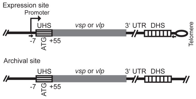
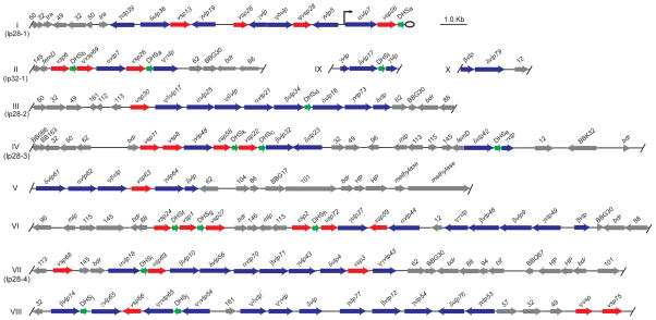
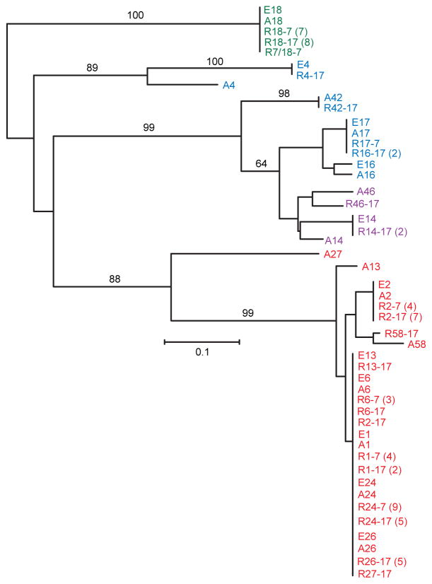
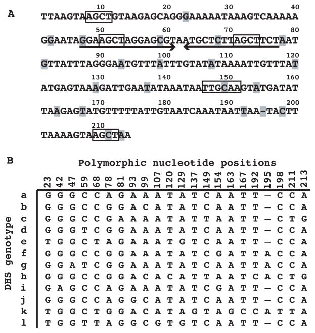
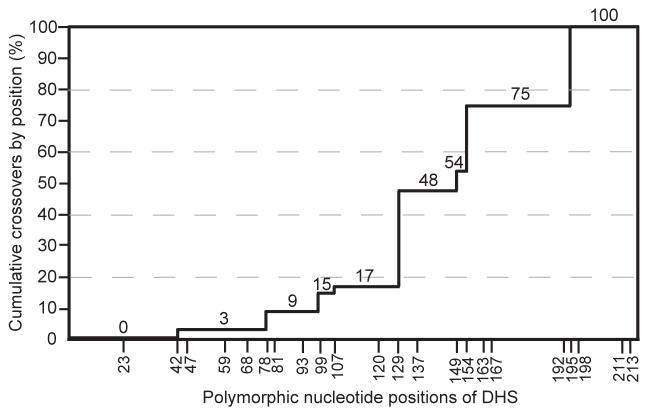
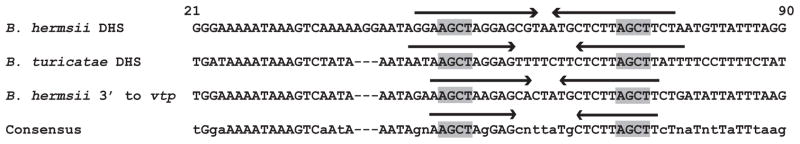
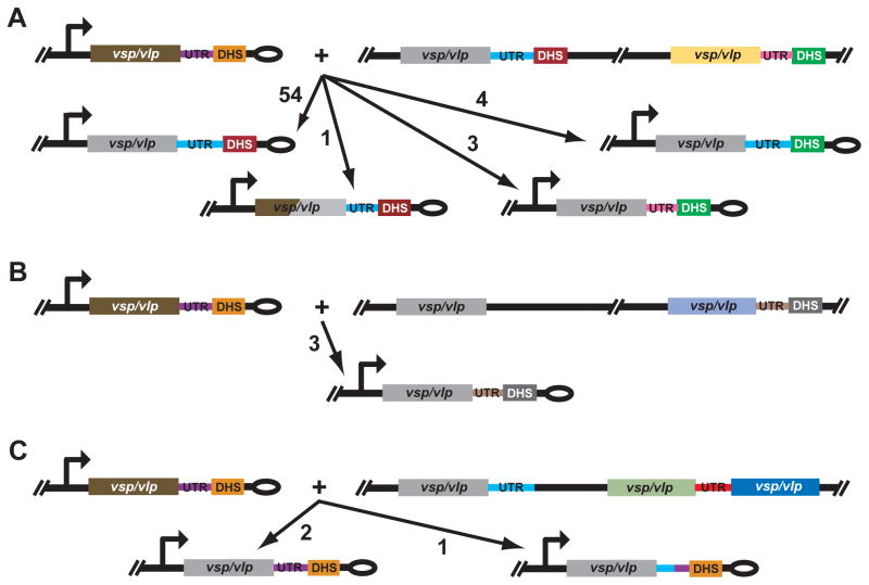
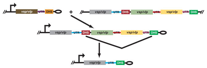
The relapsing fever agent Borrelia hermsii undergoes multiphasic antigenic variation through gene conversion of a unique expression site on a linear plasmid by an archived variable antigen gene. To further characterize this mechanism we assessed the repertoire and organization of archived variable antigen genes by sequencing approximately 85% of plasmids bearing these genes. Most archived genes shared with the expressed gene a <or= 62 nucleotide (nt) region, the upstream homology sequence (UHS), that surrounded the start codon. The 59 archived variable antigen genes were arrayed in clusters with 13 repetitive, 214 nt long downstream homology sequence (DHS) elements distributed among them. A fourteenth DHS element was downstream of the expression locus. Informative nucleotide polymorphisms in UHS regions and DHS elements were applied to the analysis of the expression site of relapse serotypes from 60 infected mice in a prospective study. For most recombinations, the upstream crossover occurred in the UHS's second half, and the downstream crossover was in the DHS's second half. Usually the closest archival DHS element was used, but occasionally a more distant DHS was employed. The downstream extragenic crossover site in B. hermsii contrasts with the upstream [corrected] extragenic crossover site for antigenic variation in African trypanosomes.


Figures

Figure 1
Figure 1
Schematic representation of expression and archival sites for vsp and vlp genes of Borrelia hermsii. The drawing is not to scale. The locations of the UHS regions that surround the start codon (ATG), the 3′ untranslated region (UTR), and DHS elements are given. By the numbering system, +1 is the transcriptional start position at the expression site. The UHS regions at the archival sites varied in length to the extent that they were >90% identical to the UHS at the expression site. The expression site is adjacent to a hair-pin telomere, indicated by the loop. The small arrows give the location of PCR primers (see text) for amplification of the expression site but not the silent site vsp or vlp gene.
Figure 2
Figure 2
Physical maps of 10 (I – X) fragments of B. hermsii linear plasmids that contain vsp genes (red arrows), vlp genes (blue arrows), and/or DHS elements (green arrows). Also shown are the locations of other open reading frames (ORF), which are indicated by gene names (e.g. femD and bdr) or by Borrelia burgdorferi gene names (e.g. BBG30) or paralogous family numbers (e.g. 50) (Casjens et al., 2000; Fraser et al., 1997). When an ORF had no discernible homology with a protein in the GenBank database, it was designated a hypothetical protein (HP). The arrowheads indicate either the direction of transcription in the case of vsp and vlp genes and other ORFs or the orientation with respect to the expression site for the DHS elements. The start and stop positions for each open reading frame or sequence element are given in Table S1 of supplementary materials. The vsp and vlp genes are further distinguished by the number of the serotype they specify (e.g. vsp6) and, in the case of vlp genes, by appending their membership in vlp sub-families α, β, γ, and δ. Pseudo genes are indicated by Ψ, and truncated or otherwise incomplete vsp or vlp sequences were not assigned a serotype number. The 12 different genotypes of the near-identical DHS elements are indicated by a letter (a – l) subscript (see Figure 5). Some fragments were identified with linear plasmids of known sizes: lp28-1, lp28-2, lp28-3, lp28-4, and lp32-1. The expression site in this example of serotype 7 is adjacent the right telomere of plasmid lp28-1; the expression site promoter is indicated by the raised arrow. Serotypes 7 and 21 are exceptional in having a silent vsp or vlp downstream of the active vlp at the expression site (Kitten and Barbour, 1990); in other serotypes only a single vsp or vlp gene is between the promoter and the sub-telomeric DHS (Restrepo et al., 1992).
Figure 3
Figure 3
UHS regions for 28 archival vsp and vlp genes of B. hermsii. Numbering of the positions is according to the transcriptional start position (+1) at the expression site; also shown are the start codon (Met) and presumed ribosomal binding sequence (RBS). Differences between the 4 sequence variants at positions +22 and 23 are shown; the counts of each variant are indicated on the right. The identification of a given archival vsp or vlp with a UHS sequence variant is given in Table S1 of supplementary materials. The 6-mer and 4-mer palindromes in the UHS are highlighted by gray. Shown below the sequence are expected (Exp) and observed (Obs) cross-over points with respect to position +24 for 28 relapses involving an infection with serotype 17, which has a GA for the expression site UHS, and relapse to a serotype whose archival vsp or vlp has another UHS sequence variant (see text). The goodness-of-fit analysis of observed and expected results with Chi square value and 2-tailed p value for 1 degree of freedom is shown. The relapse isolates for this analysis are indicated by italicized names in Table S2.
Figure 4
Figure 4
Neighbor-joining distance phylogram of aligned 3′ UTR sequences of expressed and archived vsp and vlp genes of B. hermsii. The sequences are given in Table S4. The sequences from expressed genes are distinguished between those serotypes (E) previously characterized (Burman et al., 1990; Restrepo et al., 1992) and those of relapses (R) from the current study. 3′ UTR sequences for archival genes are designated by “A”; in cases without an adjacent DHS element a maximum of 117 nucleotides was included in the alignment. The first number in the sequence name after E, R, or A indicates the serotype and, for relapses, the second number indicates the infecting serotype. Sequences with ≤ 2 nucleotide differences were grouped together; the numbers in each group of two or more are given in parentheses. The text color indicates the vsp or vlp family: red, vsp; green, α-vlp; purple, β-vlp; and blue, δ-vlp. There were no instances of a γ-vlp among the relapses. The black numbers along the branches indicate the % support from 500 bootstraps, if greater than 60%. The size marker (0.1) for the branch lengths represents nucleotide distance.
Figure 5
Figure 5. DHS elements of B. hermsii
A. The nucleotide sequence of DHSa with positions marked on the top and polymorphic positions indicated by highlighting. A large inverted repeat element is indicated by arrows, and 4-mer and 6-mer palindromes are boxed. B. Alignment of nucleotides at 22 polymorphic positions for genotypes a through l.
Figure 6
Figure 6
Percent cumulative cross-overs by polymorphic position (x-axis) of the DHS elements for 65 relapse isolates. The percent values are indicated at each level as well as on the y-axis. The sequences are given in Table S3.
Figure 7
Figure 7
Alignment of partial nucleotide sequences of DHSa of B. hermsii, DHS-like sequence in B. turicatae, and 3′ flanking region of vtp (formerly vsp33) gene of B. hermsii. Accession numbers are provided in the text. The consensus sequence is shown at the bottom. The positions are numbered according to the B. hermsii DHSa element. Long inverted repeats are indicated by arrows, and 4-mer palindromes are highlighted with gray.
Figure 8
Figure 8
Schematic representations of recombination outcomes for 68 relapse isolates of B. hermsii. The drawing is not to scale. The infecting serotype’s expressed vsp or vlp is shown as brown in panel A, B, and C; the direction of transcription is indicated by the arrow. The hair-pin telomere of the expression plasmid is denoted by an ellipse. The archival vsp or vlp gene that is the donor for each recombination is shown as gray for each recombination. Other vsp/vlp genes, their accompanying 3′ UTR sequences, and different DHS elements in the figure as denoted by other colors. The 3 panels differ in the characteristics of the archival site for the recombination. In panel A there is a DHS element adjacent to the 3′ untranslated region (UTR) for the donor archival gene and a more distant DHS downstream of another vsp or vlp and its 3′ UTR. In panel B the donor archival gene is at a distance from nearest DHS. In panel C there was not a downstream DHS element on the sequence fragment. In each panel the numbers next to the arrows with long arrowheads indicate the frequencies of each type of recombination event.
Figure 9
Figure 9
Proposed model of recombination events when a distal instead of the most proximal DHS is used. See legend for Figure 8 for description of schematic features. The model shows a two-step process in which the intervening sequence between two DHS elements in the intermediate form is deleted, thus yielding a new expressed gene near the telomere.

Comment in

Similar articles

Cited by

References

    1. Aline RF, Jr, Stuart K. Trypanosoma brucei: conserved sequence organization 3′ to telomeric variant surface glycoprotein genes. Exp Parasitol. 1989;68:57–66. - PubMed
    1. Barbour AG. Isolation and cultivation of Lyme disease spirochetes. Yale J Biol Med. 1984;57:521–525. - PMC - PubMed
    1. Barbour AG. Plasmid analysis of Borrelia burgdorferi, the Lyme disease agent. J Clin Microbiol. 1988;26:475–478. - PMC - PubMed
    1. Barbour AG. Antigenic variation by relapsing fever Borrelia species and other bacterial pathogens. In: Craig NL, Craigie R, Gellert M, Lambowitz A, editors. Mobile DNA II. Washington, D.C: ASM Press; 2002. pp. 972–994.
    1. Barbour AG, Burman N, Carter CJ, Kitten T, Bergström S. Variable antigen genes of the relapsing fever agent Borrelia hermsii are activated by promoter addition. Mol Microbiol. 1991;5:489–493. - PubMed

Publication types