. 2006 Apr;12(2):140-5.
doi: 10.1080/13550280600724319.

Dopaminergic marker proteins in the substantia nigra of human immunodeficiency virus type 1-infected brains

Affiliations

Dopaminergic marker proteins in the substantia nigra of human immunodeficiency virus type 1-infected brains

Janelle M Silvers et al. J Neurovirol. 2006 Apr.

Abstract

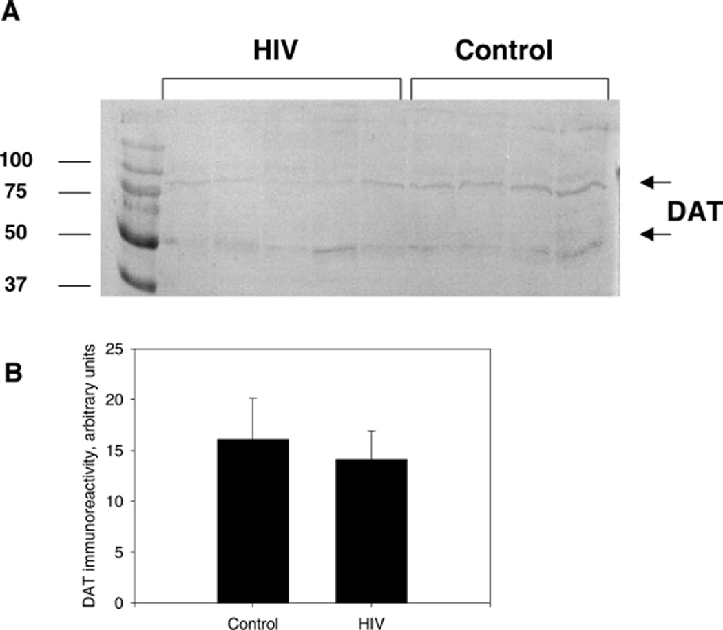
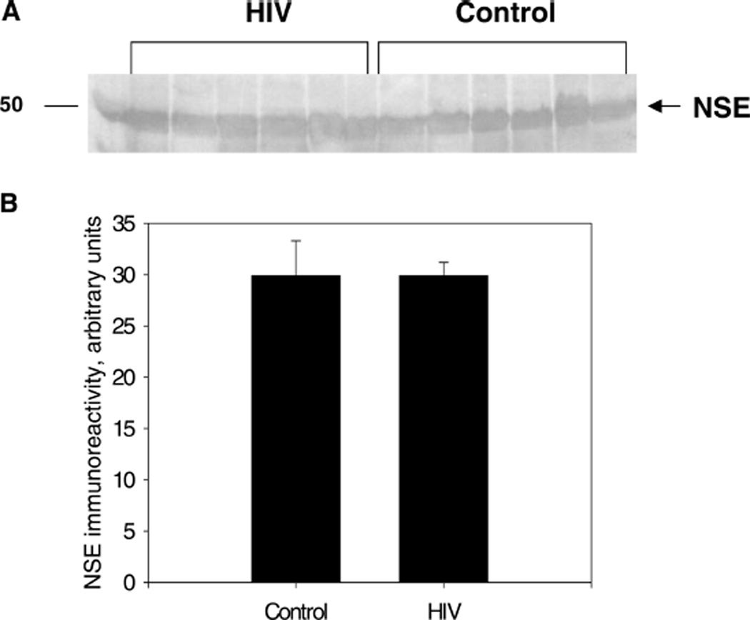
With the advent of highly active antiretroviral therapy, human immunodeficiency virus/acquired immunodeficiency syndrome (HIV/AIDS) is becoming a more chronic, manageable disease; nevertheless, the prevalence of neurological complications of AIDS is increasing. In this study, protein levels of tyrosine hydroxylase (TH) and dopamine transporter (DAT) in the substantia nigra of HIV-infected brains and -seronegative controls were determined by immunoblotting. The immunoreactivity of neuronal specific enolase (NSE) was used to assess cell loss. Although there were no changes in levels of immunoreactive DAT or NSE proteins in HIV brains, levels of immunoreactive TH were significantly reduced, relative to controls. These results suggest that decreases in TH, the rate-limiting enzyme of dopamine synthesis, may be a factor in the neurological manifestations of HIV infection.


Figures

Figure 1
Figure 1
HIV-mediated changes in availability of TH in the substantia nigra. A, Representative Western blot of HIV-1–infected brain tissues compared to seronegative controls using mouse monoclonal anti-TH antibody as the primary antibody and alkaline phosphatase–conjugated anti-mouse IgG as the secondary antibody. The molecular weight of TH is approximately 50 kDa. B, TH immunoreactivity in HIV-1–infected brain tissues compared to controls. This figure represents the slot blot data. Results presented as mean pixel density (arbitrary units) of HIV-1–infected brain tissues versus control ± SEM, n of HIV-1–infected brain tissues = 7, n of controls = 8. *HIV-1–infected brain tissues had significantly lower TH pixel density as compared to seronegative controls, P < .05.
Figure 2
Figure 2
HIV-mediated changes in availability of DAT in the substantia nigra. A, Representative Western blot of HIV-1–infected brains compared to seronegative controls using rabbit polyclonal antibody raised against amino acids 541 to 620, mapping at the C-terminus of the sodium-dependent dopamine transporter DAT of human origin as the primary antibody and alkaline phosphatase–conjugated anti-rabbit IgG as the secondary antibody. The molecular weight of DAT that is fully glycosylated is approximately 80 kDa and the bands at approximately 50 kDa are DAT with at least one less N-linked glycosylation site (see Li et al, 2004). B, DAT immunoreactivity in HIV-1–infected brain tissues compared to controls. This figure represents slot blot data. Results presented as mean pixel density (arbitrary units) of HIV-1–infected brain tissues versus control ± SEM, n of HIV-1–infected brain tissues = 7, n of controls = 8.
Figure 3
Figure 3
HIV-mediated changes in NSE density in the substantia nigra. A, Representative Western blot of HIV-1–infected brain tissues compared to seronegative controls using rabbit polyclonal anti-NSE antibody as the primary antibody and alkaline phosphatase–conjugated anti-rabbit IgG as the secondary antibody. The molecular weight of NSE is approximately 47 kDa. B, NSE immunoreactivity in HIV-1–infected brain tissues compared to controls. This figure represents slot blot data. Results presented as mean pixel density (arbitrary units) of HIV-1–infected brain tissues versus control ± SEM, n of HIV-1–infected brain tissues = 7, n of controls = 8.

References

    1. Adamson DC, Dawson TM, Zink MC, Clements JE, Dawson VL. Neurovirulent simian immunodeficiency virus infection induces neuronal, endothelial, and glial apoptosis. Mol Med. 1996;2:417–428. - PMC - PubMed
    1. Aksenov MY, Hasselrot U, Wu G, Nath A, Anderson C, Mactutus CF, Booze RM. Temporal relationships between HIV-1 Tat-induced neuronal degeneration, OX-42 immunoreactivity, reactive astrocytosis, and protein oxidation in the rat striatum. Brain Res. 2003;987:1–9. - PubMed
    1. Arendt G. Neurological manifestations of HIV-infection in the era of highly active antiretroviral therapy (HAART) Fortschr Neurol Psychiatr. 2005;73:577–586. - PubMed
    1. Berger JR, Arendt G. HIV dementia: the role of the basal ganglia and dopaminergic systems. Psychopharmacol. 2000;14:214–221. - PubMed
    1. Blanchard-Fillion B, Souza JM, Friel T, Jiang GC, Vrana K, Sharov V, et al. Nitration and inactivation of tyrosine hydroxylase by peroxynitrite. J Biol Chem. 2001;279:46017–46023. - PubMed

Publication types

Substances

LinkOut - more resources