. 2006 Jul 5;103(27):10397-10402.
doi: 10.1073/pnas.0604008103. Epub 2006 Jun 23.

TNF-alpha inhibits asbestos-induced cytotoxicity via a NF-kappaB-dependent pathway, a possible mechanism for asbestos-induced oncogenesis

Affiliations

TNF-alpha inhibits asbestos-induced cytotoxicity via a NF-kappaB-dependent pathway, a possible mechanism for asbestos-induced oncogenesis

Haining Yang et al. Proc Natl Acad Sci U S A. .

Abstract

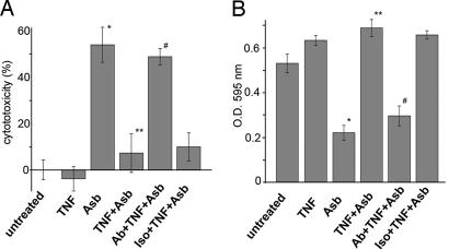
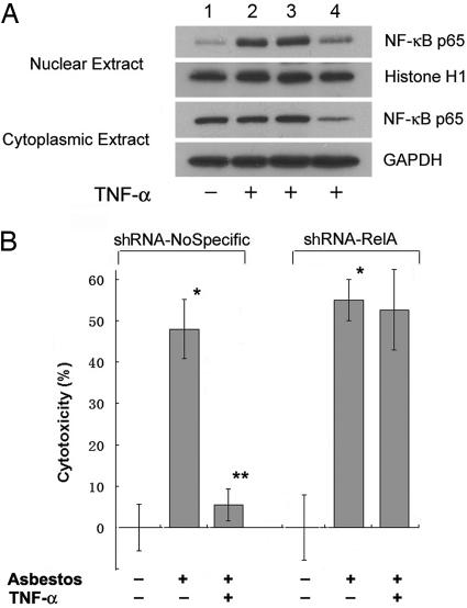
Asbestos is the main cause of human malignant mesothelioma (MM). In vivo, macrophages phagocytize asbestos and, in response, release TNF-alpha and other cytokines that contribute to carcinogenesis through unknown mechanisms. In vitro, asbestos does not induce transformation of primary human mesothelial cells (HM); instead, asbestos is very cytotoxic to HM, causing extensive cell death. This finding raised an apparent paradox: How can asbestos cause MM if HM exposed to asbestos die? We found that asbestos induced the secretion of TNF-alpha and the expression of TNF-alpha receptor I in HM. Treatment of HM with TNF-alpha significantly reduced asbestos cytotoxicity. Through numerous technical approaches, including chemical inhibitors and small interfering RNA strategies, we demonstrate that, in HM, TNF-alpha activates NF-kappaB and that NF-kappaB activation leads to HM survival and resistance to the cytotoxic effects of asbestos. Our data show a critical role for TNF-alpha and NF-kappaB signaling in mediating HM responses to asbestos. TNF-alpha signaling through NF-kappaB-dependent mechanisms increases the percent of HM that survives asbestos exposure, thus increasing the pool of asbestos-damaged HM that are susceptible to malignant transformation. Cytogenetics supported this hypothesis, showing only rare, aberrant metaphases in HM exposed to asbestos and an increased mitotic rate with fewer irregular metaphases in HM exposed to both TNF-alpha and asbestos. Our findings provide a mechanistic rationale for the paradoxical inability of asbestos to transform HM in vitro, elucidate and underscore the role of TNF-alpha in asbestos pathogenesis in humans, and identify potential molecular targets for anti-MM prevention and therapy.


Conflict of interest statement

Conflict of interest statement: No conflicts declared.

Figures

Fig. 1.
Fig. 1.
Asbestos is cytotoxic to HM in tissue culture. HM were exposed to different doses of asbestos (0.2–5.0 μg/cm2) for 24 h. (A) LDH assay. Cytotoxicity was calculated by measuring the amount of LDH released from the cytosol of asbestos-damaged cells into the supernatant. The results show that the higher the amount of asbestos in tissue culture, the higher the cytotoxicity. (B) MTT assay. The OD value of 595 nm, which reflects metabolically active cells, decreased proportionally when cells were exposed to increasing amounts of asbestos. (C) Flow cytometry assay. Cells that were negative for both annexin V and propidium iodide were considered viable. Increasing amounts of asbestos correlated with a higher percent of cell death. Columns represent the means of three separate experiments; error bars indicate SD. ∗, Significantly different compared with cells not exposed to asbestos (P < 0.05).
Fig. 2.
Fig. 2.
TNF-α and TNF-α receptor TNF-R1 are induced in HM following asbestos exposure. (A) Quantitative real-time PCR. HM were exposed to asbestos at 5.0 μg/cm2 for 6, 12, or 24 h. The levels of TNF-α mRNA expression in HM was induced ≈3.5, 4.8, or 7.2-fold, respectively, after asbestos exposure. (B) Western blot analysis. Cell culture mediums of HM were collected and concentrated by ultrafiltration. The amount of TNF-α in the medium increased significantly 48 h after asbestos exposure. (C) Western blot analysis. HM were exposed to increasing amounts of crocidolite asbestos (0.2–10.0 μg/cm2) for 24 h. Total cell extracts (40 μg) were analyzed using an anti-TNF-R1 monoclonal antibody. GAPDH (glyceraldehydes-3-phosphate dehydrogenase) was used as a loading control.
Fig. 3.
Fig. 3.
TNF-α significantly reduces asbestos cytotoxicity. HM were incubated with or without 0.5 μg/ml anti-TNF-α antiserum or normal rabbit IgG isotype control for 1 h followed by addition of TNF-α (10 ng/ml) for 24 h. Crocidolite at 5.0 μg/cm2 was added at this time point, and cell cytotoxicity was measured 24 h later. (A) LDH cytotoxicity assay. Pretreatment with TNF-α significantly reduced asbestos cytotoxicity. Addition of anti-TNF-α antiserum abolished the anticytotoxicity effect caused by TNF-α. (B) MTT assay. An OD value of 595 nm, which reflects metabolically active cells, significantly increased when HM were pretreated with TNF-α compared with HM exposed to asbestos only. TNF, TNF-α; Asb, asbestos; Ab, anti-human TNF-α antiserum; Iso, rabbit purified IgG Isotype control. Columns represent the means of three separate experiments, and bars indicate SD. ∗, Significantly different compared with HM not exposed to asbestos (P < 0.05); ∗∗, significantly different compared with HM exposed to asbestos without TNF-α pretreatment (P < 0.05); #, significantly different compared with HM exposed to TNF-α and asbestos without addition of the anti-TNF-α antiserum (P < 0.05).
Fig. 4.
Fig. 4.
TNF-α induces the activation of NF-κB. (A) TNF-α induces nuclear translocation of the p65 subunit of NF-κB in HM. Representative Western blot. Nuclear extracts (20 μg) from HM treated with TNF-α (10 ng/ml) for 0.5, 1, 2, 4, or 24 h were analyzed by Western blot using an antibody specific for the p65 subunit of NF-κB. Histone H1 was used as a loading control. (B) Representative EMSA shows that NF-κB is activated by TNF-α. An NF-κB consensus oligonucleotide was used as a probe (Materials and Methods). Five micrograms of nuclear extracts from HM treated with TNF-α (10 ng/ml) for 0.5, 1, or 2 h were used. Ctrl, control.
Fig. 5.
Fig. 5.
Inhibition of the NF-κB pathway by Bay11-7082 suppresses the anticytotoxicity effect induced by TNF-α. HM were incubated with or without 5 μM Bay11-7082 or 0.5 μg/ml anti-TNF-α antiserum for 1 h and treated with TNF-α (10 ng/ml) for 24 h. Crocidolite at 5.0 μg/cm2 was added, and cell cytotoxicity was measured 24 h later by the LDH assay. TNF, TNF-α; Asb, asbestos; Ab, anti-human TNF-α antiserum; Iso, rabbit purified Ig isotype control. Columns represent the means of three separate experiments, and bars indicate SD. ∗, Significantly different compared with HM not exposed to crocidolite (P < 0.05); ∗∗, significantly different compared with HM exposed to asbestos without TNF-α pretreatment (P < 0.05); #, significantly different compared with HM exposed to asbestos and TNF-α without the addition of anti-TNF-α antiserum or Bay 11-7082 (P < 0.05).
Fig. 6.
Fig. 6.
RNA interference assays confirm that TNF-α inhibits asbestos induced cytotoxicity by means of a NF-κB dependent mechanism. (A) Representative Western blot. The expression of NF-κB p65 in shRNA–RelA-transfected cells is markedly reduced compared with controls. Lanes: 1, uninfected HM; 2, uninfected HM treated with TNF-α; 3, HM infected with shRNA nonspecific virus and treated with TNF-α; 4, HM infected with shRNA–RelA and treated with TNF-α. Histone H1 (nuclear extract) and GAPDH (cytoplasmic extract) were used as loading controls. (B) shRNA–RelA-transfected cells and control cells were treated with TNF-α (10 ng/ml) for 24 h before exposure to asbestos (5.0 μg/cm2) for 24 h. Cell cytotoxicity was evaluated by LDH assay. The anticytotoxicity effect of TNF-α is abrogated in the shRNA–RelA-transfected cells.
Fig. 7.
Fig. 7.
Metaphases from asbestos-treated HM cell cultures. (A) Representative metaphase from an asbestos-treated HM culture. Some chromosomes have prematurely separated chromatids, and two such chromosomes are indicated by arrows. The arrowhead points to an asbestos fiber. Asbestos fibers were found in most metaphases from both HM plus asbestos cultures and in HM plus asbestos plus TNF-α cultures. (B) Tetraploid metaphase without premature separation of chromatids from asbestos-treated HM culture pretreated with TNF-α.

Similar articles

Cited by

References

    1. Mossman B. T., Bignon J., Corn M., Seaton A., Gee J. B. Science. 1990;247:294–301. - PubMed
    1. Upadhyay D., Kamp D. W. Exp. Biol. Med. (Maywood, NJ) 2003;228:650–659. - PubMed
    1. Carbone M., Kratzke R. A., Testa J. R. Semin. Oncol. 2002;29:2–17. - PubMed
    1. Pass H. I., Hahn S. M., Vogelzang N. J., Carbone M. In: Cancer Principles & Practice of Oncology. 7th Ed. Pass H. I., Vogelzang N., Carbone M., editors. Philadelphia: Lippincott Williams & Wilkins; 2005. pp. 1687–1715.
    1. Hesterberg T. W., Butterick C. J., Oshimura M., Brody A. R., Barrett J. C. Cancer Res. 1986;46:5795–5802. - PubMed

Publication types

MeSH terms