. 2006 Jul 5;103(27):10265-10270.
doi: 10.1073/pnas.0603369103. Epub 2006 Jun 23.

A burst of auxilin recruitment determines the onset of clathrin-coated vesicle uncoating

Affiliations

A burst of auxilin recruitment determines the onset of clathrin-coated vesicle uncoating

Ramiro H Massol et al. Proc Natl Acad Sci U S A. .

Abstract

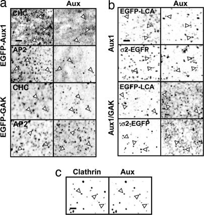
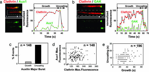
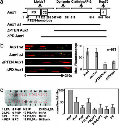
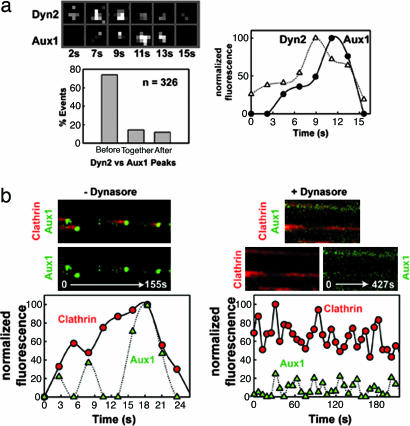
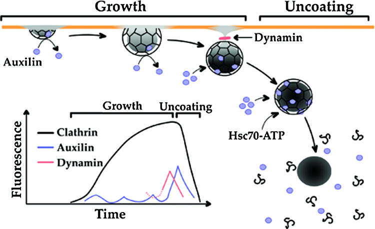
Clathrin-coated pits assemble on a membrane and pinch off as coated vesicles. The released vesicles then rapidly lose their clathrin coats in a process mediated by the ATPase Hsc70, recruited by auxilin, a J-domain-containing cofactor. How is the uncoating process regulated? We find that during coat assembly small and variable amounts of auxilin are recruited transiently but that a much larger burst of association occurs after the peak of dynamin signal, during the transition between membrane constriction and vesicle budding. We show that the auxilin burst depends on domains of the protein likely to interact with lipid head groups. We conclude that the timing of auxilin recruitment determines the onset of uncoating. We propose that, when a diffusion barrier is established at the constricting neck of a fully formed coated pit and immediately after vesicle budding, accumulation of a specific lipid can recruit sufficient auxilin molecules to trigger uncoating.


Conflict of interest statement

Conflict of interest statement: No conflicts declared.

Figures

Fig. 1.
Fig. 1.
Auxilins are present in clathrin-coated structures. Shown are confocal sections of selected cells (a and b) or of a clathrin-coated vesicle sample (c). Arrows point to examples of colocalization between auxilins and the indicated markers. (a) U373mg astrocytes stably expressing EGFP-Aux1 or EGFP-GAK were stained with antibodies specific for clathrin heavy chain or β1/β2-subunits of AP-1 and AP-2 complexes. EGFP-tagged Aux1 and GAK also gave a strong, diffuse signal, probably originating from excess auxilin at the plasma membrane and/or in the cytosol. (b) U373mg astrocytes (top two rows) and BSC1 cells (bottom two rows) stably expressing EGFP-LCA or σ2-EGFP were stained with antibodies specific for Aux1 or Aux1/GAK. (c) Calf brain clathrin-coated vesicles were stained with antibodies specific for clathrin heavy chain and Aux1/GAK. (Scale bars: 2 μm.)
Fig. 2.
Fig. 2.
Aux1 or GAK are synchronously recruited at a late stage during coated vesicle formation. (a) Confocal time series acquired every ≈2 s from U373mg astrocytes stably expressing EGFP-Aux1 together with tomato-LCa (expressed transiently for 24 h); examples of clathrin-coated structures are shown in the kymograph view (Left); the fluorescence intensity plot (Right) shows that Aux1 is recruited transiently to the clathrin spot in small and variable amounts during the growth phase and in a significantly larger burst at the onset of uncoating. (b) As in a, but cells transiently coexpressing EGFP-GAK and tomato-LCa (16 h). (c) Bar plot showing that the most prominent Aux1 burst occurs during the uncoating phase. Similar results were obtained with all other cells expressing Aux1 or GAK (data not shown). (d) Scatter plot of the maximum fluorescence intensities of Aux1 and clathrin recruited to a given spot shows no correlation (r = 0.08) of the number of auxilin molecules recruited with the size of the coated vesicle. (e) Scatter plot shows no correlation (r = 0.2) between the durations of the uncoating and growth phases of a coated vesicle (n = 196) in cells expressing EGFP-Aux1.
Fig. 3.
Fig. 3.
The PTEN homology region of Aux1 is required for the major recruitment to clathrin-coated structures. (a) Domain organization of WT and mutant forms of Aux1 used in our experiments. The domain organization of Aux1 and GAK are similar, with an additional Ser/Thr kinase located at the N terminus of GAK (data not shown). (b) Confocal time series acquired every 2 s from U373mg astrocytes stably expressing WT EGFP-Aux1 or the corresponding auxilin deletion mutants together with tomato-LCa (all expressed transiently for 24 h). Selected examples are presented as equally normalized kymographs (Left). The maximum amount of WT and truncated auxilin recruited to a given clathrin coat is shown. (c) Aux1 interacts with specific phosphoinositides in vitro. Shown is a representative chemiluminescence image obtained from the lipid–protein overlay assay using Aux1 purified from insect cells. The strip contained the following lipids: lysophosphatidic acid (LPA), lysophosphocholine (LPC), PI, PI (3)-phosphate (PI3P), PI (4)-phosphate (PI4P), PI (5)phosphate (PI5P), phosphatidylethanolamine (PE), phosphatidylcholine (PC), sphingosine-1-phosphate (S1P), PI (3, 4)-biphosphate (PI3,4P2), PI (3, 5)-biphosphate (PI3,5P2), PI (4, 5)-biphosphate (PI4,5P2), PI (3, 4, 5)-triphosphate (PI3,4,5P3), phosphatidic acid (PA), and phosphatidylserine (PS). The position of the spots (red circles) is shown. The bar plot shows the binding (average ± SE) of Aux1 determined in nine independent experiments (five using Aux1 purified from four bacterial preparations and four from two insect cell preparations).
Fig. 4.
Fig. 4.
Aux1 recruitment follows dynamin peak and requires normal dynamin function. (a) Selected frames from a time series acquired every 2 s of a small area of U373mg astrocytes expressing EGFP-Aux1 stably and Dyn2-mRFP transiently (24 h). Normalized fluorescence intensity as a function of time of the selected coated pit is shown. A bar plot shows the distribution of occurrences of Aux1 major bursts in relation to the time of appearance of Dyn2 peaks (n = 326). These data were obtained by first tracking the auxilin bursts, then extending the mask back in time for an additional five time frames (≈10 s). Finally, the relative timing corresponding to the appearance of the maximum fluorescence signals for Aux1 and Dyn2 was determined automatically. Accuracy of the method was verified manually on a subset of the observations (see additional examples in Fig. 12b). (b) Dynasore, a chemical inhibitor of dynamin, prevents the appearance of the auxilin burst. U373mg astrocytes stably expressing EGFP-Aux1 (green) and transiently (24 h) expressing tomato-LCa (red) were incubated with medium containing 0.5% DMSO or 80 μM dynasore/0.5% DMSO at 37°C for 5 min. After this treatment, confocal time series were recorded every 2 s (− Dynasore) or 6 s (+ Dynasore), and examples are displayed as kymograph views (Top and Middle) and as plots of normalized fluorescence intensities as a function of time (Bottom).
Fig. 5.
Fig. 5.
Model for the recruitment of auxilin. The principal stages leading to the auxilin burst and its relation to the onset of clathrin coat disassembly are shown. The plasma membrane is represented by the orange line; the membrane containing the lipid signal used to recruit auxilin is shown in gray. During the growth phase, cargo, clathrin, adaptors, and other coat components continuously accumulate. Continued growth results in deep membrane invaginations, until membrane constriction and fission coordinated by a final burst of dynamin occur. After budding, the coated vesicle moves away from the membrane, and uncoating rapidly ensues. Small and variable amounts of auxilin associate to and dissociate from growing pits, while a large burst of auxilin recruitment starts during membrane constriction and ends with the onset of uncoating. At this stage, sufficient amounts of ATP-bound Hsc70 are captured by auxilin to drive coat disassembly. In this model, a lipid species (indicated by the gray shadowing of the membrane), generated within the coated membrane and perhaps recognized by the PTEN-like domain of auxilin, acts as a recruiting signal for auxilin. Before vesicle budding, the underlying membrane remains connected to the plasma membrane, and any such lipid will rapidly diffuse into the surrounding membrane. It will therefore fail to reach the concentration threshold needed to capture enough auxilin to ensue coat disassembly.

Similar articles

Cited by

References

    1. Ehrlich M., Boll W., Van Oijen A., Hariharan R., Chandran K., Nibert M. L., Kirchhausen T. Cell. 2004;118:591–605. - PubMed
    1. Lemmon S. K. Curr. Biol. 2001;11:R49–R52. - PubMed
    1. Jiang J., Prasad K., Lafer E. M., Sousa R. Mol. Cell. 2005;20:513–524. - PMC - PubMed
    1. Greener T., Zhao X., Nojima H., Eisenberg E., Greene L. E. J. Biol. Chem. 2000;275:1365–1370. - PubMed
    1. Jiang J. W., Taylor A. B., Prasad K., Ishikawa-Brush Y., Hart P. J., Lafer E. M., Sousa R. Biochemistry. 2003;42:5748–5753. - PubMed

Publication types