. 2006 Aug;141(4):1533-43.
doi: 10.1104/pp.106.082198. Epub 2006 Jun 23.

Cytosolic triacylglycerol biosynthetic pathway in oilseeds. Molecular cloning and expression of peanut cytosolic diacylglycerol acyltransferase

Affiliations

Cytosolic triacylglycerol biosynthetic pathway in oilseeds. Molecular cloning and expression of peanut cytosolic diacylglycerol acyltransferase

Saikat Saha et al. Plant Physiol. 2006 Aug.

Abstract

Triacylglycerols (TAGs) are the most important storage form of energy for eukaryotic cells. TAG biosynthetic activity was identified in the cytosolic fraction of developing peanut (Arachis hypogaea) cotyledons. This activity was NaF insensitive and acyl-coenzyme A (CoA) dependent. Acyl-CoA:diacylglycerol acyltransferase (DGAT) catalyzes the final step in TAG biosynthesis that acylates diacylglycerol to TAG. Soluble DGAT was identified from immature peanuts and purified by conventional column chromatographic procedures. The enzyme has a molecular mass of 41 +/- 1.0 kD. Based on the partial peptide sequence, a degenerate probe was used to obtain the full-length cDNA. The isolated gene shared less than 10% identity with the previously identified DGAT1 and 2 families, but has 13% identity with the bacterial bifunctional wax ester/DGAT. To differentiate the unrelated families, we designate the peanut gene as AhDGAT. Expression of peanut cDNA in Escherichia coli resulted in the formation of labeled TAG and wax ester from [14C]acetate. The recombinant E. coli showed high levels of DGAT activity but no wax ester synthase activity. TAGs were localized in transformed cells with Nile blue A and oil red O staining. The recombinant and native DGAT was specific for 1,2-diacylglycerol and did not utilize hexadecanol, glycerol-3-phosphate, monoacylglycerol, lysophosphatidic acid, and lysophosphatidylcholine. Oleoyl-CoA was the preferred acyl donor as compared to palmitoyl- and stearoyl-CoAs. These data suggest that the cytosol is one of the sites for TAG biosynthesis in oilseeds. The identified pathway may present opportunities of bioengineering oil-yielding plants for increased oil production.


Figures
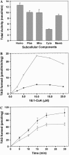
Figure 1.
Figure 1.
Generation of TAG. A, Fresh developing peanut cotyledons (50 g) were used for obtaining various subcellular fractions by differential centrifugation. TAG formation was monitored using [14C]oleoyl-CoA in the absence of exogenously added acyl acceptors. Values are mean ± sd of three independent determinations. Homo, Homogenate; Plas, plastidal fraction; Mito, mitochondrial fraction; Cyto, cytosolic fraction; Memb, membrane fraction. B, Acylation of [14C]oleoyl-CoA into TAG was carried out for 15 min at 30°C in the presence of 50 μm 1,2-DAG in cytosolic (28 to 36 μg; •) and in membrane (40 to 45 μg; ○) fractions of developing peanut cotyledons. Each point is the average of two independent experiments. C, Time-dependent formation of TAG. Incorporation of [14C]oleoyl-CoA was performed with cytosolic fraction of developing peanut cotyledon into TAG in the absence of added acyl acceptors. The formation of TAG was monitored either in the presence (○) or in the absence (•) of 20 mm NaF. Each point is mean ± sd of four independent determinations.
Figure 2.
Figure 2.
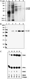
Purification of cytosolic DGAT from developing peanut cotyledons. A, SDS-PAGE protein profile. Samples from each purification step were separated on a 12% (w/v) SDS-PAGE and stained with silver nitrate. Lane 1 is standard molecular mass marker, and lanes 2 to 6 correspond to the pooled active fractions from steps 1 to 5 (Table I). B, Western-blot analysis. Samples from each step of purification were separated by 12% (w/v) SDS-PAGE and electroblotted onto a nitrocellulose membrane, followed by probing with anti-DGAT antibodies from R. glutinis. Lane 1, Prestained protein molecular mass marker. Lanes 2 to 6 correspond to the pooled active fractions from steps 1 to 5 (Table I). C, Autoradiography of the reaction products formed at each step of the purification. DGAT assays were performed using [14C]oleoyl-CoA and DAG for 15 min. The products formed were separated on a TLC using petroleum ether:diethyl ether:acetic acid (70:30:1, v/v). Lanes 1 to 5 correspond to the active fractions from steps 1 to 5 of purification Table I.
Figure 3.
Figure 3.
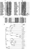
DGAT gene sequence analyses. A, Hydropathy plot of cytosolic peanut DGAT. No transmembrane domain was predicted. B, Alignment of AhDGAT and homolog genes. Sequence alignment was performed using the http://www.ch.embnet.org/software/BOX_form.html program. AhDGAT, peanut DGAT (AY875644); G. max, soybean (BM187962; nucleotide sequence was retrieved from the National Center for Biotechnology Information (NCBI) and then translated; longest reading frame was selected for alignment); At1g48300, Arabidopsis 1g48300 (AAD49767); O. sativa, rice (XP_475575). Conserved amino acids are shaded black.
Figure 4.
Figure 4.
Conserved acyltransferase domains across acyltransferase families and phylogenetic analysis. Sequence alignment of peanut DGAT (AY875644) and other known acyltransferase family members was performed using the http://www.ch.embnet.org/software/BOX_form.html sequence alignment program. Identical amino acid residues are highlighted in black. Conserved acyltransferase domains are underlined. Catalytic site conserved residues are marked with stars. A, Alignment of the catalytic domains of members of the acyltransferase family. Sequences aligned are designated as follows: E. coli GPAT and LPEAT, EcGPAT and EcLPEAT; Mus musculus GPAT and LPAAT, MmGPAT and MmLPAAT; S. cerevisiae GPAT1, GPAT2, and LPAAT are ScGAT1, ScGAT2, and ScLPAAT; Homo sapiens LPAAT1 to 6 and DHAPT are HsLPAAT1 to 6 and HsDHAPT; Arabidopsis GPAT, AtGPAT; A. calcoaceticus wax synthase/DGAT, AcWS/DGAT; and peanut DGAT, AhDGAT. B, Similar alignment of catalytic domain of known acyltransferase with yet another domain in peanut DGAT. C, Active site of catalytic residues of known DGATs aligned with AhDGAT. Designation of the sequences aligned is as follows: M. musculus DGAT1, MmDGAT1; HARGP1; Nicotiana tabacum DGAT1, NtDGAT1; Caenorhabditis elegans DGAT1, CeDGAT1; Arabidopsis DGAT1, AtDGAT1; and rapeseed DGAT1, BnDGAT1. D, Phylogenetic tree of different DGAT family members. Cytosolic DGAT from peanut (AhDGAT; AY875644) was added to the assembly and a similarity tree was constructed. G. max, soybean (BM187962); O. sativa, rice (XP_475575); At1g48300, Arabidopsis 1g48300 (AAD49767); Rv3130c, M. tuberculosis H37Rv DGAT (NP_217646); AcDGAT, A. calcoaceticus WS/DGAT (AF529086); MmDGAT1, M. musculus DGAT1 (NP_034176); NtDGAT1, N. tabacum DGAT1 (AF129003); BnDGAT1, rapeseed DGAT1 (AF164434); AtDGAT1, Arabidopsis DGAT1 (AF051849); ScDGAT2, S. cerevisiae DGAT2 (YOR245c); HsDGAT2, H. sapiens DGAT2 (NP_115953); MrDGAT2b, M. ramanniana DGAT2b (AF391090); MrDGAT2a, M. ramanniana DGAT2a (AF391089); CeDGATa, C. elegans DGATa (CAB04533); CeDAGT2b, C. elegans DGAT2b (AAB04969); AtPDGAT, Arabidopsis PDGAT (NP_171897); and ScPDGAT, S. cerevisiae PDGAT (CAA54576). GenBank/NCBI accession numbers are given (in parentheses).
Figure 5.
Figure 5.
Induction of TAG synthesis in E. coli overexpressing AhDGAT. A, Time course of acetate labeling. Vector-transformed and recombinant E. coli cells expressing soluble peanut DGAT were labeled with [14C]acetate for various time points. The labeling experiments were repeated three times and a typical phosphor image is shown. B, Time course of recombinant DGAT activity. Enzyme activity was done using 18,000g supernatant of E. coli BL21 (DE3) cells overexpressing AhDGAT. C and D, Immunoblot analyses of the expressed AhDGAT. Induced (+IPTG) and uninduced (−IPTG) recombinant cell lysates were resolved in 12% (w/v) SDS-PAGE, transferred onto a nitrocellulose membrane, and probed with anti-His-tag (C) and peptide anti-RgDGAT (D) antibodies. E, Alignment of R. glutinis DGAT peptide sequence with deduced amino acid sequence of peanut DGAT.
Figure 6.
Figure 6.
Purification of recombinant AhDGAT. A, SDS-PAGE profile of recombinant DGAT purification. Samples from each fraction were separated by 12% (w/v) SDS-PAGE and stained with Coomassie Blue R-250. Lane 1 represents vector IPTG uninduced; lane 2, vector IPTG induced; lane 3, molecular mass marker; lane 4, AhDGAT IPTG uninduced; and lane 5, AhDGAT IPTG induced. Lanes 6 to 7 correspond to 250 mm imidazole eluted fractions from Ni-nitrilotriacetic acid agarose chromatography. B, Time course of DGAT activity using purified recombinant enzyme. Lane 1 represents no-enzyme control. Lanes 2 to 6 represent 0, 5, 10, 15, and 30 min, respectively.
Figure 7.
Figure 7.
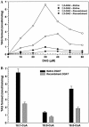
Substrate specificity of DGAT. Enzyme activity was measured under standard assay conditions using active fraction from the last step of the purification of peanut cytosolic enzyme (native) or Ni-nitrilotriacetic acid agarose chromatography-purified recombinant DGAT from E. coli cells (recombinant). Assays were performed as described in “Materials and Methods.” A, Effect of 1,2-DAG (white circles and squares) and 1,3-DAG (black circles and squares) on DGAT activity. Substrate specificity was determined with a fixed concentration of labeled oleoyl-CoA and varying concentrations of DAG. Each point is the average of two determinations. B, Acyl donor specificity of DGAT was determined using native and purified recombinant enzyme. Values are expressed as mean ± sd of three determinations.
Figure 8.
Figure 8.
Gel image of RT-PCR for AhDGAT expression. A, One microgram of total RNA from four different seed development stages based on DAF (lane 2, 8–15 DAF; lane 3, 16–24 DAF; lane 4, 25–30 DAF; lane 5, 30–35 DAF), leaf (lane 6), and root (lane 7) were used for cDNA synthesis. Transcripts shown were obtained after 30 cycles using cDNA as template. Lane 1 is negative control referred as −RT; Actin2 was used as a positive control. B, Tissue-specific expression of soluble DGAT in peanut. Lanes 1 to 4 represent 8 to 15, 16 to 24, 25 to 30, and 30 to 35 DAF, respectively. Lane 5 represents soluble fraction from peanut seedlings.

Similar articles

Cited by

References

    1. Bouvier-Nave P, Benveniste P, Oelkers P, Sturley SL, Schaller H (2000) Expression in yeast and tobacco of plant cDNAs encoding acyl CoA:diacylglycerol acyltransferase. Eur J Biochem 267: 85–96 - PubMed
    1. Broun P, Gettner S, Somerville C (1999) Genetic engineering of plant lipids. Annu Rev Nutr 19: 197–216 - PubMed
    1. Cases S, Smith SJ, Zheng Y-W, Myers HM, Lear SR, Sande E, Novak S, Collins C, Welch CB, Lusis AJ, et al (1998) Identification of a gene encoding an acyl CoA:diacylglycerol acyltransferase, a key enzyme in triacylglycerol synthesis. Proc Natl Acad Sci USA 95: 13018–13023 - PMC - PubMed
    1. Dahlqvist A, Stahl U, Lenman M, Banas A, Lee M, Sandager L, Ronne H, Stymne S (2000) Phospholipid:diacylglycerol acyltransferase: an enzyme that catalyzes the acyl-CoA-independent formation of triacylglycerol in yeast and plants. Proc Natl Acad Sci USA 97: 6487–6492 - PMC - PubMed
    1. Daniel J, Deb C, Dubey VS, Sirakova TD, Abomoelak B, Morbidoni HR, Kolattukudy PE (2004) Induction of a novel class of diacylglycerol acyltransferases and triacylglycerol accumulation in Mycobacterium tuberculosis as it goes into a dormancy-like state in culture. J Bacteriol 186: 5017–5030 - PMC - PubMed

Publication types

MeSH terms

Substances

Associated data

LinkOut - more resources