

Detection of the bcl-2 t(14;18) translocation and proto-oncogene expression in primary intraocular lymphoma
- PMID: 16799010
- PMCID: PMC1945012
- DOI: 10.1167/iovs.05-1312
Detection of the bcl-2 t(14;18) translocation and proto-oncogene expression in primary intraocular lymphoma
Abstract
Purpose: Primary intraocular lymphoma (PIOL) is a diffuse large B cell lymphoma that initially infiltrates the retina, vitreous, or optic nerve head, with or without central nervous system involvement. This study examined the expression of the bcl-2 t(14;18) translocation, the bcl-10 gene, and high expression of bcl-6 mRNA in PIOL cells.
Methods: Microdissection and PCR analysis were used to examine vitreous specimens in patients with PIOL for the presence of bcl-2 t(14;18) translocations, the bcl-10 gene, and expression of bcl-6 mRNA. A medical record review was also conducted to determine whether the bcl-2 t(14;18) translocation correlated with prognosis.
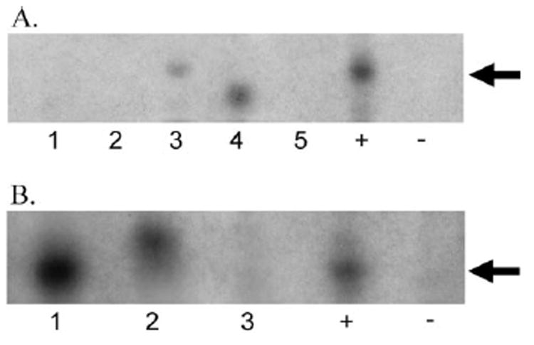
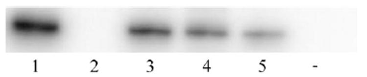
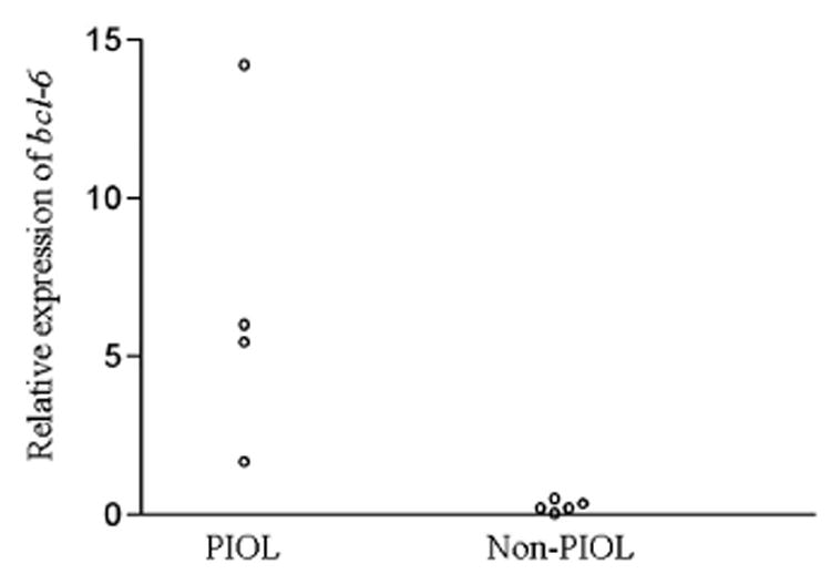
Results: Forty of 72 (55%) PIOL patients expressed the bcl-2 t(14;18) translocation at the major breakpoint region. Fifteen of 68 (22%) patients expressed the translocation at the minor cluster region. The bcl-10 gene was detected in 6 of 26 (23%) patients, whereas 4 of 4 (100%) PIOL patients expressed higher levels of bcl-6 mRNA compared with inflammatory lymphocytes. An analysis of clinical outcome in 23 PIOL patients revealed no significant association between bcl-2 t(14;18) translocations and survival or relapse. However, patients with the translocation were significantly younger.
Conclusions: PIOL has unique molecular patterns of bcl-2, bcl-10, and bcl-6 when compared with other systemic lymphomas. This study lays the foundation for future studies aimed at exploring the genotypic classification of PIOL based on the quantitative molecular framework of gene expression profiling, with the goal of providing useful adjuncts to the pathologic diagnosis of this complex disease.
Figures

References
-
- Chan CC, Buggage RR, Nussenblatt RB. Intraocular lymphoma. Curr Opin Ophthalmol. 2002;13:411–418. - PubMed
-
- Hormigo A, DeAngelis LM. Primary ocular lymphoma: clinical features, diagnosis, and treatment. Clin Lymphoma. 2003;4:22–29. - PubMed
-
- Paulus W. Classification, pathogenesis, and molecular pathology of primary CNS lymphomas. J Neurooncol. 1999;43:203–208. - PubMed
-
- Hoffman PM, McKelvie P, Hall AJ, Stawell RJ, Santamaria JD. Intraocular lymphoma: a series of 14 patients with clinicopathological features and treatment outcomes. Eye. 2003;17:513–521. - PubMed
-
- Hochberg FH, Miller DC. Primary central nervous system lymphoma. J Neurosurg. 1988;68:835–853. - PubMed
Publication types
MeSH terms
Substances
Grants and funding
LinkOut - more resources
Full Text Sources
Medical
Research Materials
