. 2006 Jul;47(7):2976-84.
doi: 10.1167/iovs.05-1346.

Accommodative lens refilling in rhesus monkeys

Affiliations

Accommodative lens refilling in rhesus monkeys

Steven A Koopmans et al. Invest Ophthalmol Vis Sci. 2006 Jul.

Erratum in

  • Invest Ophthalmol Vis Sci. 2006 Aug;47(8):3246. Vilipuru, Abhiram S [corrected to Vilupuru, Abhiram S]

Abstract

Purpose: Accommodation can be restored to presbyopic human eyes by refilling the capsular bag with a soft polymer. This study was conducted to test whether accommodation, measurable as changes in optical refraction, can be restored with a newly developed refilling polymer in a rhesus monkey model. A specific intra- and postoperative treatment protocol was used to minimize postoperative inflammation and to delay capsular opacification.

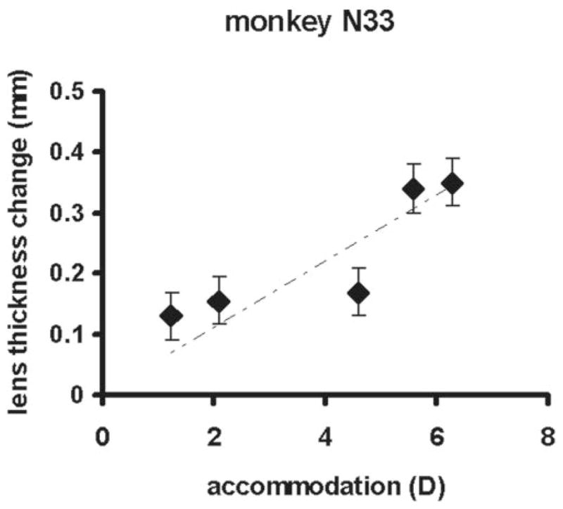
Methods: Nine adolescent rhesus monkeys underwent refilling of the lens capsular bag with a polymer. In the first four monkeys (group A) the surgical procedure was followed by two weekly subconjunctival injections of corticosteroids. In a second group of five monkeys (group B) a treatment intended to delay the development of capsular opacification was applied during the surgery, and, in the postoperative period, eye drops and two subconjunctival injections of corticosteroids were applied. Accommodation was stimulated with carbachol iontophoresis or pilocarpine and was measured with a Hartinger refractometer at regular times during a follow-up period of 37 weeks in five monkeys. In one monkey, lens thickness changes were measured with A-scan ultrasound.

Results: In group A, refraction measurement was possible in one monkey. In the three other animals in group A, postoperative inflammation and capsular opacification prevented refraction measurements. In group B, the maximum accommodative amplitude of the surgically treated eyes was 6.3 D. In three monkeys the accommodative amplitude decreased to almost 0 D after 37 weeks. In the two other monkeys, the accommodative amplitude remained stable at +/-4 D during the follow-up period. In group B, capsular opacification developed in the postoperative period, but refraction measurements could still be performed during the whole follow-up period of 37 weeks.

Conclusions: A certain level of accommodation can be restored after lens refilling in adolescent rhesus monkeys. During the follow-up period refraction measurements were possible in all five monkeys that underwent the treatment designed to prevent inflammation and capsular opacification.


Figures

Figure 1
Figure 1
Refilled monkey lens just after refilling of the capsular bag. The anterior chamber maintainer (ACM) is visible in the corneal paracentesis. The plug closing the capsulorrhexis is visible at the lens periphery.
Figure 2
Figure 2
(A) Hartinger coincidence refractometer. A charge-coupled device (CCD) camera is mounted in front of the eyepiece. The cone on the right side of the equipment is placed in front of the eye under investigation. (B, C) View through the eyepiece of a Hartinger refractometer. On the left a scale is visible that indicates the axis in degrees on which the refraction is being measured. The examiner turns a knob on the outside of the equipment until the two sets of three lines are aligned. Then the refraction can be read from the right scale. (B) Image photographed through the eyepiece of the instrument when measuring a nonsurgical eye. (C) A similar image of a surgical eye 4 weeks after surgery. The visibility of the vernier lines depends on the optical quality of the eye.
Figure 3
Figure 3
Pre- and postoperative carbachol-induced accommodation for four monkeys in group B: (A) 112, (B) 114, (C) 64, and (D) 109. The preoperative accommodative amplitude was measured 3 weeks after the total iridectomy and 3 weeks before surgery. The postoperative measurements shown are those at the follow-up time when the postoperative accommodative amplitude was at its maximum. Carbachol was applied iontophoretically to the temporal and nasal cornea for 4 seconds each (down arrows). Refraction was then measured three times at 2-minute intervals with a Hartinger coincidence refractometer. Accommodation is the difference between the present refraction and the refraction at the start of the experiment. Once the accommodation did not increase for three successive 2-minute periods, carbachol was again iontophoretically applied for a further 4 seconds (up arrows). Refraction was again measured until no further increase of accommodation occurred at three successive measurements. The preoperative accommodative amplitude in monkey 109 is unlikely to be the maximum accommodative amplitude in this monkey.
Figure 4
Figure 4
Maximum pharmacologically induced accommodative amplitude of the left (surgical) eye and of the right (nonsurgical) eye in the postoperative period in the five monkeys in group B: (A) 112, (B) 114, (C) 64, (D) 109, and (E) N33. Standard deviations of the accommodative amplitude measurements are smaller than the symbol size. In monkey N33 only the left (surgical) eye was measured.
Figure 5
Figure 5
(A–E) Pre- and postoperative baseline refraction of the left eyes of monkeys in group B as measured before pharmacological stimulation of accommodation: (A) 112, (B) 114, (C) 64, (D) 109, and (E) N33.
Figure 6
Figure 6
Lens thickness change measured with A-scan after pilocarpine stimulated accommodation at several dates during the follow-up period. A linear regression line was fitted through the data points. The slope of the regression line amounts to 0.05 mm/D. Error bars, SD.

Similar articles

Cited by

References

    1. Von Helmholtz H. Ueber die Akkommodation des Auges. Albrecht von Graefes Arch Klin Ophthalmol. 1855;1:1–74.
    1. Duane A. Studies in monocular and binocular accommodation with their clinical applications. Am J Ophthalmol. 1922;5:867–877. - PMC - PubMed
    1. Glasser A, Campbell MCW. Presbyopia and the optical changes in the human crystalline lens with age. Vision Res. 1998;38:209–229. - PubMed
    1. Soergel F, Meyer C, Eckert G, et al. Spectral analysis of viscoelasticity of the human lens. J Refract Surg. 1999;15:714–716. - PubMed
    1. Koopmans SA, Terwee T, Barkhof J, Haitjema H, Kooijman AC. Polymer refilling of presbyopic human lenses in vitro restores the ability to undergo accommodative changes. Invest Ophthalmol Vis Sci. 2003;44:250–257. - PubMed

Substances