

Sema4D/plexin-B1 activates GSK-3beta through R-Ras GAP activity, inducing growth cone collapse
- PMID: 16799460
- PMCID: PMC1500830
- DOI: 10.1038/sj.embor.7400737
Sema4D/plexin-B1 activates GSK-3beta through R-Ras GAP activity, inducing growth cone collapse
Abstract
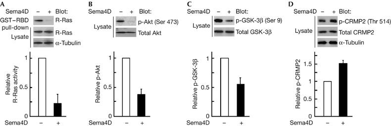
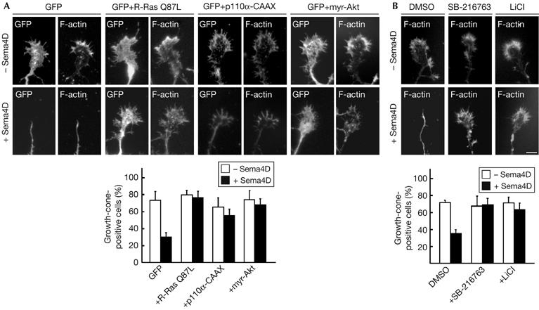
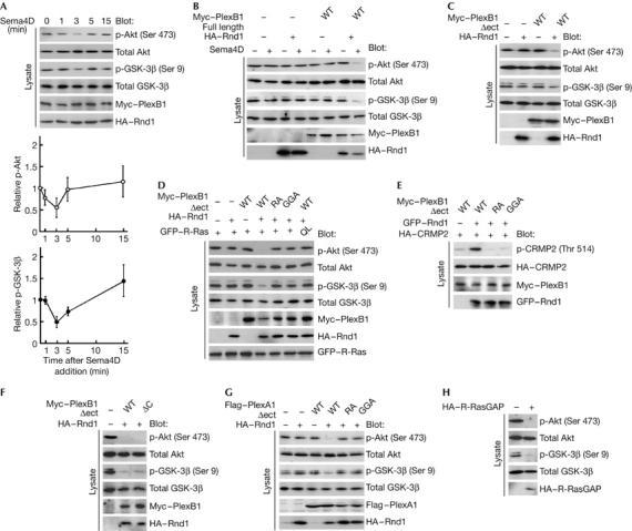
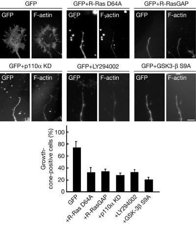
Plexins are receptors for the axonal guidance molecules known as semaphorins, and the semaphorin 4D (Sema4D) receptor plexin-B1 induces repulsive responses by functioning as an R-Ras GTPase-activating protein (GAP). Here we characterized the downstream signalling of plexin-B1-mediated R-Ras GAP activity, inducing growth cone collapse. Sema4D suppressed R-Ras activity in hippocampal neurons, in parallel with dephosphorylation of Akt and activation of glycogen synthase kinase (GSK)-3beta. Ectopic expression of the constitutively active mutant of Akt or treatment with GSK-3 inhibitors suppressed the Sema4D-induced growth cone collapse. Constitutive activation of phosphatidylinositol-3-OH kinase (PI(3)K), an upstream kinase of Akt and GSK-3beta, also blocked the growth cone collapse. The R-Ras GAP activity was necessary for plexin-B1-induced dephosphorylation of Akt and activation of GSK-3beta and was also required for phosphorylation of a downstream kinase of GSK-3beta, collapsin response mediator protein-2. Plexin-A1 also induced dephosphorylation of Akt and GSK-3beta through its R-Ras GAP activity. We conclude that plexin-B1 inactivates PI(3)K and dephosphorylates Akt and GSK-3beta through R-Ras GAP activity, inducing growth cone collapse.
Figures
Similar articles
-
Semaphorin 4D/Plexin-B1 stimulates PTEN activity through R-Ras GTPase-activating protein activity, inducing growth cone collapse in hippocampal neurons.J Biol Chem. 2010 Sep 3;285(36):28200-9. doi: 10.1074/jbc.M110.147546. Epub 2010 Jul 7. J Biol Chem. 2010. PMID: 20610402 Free PMC article.
-
Molecular dissection of the semaphorin 4D receptor plexin-B1-stimulated R-Ras GTPase-activating protein activity and neurite remodeling in hippocampal neurons.J Neurosci. 2004 Dec 15;24(50):11473-80. doi: 10.1523/JNEUROSCI.3257-04.2004. J Neurosci. 2004. PMID: 15601954 Free PMC article.
-
Semaphorin 4D/Plexin-B1-mediated R-Ras GAP activity inhibits cell migration by regulating beta(1) integrin activity.J Cell Biol. 2006 May 22;173(4):601-13. doi: 10.1083/jcb.200508204. Epub 2006 May 15. J Cell Biol. 2006. PMID: 16702230 Free PMC article.
-
R-ras as a key player for signaling pathway of plexins.Mol Neurobiol. 2005 Dec;32(3):217-22. doi: 10.1385/MN:32:3:217. Mol Neurobiol. 2005. PMID: 16385138 Review.
-
GTPases in semaphorin signaling.Adv Exp Med Biol. 2007;600:12-23. doi: 10.1007/978-0-387-70956-7_2. Adv Exp Med Biol. 2007. PMID: 17607943 Review.
Cited by
-
The role of semaphorin 4D in tumor development and angiogenesis in human breast cancer.Onco Targets Ther. 2016 Sep 26;9:5737-5750. doi: 10.2147/OTT.S114708. eCollection 2016. Onco Targets Ther. 2016. PMID: 27729799 Free PMC article.
-
Plexin-B1 signalling promotes androgen receptor translocation to the nucleus.Oncogene. 2016 Feb 25;35(8):1066-72. doi: 10.1038/onc.2015.160. Epub 2015 May 18. Oncogene. 2016. PMID: 25982277
-
Ephexin4-mediated promotion of cell migration and anoikis resistance is regulated by serine 897 phosphorylation of EphA2.FEBS Open Bio. 2013 Jan 21;3:78-82. doi: 10.1016/j.fob.2013.01.002. Print 2013. FEBS Open Bio. 2013. PMID: 23772378 Free PMC article.
-
Stimulating neuroregeneration as a therapeutic drug approach for traumatic brain injury.Br J Pharmacol. 2009 Jul;157(5):675-85. doi: 10.1111/j.1476-5381.2009.00220.x. Epub 2009 May 5. Br J Pharmacol. 2009. PMID: 19422372 Free PMC article. Review.
-
Exploring Optic Nerve Axon Regeneration.Curr Neuropharmacol. 2017;15(6):861-873. doi: 10.2174/1570159X14666161227150250. Curr Neuropharmacol. 2017. PMID: 28029073 Free PMC article. Review.
References
-
- Barberis D, Artigiani S, Casazza A, Corso S, Giordano C, Love CA, Jones EY, Comoglio PM, Tamagnone L (2004) Plexin signaling hampers integrin-based adhesion, leading to Rho-kinase independent cell rounding, and inhibiting lamellipodia extension and cell motility. FASEB J 18: 592–594 - PubMed
-
- Brown M, Jacobs T, Eickholt B, Ferrari G, Teo M, Monfries C, Qi RZ, Leung T, Lim L, Hall C (2004) α2-Chimaerin, cyclin-dependent kinase 5/p35, and its target collapsin response mediator protein-2 are essential components in semaphorin 3A-induced growth-cone collapse. J Neurosci 24: 8994–9004 - PMC - PubMed
-
- Cantley LC (2002) The phosphoinositide 3-kinase pathway. Science 296: 1655–1657 - PubMed
-
- Chadborn NH, Ahmed AI, Holt MR, Prinjha R, Dunn GA, Jones GE, Eickholt BJ (2005) PTEN couples Sema3A signaling to growth cone collapse. J Cell Sci 119: 951–957 - PubMed
Publication types
MeSH terms
Substances
LinkOut - more resources
Full Text Sources
Other Literature Sources
Molecular Biology Databases
Research Materials
Miscellaneous
