. 2006 Aug;7(8):875-82.
doi: 10.1038/ni1359. Epub 2006 Jun 25.

Ancient evolutionary origin of diversified variable regions demonstrated by crystal structures of an immune-type receptor in amphioxus

Affiliations

Ancient evolutionary origin of diversified variable regions demonstrated by crystal structures of an immune-type receptor in amphioxus

José A Hernández Prada et al. Nat Immunol. 2006 Aug.

Abstract

Although the origins of genes encoding the rearranging binding receptors remain obscure, it is predicted that their ancestral forms were nonrearranging immunoglobulin-type domains. Variable region-containing chitin-binding proteins (VCBPs) are diversified immune-type molecules found in amphioxus (Branchiostoma floridae), an invertebrate that diverged early in deuterostome phylogeny. To study the potential evolutionary relationships between VCBPs and vertebrate adaptive immune receptors, we solved the structures of both a single V-type domain (to 1.15 A) and a pair of V-type domains (to 1.85 A) from VCBP3. The deduced structures show integral features of the ancestral variable-region fold as well as unique features of variable-region pairing in molecules that may reflect characteristics of ancestral forms of diversified immune receptors found in modern-day vertebrates.


Figures

Figure 1
Figure 1
Structural comparison of the VCBP3 domain fold and packing interactions with antigen receptors. (a) Among V-set immunoglobulin domains, VCBP3 V1 is most similar to a TCR Vδ domain (in salmon superimposed on V1, in cyan); the CC′ loop is curled over the front sheet (A′GFCC′C″) in V1. (b) VCBP3 V1 (cyan) superimposed on V2 (gold). The FG loops and G strands, encoded by J region–like elements in VCBPs, are nearly identical, whereas the BC and CC′ loops of V1 are longer than those in V2.
Figure 2
Figure 2
Noncanonical interactions in the core of VCBP3 V1 solved by multiple-wavelength anomalous dispersion and refined to 1.15 Å. The side chain of Trp46 and the intrachain disulfide bond (Cys27-Cys110) are in the core of VCBP3 V1. Noncanonical interactions mediated by π electrons and H atoms apparent in the FoFc electron density are presented at the level of 3σ (red) or 2σ (white) and are superimposed onto the final model (carbon, yellow; sulfur, orange; nitrogen, blue; oxygen, red). The 2FoFc electron density calculated from phases to 1.15 Å is contoured at the level of 2σ (green-blue).
Figure 3
Figure 3
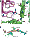
Packing interactions in antigen receptors and paired V-type immunoglobulin domains in VCBP3. (a) The three-layer packing interaction characteristic of the interface between V domains of antigen receptors. TCR Vα (gold and gray) interacts with the corresponding Vβ chain (magenta) by a front-sheet–to–front-sheet interaction in which Vα side chains (cyan) from the highly twisted edge strands C′ and G fold over the central strands of the Vb front sheet to form an inner layer at the core of the three-layer interface between the domains. (b) A three-layer packing interaction at the interface between VCBP3 V1 and V2 in which residues form an inner layer and the C′ loop folds over the central strands of the front sheet. VCBP3 V1 is gold (β-sheet), gray (loop regions) and red (helical regions); VCBP3 V2 is magenta; and side chains of Pro43, Leu104 and Phe106 (a) or Leu42, Leu122 and Ala124 (b) are cyan. View is rotated 180° about a vertical axis in the plane of the page compared with Figure 1.
Figure 4
Figure 4
Structural elements at the V-domain interface. (a) V domain–packing interactions in VCBP3, including the H-bond network formed between residues across the interface between V1 and V2 of VCBP3. Interactions between side chains of one V domain and main-chain atoms of the opposing V domain form the outer layer of the three-layer packing arrangement noted in antigen receptors and VCBP3. The guanidinium group of Arg106 from VCBP3 V1 forms H-bond interactions with main-chain atoms from V2. Carbon atoms are magenta for V1 and green for V2; nitrogen atoms are blue; oxygen atoms are red. (b) Conformational similarities to J regions of antigen receptors: structural comparison of the G strands in antigen receptors (cyan) with VCBP3 V1 (gold). The sequence motif FGXGTXLXV is highly conserved in J gene segments of antigen receptors and corresponds to G-strand residues critical in V-domain packing. In VCBP3, the G strands of V1 and V2 are in an extended β-strand geometry, whereas antigen receptors have a conserved β-bulge at the position corresponding to FGX–(β-bulge)–G.
Figure 5
Figure 5
Crystal structure of VCBP3 V1-V2 solved by single-wavelength anomalous dispersion and refined to 1.85 Å. (a,b) Secondary structure (β-strands, gold; loop regions, gray; helices, red). The G strand and FG loop encoded by a J gene segment-like element is cyan. Loops corresponding to CDR regions in TCR and immunoglobulin: BC loop, CDR1; C′Cx02033;, CDR2; FG, CDR3. (c,d) The molecular surface of VCBP3 V1·V2. The molecular surface of V1 is magenta and that of V2 is violet. Polymorphic residues in VCBP sequences (gold) form a contiguous patch of solvent-exposed hypervariable residues (outlined by a green dashed rectangle). The views in b,d are rotated 180° about a vertical axis in plane of the page with respect to a,c.
Figure 6
Figure 6
The distribution of known binding sites in V domains occupying alternative surfaces in the V-type fold in the TCR, a V-type viral receptor and the contiguous region of hypervariation in VCBPs. The main-chain atoms of a Vδ domain (PDB accession number 1TVD:B; red), the human coxsackie and adenovirus receptor D1 (PDB accession number 1EAJ:B; blue) and VCBP3 V1 (gold) are superimposed. The main-chain and side-chain atoms of the CDR loops in Vδ and the front-sheet residues involved in binding to the adenovirus knob fiber protein (PDB accession number 1KAC) are presented. The residues corresponding to the VCBP hypervariable edge strand residues are spheres. (a) V domains are oriented with the CDRs on the top surface and the front sheets, used in viral receptor interactions, facing right. The edge of VCBP3 V1 is oriented toward the viewer. (b) The orientation of V domains is rotated 90° around a horizontal axis on the plane of the page compared with a. The highly conserved V-type domain supports a particularly extensive range of different recognition-interaction functions.

Comment in

References

    1. Orengo CA, Thornton JM. Protein families and their evolution—a structural perspective. Annu Rev Biochem. 2005;74:867–900. - PubMed
    1. Bork P, Holm L, Sander C. The immunoglobulin fold. Structural classification, sequence patterns and common core. J Mol Biol. 1994;242:309–320. - PubMed
    1. Barclay AN. Membrane proteins with immunoglobulin-like domains–a master super-family of interaction molecules. Semin Immunol. 2003;15:215–223. - PubMed
    1. Hunkapiller T, Hood L. Diversity of the immunoglobulin gene superfamily. Adv Immunol. 1989;44:1–62. - PubMed
    1. Eason DD, et al. Mechanisms of antigen receptor evolution. Semin Immunol. 2004;16:215–226. - PubMed

Publication types

MeSH terms

Substances