. 2004 Feb;1(1):47-55.
doi: 10.1017/S1740925X04000079.

Remyelination of spinal cord axons by olfactory ensheathing cells and Schwann cells derived from a transgenic rat expressing alkaline phosphatase marker gene

Affiliations

Remyelination of spinal cord axons by olfactory ensheathing cells and Schwann cells derived from a transgenic rat expressing alkaline phosphatase marker gene

Yukinori Akiyama et al. Neuron Glia Biol. 2004 Feb.

Abstract

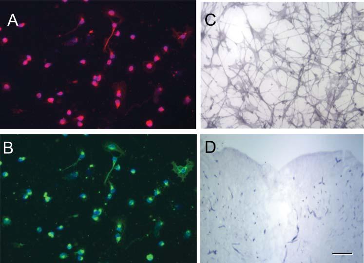
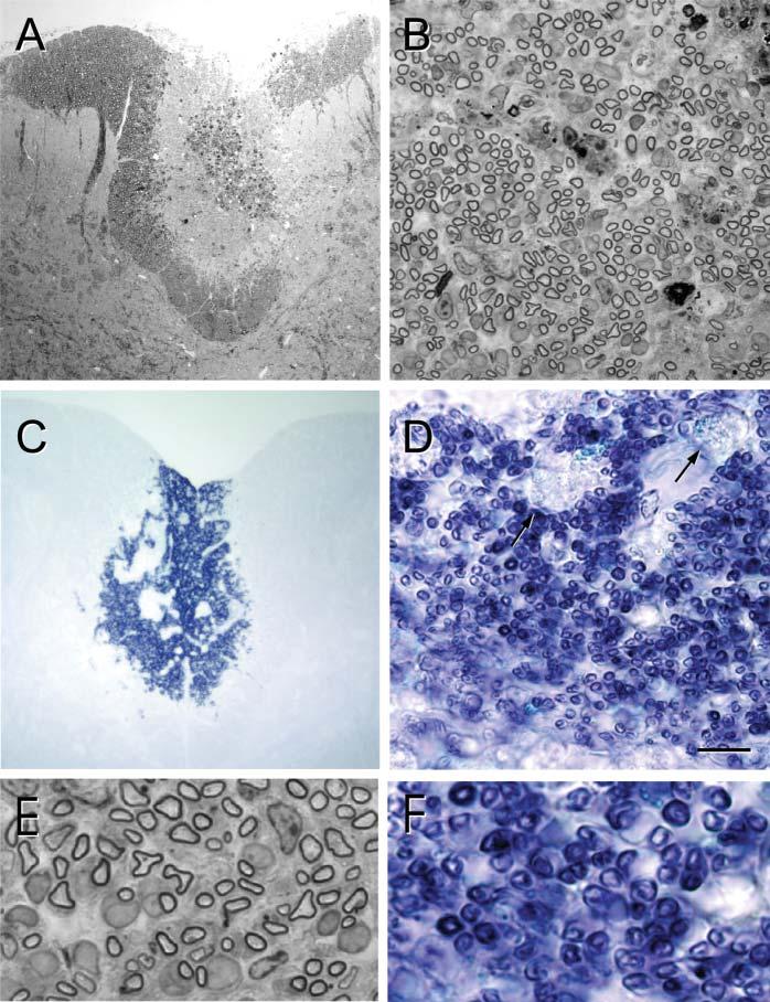
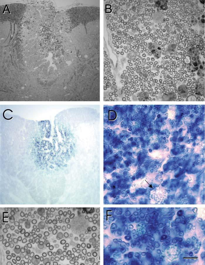
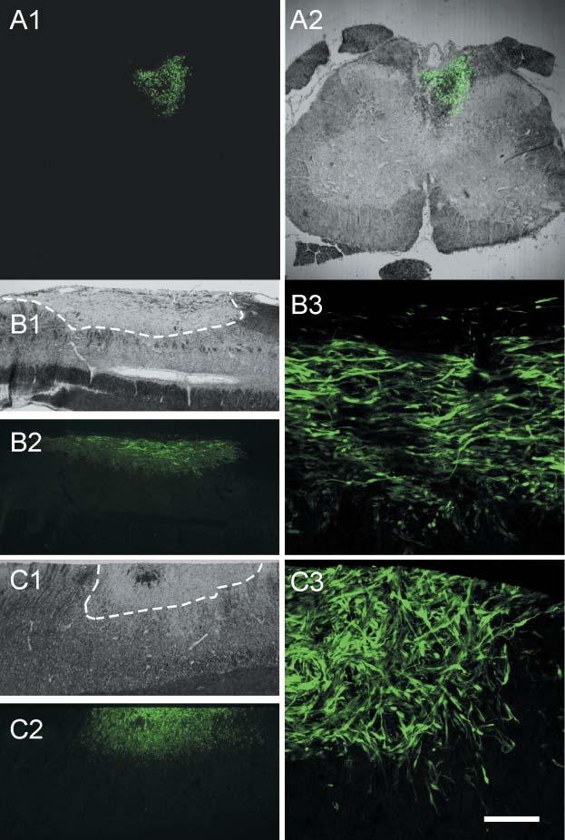
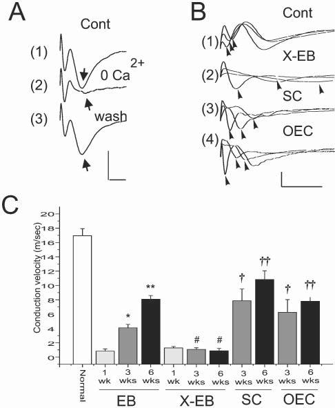
Transplantation of cell suspensions containing olfactory ensheathing cells (OECs) has been reported to remyelinate demyelinated axons in the spinal cord with a Schwann cell (SC)-like pattern of myelination. However, questions have been raised recently as to whether OECs can form SC-like myelin. To address this issue we prepared SCs and OECs from transgenic rats in which a marker gene, human placental alkaline phosphatase (hPAP), is linked to the ubiquitously active promoter of the R26 gene. SCs were prepared from the sciatic nerve and OECs from the outer nerve-fiber layer of the olfactory bulb. Positive S100 and p75 immunostaining indicated that >95% of cells in culture displayed either SC or OEC phenotypes. Suspensions of either SCs or OECs were transplanted into an X-irradiation/ethidium bromide demyelinating lesion in the spinal cord. We observed extensive SC-like remyelination following either SC or OEC transplantation 3 weeks after injection of the cells. Alkaline phosphatase (ALP) chromagen reaction product was associated clearly with the myelin-forming cells. Thus, cell suspensions that are enriched in either SCs or OECs result in peripheral-like myelin when transplanted in vivo.


Figures

Fig. 1
Fig. 1
Phentotypic characterization of OECs and hPAP expression in culture. (A,B) Dissociated OECs were p75+ (red) and S100+ (green). The nuclei were stained with the Hoechst method and >95% of cells were p75+. (C) ALP-reaction product can be seen in OECs that were maintained in culture for 5 days. (D) A coronal section through a demyelinated lesion (X–EB) site after ALP chromagen reaction. Note that aside from minor staining in blood vessels, the central lesion site showed little reaction product. Scale bar: 20 μm in A-C; 300 μm in D.
Fig. 2
Fig. 2
Remyelination after transplantation of hPAP-SCs into an XEB lesion of the dorsal funiculus. (A) Low-power, plastic-embedded-section of the dorsal spinal cord 3 weeks after hPAP-SC transplantation. Note the centrally positioned lesion site (lighter region). (B) Higher power micrograph shows extensive myelinated profiles throughout the transplantation site. (C) In adjacent, frozen sections reacted for ALP, blue reaction product can be observed in the transplantation site. (D) Higher power of this area shows numerous blue profiles that are characteristic of myelinated axons. Note that the reaction product is minimal in macrophages (arrows). (E) Higher-power images of plastic-embedded sections show myelinated axons, many of which are associated with large cytoplasmic and nuclear surrounds that are characteristic of SC myelination. (F) These profiles are associated with hPAP reaction product. Scale bar: 200 μm in A,C; 25 μm in B,D; 7 μm in E,F.
Fig. 3
Fig. 3
Remyelination after transplantation of hPAP-OECs into an XEB dorsal funiculus lesion. (A) Low-power, plastic-embedded section of the dorsal spinal cord 3 weeks after hPAP-OEC transplantation. (B) A higher power micrograph shows extensive myelinated profiles throughout the transplantation site. (C) In adjacent, frozen sections reacted for ALP, blue reaction product can be observed in the transplantation site. (D) Higher power of this area shows numerous blue profiles that are characteristic of myelinated axons and similar to that observed following SC transplantation (Fig. 2). (E) Higher power images of plastic-embedded sections shows myelinated axons, many of which are associated with large cytoplasmic and nuclear surrounds characteristic of peripheral myelination. (F) These profiles are associated with hPAP reaction product. Scale bar: 200 μm in A,C; 25 μm in B,D; 7 μm in E,F.
Fig. 4
Fig. 4
Distribution of GFP-expressing mouse SCs and OECs transplanted into an XEB dorsal funiculus lesion. (A1) Coronal section of the spinal cord from a rat that was transplanted with SCs from GFP-expressing mice 3 weeks after transplantation. (A2) Superimposition of GFP fluorescent and DIC images. Note that donor SCs that express GFP are localized only in the dorsal funiculus. (B) Sagittal sections through the lesion site showing the distribution of transplanted SCs. The lesion site is within the stippled area in B1. The transplanted cells extend throughout the lesion site (B2). Higher-power micrograph (B3) shows clusters of transplanted cells. (C) Distribution of OECs in the lesion site. C1 outlines the lesion site, and distribution of cells in low and higher power micrographs are shown in C2 and C3, respectively. Scale bar: 500 μm in A1,A2; 1 mm in B1,B2,C1,C2; 50 μm in B3,C3.
Fig. 5
Fig. 5
Conduction velocity of demyelinated and remyelinated axons, recorded in vivo. (A) Compound action potentials recorded from the dorsal column (near the midline and within 100 μm of the surface) with a glass microlectrode in vivo following stimulation of the dorsal column surface near the midline. Note the early and late negativities. Washing the surface of the spinal cord with a Ca2+-free, high (6 mM) Mg2+ Krebs' solution (2) reversibly (3) eliminates the second negativity (arrow), indicating its synaptic nature. Thus, the first negativity corresponds to conducting dorsal column axons. (B) Superimposed compound action potentials recorded at 1.0 mm increments longitudinally along the dorsal columns in normal (1), X–EB lesion (2) and 3 weeks after transplantation of either SCs (3) or OECs (4). (C) Conduction velocity (error bars indicate S.E.M.) of dorsal column axons from control animals (normal), and 1, 3 and 6 weeks after EB injection without prior X-irradiation, after X–EB lesion induction, and 3 and 6 weeks after SC and OEC transplantation into the X–EB lesion. * P<0.1, ** P<0.01, †P <0.05, †† P<0.05, # not statistically significant. Scals bars: A, 2 mV, 1 msec; B, 1 mV; 5 msec.

References

    1. Akiyama Y, Radtke C, Kocsis JD. Remyelination of the rat spinal cord by transplantation of identified bone marrow stromal cells. Journal of Neuroscience. 2002;22:6623–6630. - PMC - PubMed
    1. Au W, Treloar HB, Greer CA. Sublaminar organization of the mouse olfactory bulb nerve layer. Journal of Comparative Neurology. 2002;446:68–80. - PubMed
    1. Au W, Roskams AJ. Olfactory ensheathing cells of the lamina propria in vivo and in vitro. Glia. 2003;3:224–236. - PubMed
    1. Barnett SC, Alexander CL, Iwashita Y, Gilson JM, Crowther J, Clark L, et al. Identification of a human olfactory ensheathing cell that can effect transplant-medicated remyelination of demyelinated CNS axons. Brain. 2000;123:1581–1588. - PubMed
    1. Baron-Van Evercooren A, Gansmuller A, Duhamel E, Pascal F, Gumpel M. Repair of a myelin lesion by Schwann cells transplanted into the adult mouse spinal cord. Journal of Neuroimunology. 1992;40:235–242. - PubMed

LinkOut - more resources