. 2006 Jun 26:3:38.
doi: 10.1186/1742-4690-3-38.

Wild-type and central DNA flap defective HIV-1 lentiviral vector genomes: intracellular visualization at ultrastructural resolution levels

Affiliations

Wild-type and central DNA flap defective HIV-1 lentiviral vector genomes: intracellular visualization at ultrastructural resolution levels

Nathalie J Arhel et al. Retrovirology. .

Abstract

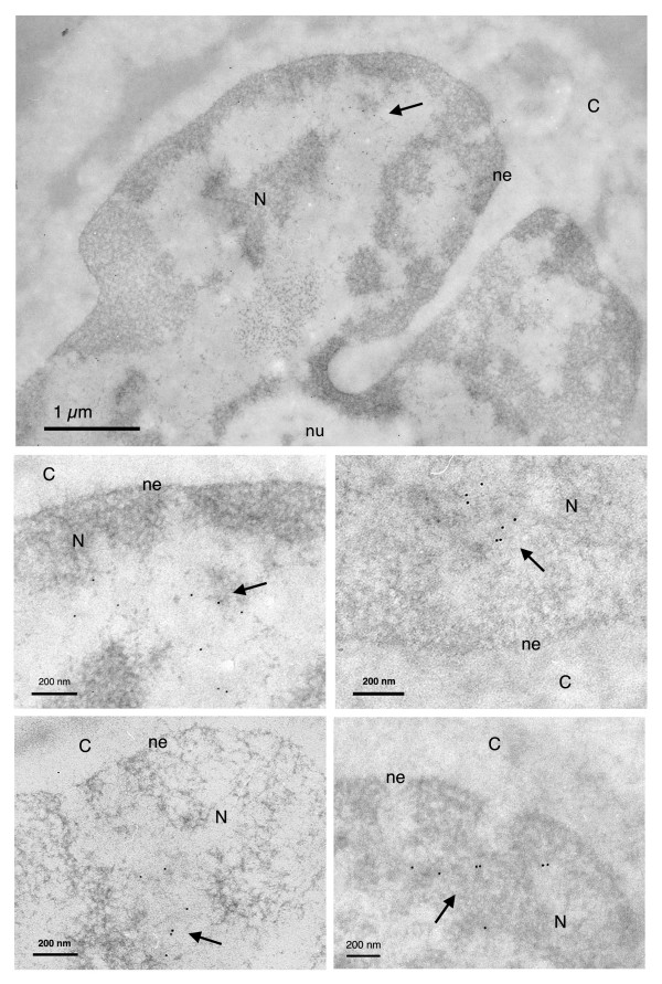
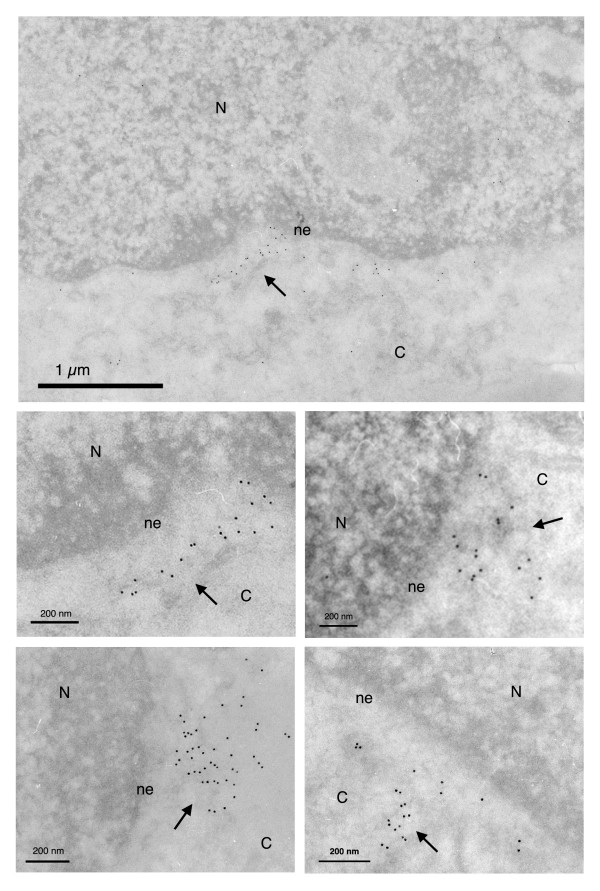
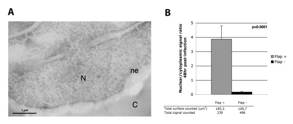
HIV-1 and other lentiviruses have the unique ability among retroviruses to efficiently replicate in non-dividing cells as a result of the active nuclear import of their DNA genome across an interphasic nuclear membrane. Previous work has shown that a three-stranded DNA structure synthesized during HIV-1 reverse transcription, called the central DNA flap, acts as a cis-determinant of HIV-1 genome nuclear import. Concordantly, DNA Flap re-insertion in lentiviral-derived gene therapy vectors stimulates gene transfer efficiencies and complements the level of nuclear import to wild-type levels quantitatively indistinguishable from wild-type virus in all cell types and tissues examined so far. In order to define the precise nature of the replicative defect of DNA flap mutant viruses, we carried out in situ DNA hybridization experiments with electron microscopy to determine the subcellular localization of DNA flap mutant and wild-type HIV-1 genomes. We found that Flap defective DNA genomes accumulate at the cytoplasmic face of the nuclear membrane with no overlap across the nuclear membrane, whereas wild-type genomes localize throughout the nuclear compartment. These data provide an unequivocal confirmation of the role of the DNA flap in HIV-1 nuclear import and further establish that the DNA flap controls a step that immediately precedes translocation through the nuclear pore. Further, the widespread distribution of wild-type genomes within the open chromatin confirms the recent genome-wide mapping of HIV-1 cDNA integration sites and points to an as-yet poorly understood step of intranuclear transport of HIV-1 pre-integration complexes.


Figures

Figure 1
Figure 1
Ultrastructural subcellular localization of HIV-1 derived vector genomes including the central DNA Flap (Flap +). Electron micrographs showing MT4 cells 48 hr following transduction with the TRIP Flap+ vector. Vector DNA genomes including the DNA Flap are found predominantly within the nucleus. N = nucleus; ne = nuclear envelope; nu = nucleolus; C = cytoplasm. Images show one low and four high magnification micrographs. The first high magnification micrograph is an enlargement from the low magnification image. The other three are taken from other independent experiments. All are highly representative of the data obtained. Arrows point to clusters of immunogold labeled vector DNA.
Figure 2
Figure 2
Ultrastructural subcellular localization of HIV-1 derived vector genomes without the central DNA Flap (Flap -). MT4 cells 48 hr post-transduction with the HR Flap- vector. DNA Flap defective vector genomes localize on the cytoplasmic side of the nuclear membrane. N = nucleus; ne = nuclear envelope; C = cytoplasm. Images show one low and four high magnification micrographs. The first high magnification micrograph is an enlargement from the low magnification image. The other three are taken from other independent experiments. All are highly representative of the data obtained. Arrows point to clusters of immunogold labeled vector DNA.
Figure 3
Figure 3
Quantification of intracellular vector genome detection. (A) Electron micrograph of control non-transduced cells showing minimal background signal. (B) DNA hybridization signals from 4 independent cell population infections were counted and represented as total nuclear over cytoplasmic signal ratio. All cell areas containing DNA hybridization signal were systematically photographed (about 150–200 photos) and signal was carefully quantified, each time with equal surface of nuclear and cytoplasmic compartments. The p value (Mann-Whitney test) shows the results are highly statistically relevant.

References

    1. Charneau P, Clavel F. A single-stranded gap in human immunodeficiency virus unintegrated linear DNA defined by a central copy of the polypurine tract. J Virol. 1991;65:2415–2421. - PMC - PubMed
    1. Charneau P, Alizon M, Clavel F. A second origin of DNA plus-strand synthesis is required for optimal human immunodeficiency virus replication. J Virol. 1992;66:2814–2820. - PMC - PubMed
    1. Charneau P, Mirambeau G, Roux P, Paulous S, Buc H, Clavel F. HIV-1 reverse transcription. A termination step at the center of the genome. J Mol Biol. 1994;241:651–662. doi: 10.1006/jmbi.1994.1542. - DOI - PubMed
    1. Zennou V, Petit C, Guetard D, Nerhbass U, Montagnier L, Charneau P. HIV-1 genome nuclear import is mediated by a central DNA Flap. Cell. 2000;101:173–185. doi: 10.1016/S0092-8674(00)80828-4. - DOI - PubMed
    1. Sirven A, Pflumio F, Zennou V, Titeux M, Vainchenker W, Coulombel L, Dubart-Kupperschmitt A, Charneau P. The human immunodeficiency virus type-1 central DNA Flap is a crucial determinant for lentiviral vector nuclear import and gene transduction of human hematopoietic stem cells. Blood. 2000;96:4103–4110. - PubMed

LinkOut - more resources