

Expression of prostasin and protease nexin-1 in rhesus monkey (Macaca mulatta) endometrium and placenta during early pregnancy
- PMID: 16801525
- PMCID: PMC3957810
- DOI: 10.1369/jhc.6A7005.2006
Expression of prostasin and protease nexin-1 in rhesus monkey (Macaca mulatta) endometrium and placenta during early pregnancy
Abstract
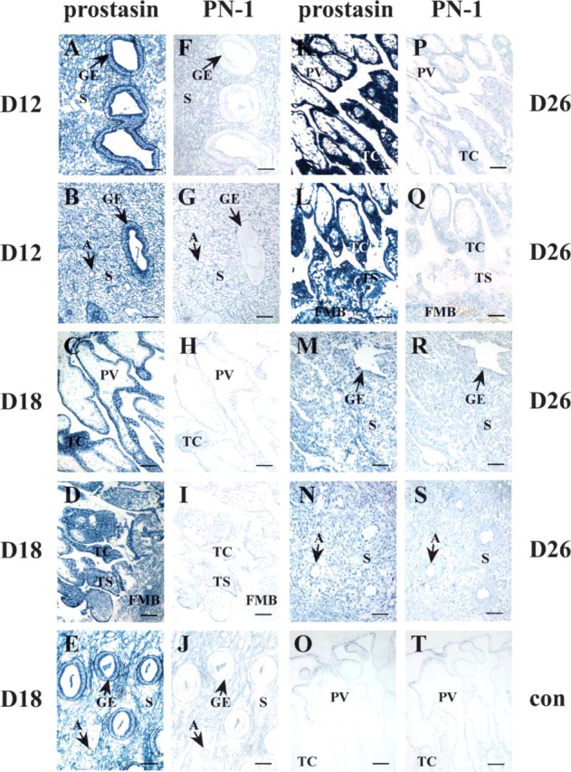
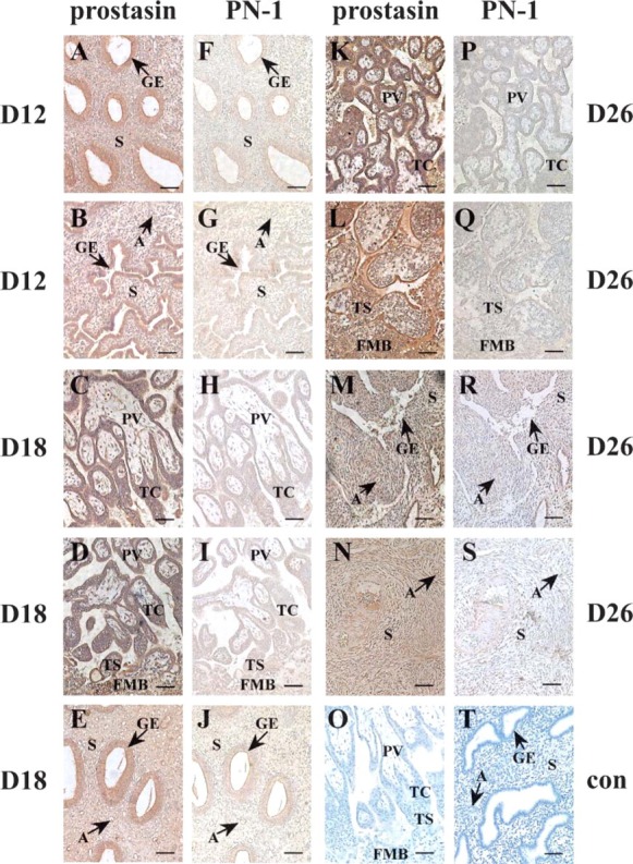
Serine proteases have been documented to play key roles in uterine matrix turnover and trophoblastic invasion during implantation. Roles of prostasin serine protease in these processes, however, are currently unclear. The present study was first conducted to investigate the colocalization of prostasin and its cognate serpin, protease nexin-1 (PN-1), in rhesus monkey endometrium and placenta on days 12, 18, and 26 of pregnancy by using in situ hybridization (ISH) and immunohistochemistry. With ISH, expression of prostasin mRNA was intensely localized in the glandular epithelium on days 12 and 18 and in the placental villi, trophoblastic column, trophoblastic shell, and fetal-maternal border on days 18 and 26. With the progress of pregnancy, expression level in the glandular epithelium was significantly decreased, and the accumulation in the placental compartments was further increased. In addition, the stroma and arterioles exhibited modest levels of prostasin signals. However, expression level of PN-1 in these compartments on adjacent sections in the three stages of early pregnancy was weak or below the level of detection. Prostasin protein expression in the endometrium was found to be consistent with the distribution patterns revealed in the ISH experiments. It may be suggested from these results that prostasin is involved in endometrial epithelial morphology establishment, tissue remodeling, and trophoblastic invasion during early pregnancy. The cognate serpin PN-1 was not coordinately expressed along with prostasin, creating a tissue environment favorable for proteolytic activities of prostasin during early pregnancy events.
Figures
Similar articles
-
Expression of prostasin serine protease and protease nexin-1 (PN-1) in rhesus monkey ovary during menstrual cycle and early pregnancy.J Histochem Cytochem. 2007 Dec;55(12):1237-44. doi: 10.1369/jhc.7A7232.2007. Epub 2007 Sep 6. J Histochem Cytochem. 2007. PMID: 17827166
-
Expression of proteasome subunits low molecular mass polypeptide (LMP) 2 and LMP7 in the endometrium and placenta of rhesus monkey (Macaca mulatta) during early pregnancy.Biol Reprod. 2004 Oct;71(4):1317-24. doi: 10.1095/biolreprod.104.030213. Epub 2004 Jun 16. Biol Reprod. 2004. PMID: 15201202
-
Expression of adamalysin 19/ADAM19 in the endometrium and placenta of rhesus monkey (Macaca mulatta) during early pregnancy.Mol Hum Reprod. 2005 Jun;11(6):429-35. doi: 10.1093/molehr/gah183. Epub 2005 May 18. Mol Hum Reprod. 2005. PMID: 15901844
-
Involvement of molecules related to angiogenesis, proteolysis and apoptosis in implantation in rhesus monkey and mouse.Contraception. 2005 Apr;71(4):249-62. doi: 10.1016/j.contraception.2004.12.008. Contraception. 2005. PMID: 15792644 Review.
-
SERPINE2/Protease Nexin-1 in vivo multiple functions: Does the puzzle make sense?Semin Cell Dev Biol. 2017 Feb;62:160-169. doi: 10.1016/j.semcdb.2016.08.012. Epub 2016 Aug 18. Semin Cell Dev Biol. 2017. PMID: 27545616 Review.
Cited by
-
Urinary serine proteases and activation of ENaC in kidney--implications for physiological renal salt handling and hypertensive disorders with albuminuria.Pflugers Arch. 2015 Mar;467(3):531-42. doi: 10.1007/s00424-014-1661-5. Epub 2014 Dec 9. Pflugers Arch. 2015. PMID: 25482671 Review.
-
Prostasin inhibits cell invasion in human choriocarcinomal JEG-3 cells.Histochem Cell Biol. 2009 Dec;132(6):639-46. doi: 10.1007/s00418-009-0652-7. Epub 2009 Oct 22. Histochem Cell Biol. 2009. PMID: 19847458
-
Spatiotemporal expression of the serine protease inhibitor, SERPINE2, in the mouse placenta and uterus during the estrous cycle, pregnancy, and lactation.Reprod Biol Endocrinol. 2010 Oct 27;8:127. doi: 10.1186/1477-7827-8-127. Reprod Biol Endocrinol. 2010. PMID: 20977773 Free PMC article.
-
Spatiotemporal expression of SERPINE2 in the human placenta and its role in extravillous trophoblast migration and invasion.Reprod Biol Endocrinol. 2011 Aug 2;9:106. doi: 10.1186/1477-7827-9-106. Reprod Biol Endocrinol. 2011. PMID: 21806836 Free PMC article.
-
Embryo-derived trypsin-induced calcium entry is inhibited by endometrial infertility factor, LEFTY2.Front Cell Dev Biol. 2025 May 29;13:1499339. doi: 10.3389/fcell.2025.1499339. eCollection 2025. Front Cell Dev Biol. 2025. PMID: 40510743 Free PMC article.
References
-
- Andreasen D, Vuagniaux G, Fowler-Jaeger N, Hummler E, Rossier BC. (2006) Activation of epithelial sodium channels by mouse channel activating proteases (mCAP) expressed in Xenopus oocytes requires catalytic activity of mCAP3 and mCAP2 but not mCAP1. J Am Soc Nephrol 17:968–976 - PubMed
-
- Bhagwandin VJ, Hau LW, Mallen-St Clair J, Wolters PJ, Caughey GH. (2003) Structure and activity of human pancreasin, a novel tryptic serine peptidase expressed primarily by the pancreas. J Biol Chem 278:3363–3371 - PubMed
-
- Carson DD, Bagchi I, Dey SK, Enders AC, Fazleabas AT, Lessey BA, Yoshinaga K. (2000) Embryo implantation. Dev Biol 223:217–237 - PubMed
-
- Chen LM, Chai KX. (2002) Prostasin serine protease inhibits breast cancer invasiveness and is transcriptionally regulated by promoter DNA methylation. Int J Cancer 97:323–329 - PubMed
-
- Chen LM, Hodge GB, Guarda LA, Welch JL, Greenberg NM, Chai KX. (2001a) Down-regulation of prostasin serine protease: a potential invasion suppressor in prostate cancer. Prostate 48:93–103 - PubMed
Publication types
MeSH terms
Substances
Grants and funding
LinkOut - more resources
Full Text Sources
Miscellaneous
