. 2006 Jul 5;103(27):10271-10276.
doi: 10.1073/pnas.0601058103. Epub 2006 Jun 26.

Distinct structural and mechanical properties of the nuclear lamina in Hutchinson-Gilford progeria syndrome

Affiliations

Distinct structural and mechanical properties of the nuclear lamina in Hutchinson-Gilford progeria syndrome

Kris Noel Dahl et al. Proc Natl Acad Sci U S A. .

Abstract

The nuclear lamina is a network of structural filaments, the A and B type lamins, located at the nuclear envelope and throughout the nucleus. Lamin filaments provide the nucleus with mechanical stability and support many basic activities, including gene regulation. Mutations in LMNA, the gene encoding A type lamins, cause numerous human diseases, including the segmental premature aging disease Hutchinson-Gilford progeria syndrome (HGPS). Here we show that structural and mechanical properties of the lamina are altered in HGPS cells. We demonstrate by live-cell imaging and biochemical analysis that lamins A and C become trapped at the nuclear periphery in HGPS patient cells. Using micropipette aspiration, we show that the lamina in HGPS cells has a significantly reduced ability to rearrange under mechanical stress. Based on polarization microscopy results, we suggest that the lamins are disordered in the healthy nuclei, whereas the lamins in HGPS nuclei form orientationally ordered microdomains. The reduced deformability of the HGPS nuclear lamina possibly could be due to the inability of these orientationally ordered microdomains to dissipate mechanical stress. Surprisingly, intact HGPS cells exhibited a degree of resistance to acute mechanical stress similar to that of cells from healthy individuals. Thus, in contrast to the nuclear fragility seen in lmna null cells, the lamina network in HGPS cells has unique mechanical properties that might contribute to disease phenotypes by affecting responses to mechanical force and misregulation of mechanosensitive gene expression.


Conflict of interest statement

Conflict of interest statement: No conflicts declared.

Figures

Fig. 1.
Fig. 1.
Reduced dynamic exchange of A type lamins in HGPS cells. (A) WT and HGPS cells expressing either WT or Δ50 GFP-lamin A were imaged before and during recovery after bleaching of a part of the nucleus. Images are shown by using intensity pseudocolors. Contrast was adjusted in the 60-s frame to visually normalize for total loss of fluorescence during imaging. (Scale bar: 5 μm.) (BD) Quantitative analysis of fluorescence recovery after photobleaching recovery in WT and HGPS cells. Kinetics of recovery of the fluorescence signal in the whole bleached area in cells expressing either WT or Δ50 GFP-lamin A (B), WT lamin C (C), or WT lamin B (D). The statistical significance of the difference between the two recovery curves is indicated. Values represent means from at least 10 cells each from three experiments. Standard errors were between 10% and 20%.
Fig. 2.
Fig. 2.
Reduced extractability of A type lamins in HGPS cells. (A) Western blot analysis of isolated nuclei from HeLa cells expressing either WT or Δ50 GFP-lamin A after extraction with increasing concentrations of NaCl. The samples were probed with anti-GFP and anti-lamin A/C antibody. Δ50 GFP-lamin A is less extractable than the WT protein and reduces the extractability of endogenous WT lamin A and lamin C. (B) Western blot analysis of isolated nuclei from control and HGPS fibroblasts after extraction with increasing concentrations of NaCl. Endogenous Δ50 lamin A is less extractable than WT protein in HGPS. Also the extractability of WT lamin A and lamin C is reduced in HGPS versus WT cells.
Fig. 3.
Fig. 3.
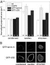
Swelling and shrinking behavior of isolated nuclei. (A) Quantitative analysis of swelling and shrinking. The projected area of nuclei was calculated and normalized. All fibroblast data (WT and HGPS) was normalized such that the condensed WT nucleus average was 1. All HeLa data (GFP-lamin A or GFP-Δ50) was normalized such that the condensed GFP-lamin A nuclear average was 1. Swollen HGPS or Δ50 GFP-lamin A nuclei were slightly larger than their respective control nuclei (Student t test; P = 0.023, HGPS; P = 0.003, Δ50 GFP-lamin A). Differences between shrunken control and HGPS or Δ50 lamin A transfected nuclei were all significant at P < 0.001 in a Student t test. Numbers of measured nuclei for each condition are indicated. Graphed values represent averages ± SEM of the projected area. (B) Differential behavior seen in nuclei isolated from HeLa cells transfected with GFP-lamin A or Δ50 GFP-lamin A in their condensed (high divalent salt), swollen (near-zero salt) and shrunken (swollen with added divalent salt) states.
Fig. 4.
Fig. 4.
MPA of isolated nuclei. (A) The creep compliance, a function of the increased deformation into the micropipette at a step pressure, is plotted versus time. Data points from one experiment are overlaid with lines, which are fits of the combined data (a modified form of this data are in Table 1). HGPS and WT nuclei show no difference in chromatin mechanics on this measurement length scale. The slope of the creep compliance for the lamina-dominated measurement is higher in WT than HGPS nuclei, indicating that WT deforms more easily over time. (B and C) Control nuclei buckled relatively smoothly outside the pipette as aspiration increased (B), indicating that mechanical stresses were distributed across the entire lamina. In striking contrast, the lamina of HGPS nuclei collapsed along long prominent fault lines, appearing crumpled or folded (C).
Fig. 5.
Fig. 5.
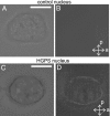
Polarized light microscopy. (A and B) Isolated WT nuclei (shown in brightfield in A) showed no detectable birefringence (B) at any angle, suggesting completely isotropic orientation of lamin filaments. (C and D) As isolated HGPS nuclei (brightfield; C) were rotated with respect to the crossed polarizers, a bright birefringent pattern was detected at the nuclear periphery possibly due to filament alignment (D). (Scale bars: 4 μm.)
Fig. 6.
Fig. 6.
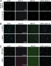
In vivo pressure assay. Immunofluorescence microscopy of WT (A and C) and HGPS (C) primary dermal fibroblasts and lmna+/+ and lmna −/− MEFs (B) after exposure to 700-kPa pressure. Cells were stained with DAPI and antibodies against the indicated proteins in the absence of detergents. (Scale bar: 80 μm.) The percentage of staining-positive cells is indicated. n ≥ 300 per condition.
Fig. 7.
Fig. 7.
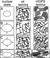
Model of HGPS nuclear lamina ultrastructure and responses to force. Isotropic filaments in WT nuclei can expand reversibly and easily (AC, wt lamina). However, the order in the HGPS lamina (A, HGPS lamina) is lost when surface area increases, because lamins can occupy a larger space (B, HGPS lamina). Upon sudden compression, there is insufficient time to return to the ordered state, and the lamina forms a jammed state (C, HGPS lamina). Isotropic filaments can realign easily along any director of force and redistribute the force evenly (D, wt lamina). Microdomains tend to move as whole domains and align their preferred orientation to be parallel with the director of the force (D, HGPS lamina).

References

    1. DeBusk F. L. J. Pediatr. 1972;80:697–724. - PubMed
    1. Uitto J. Trends. Mol. Med. 2002;8:155–157. - PubMed
    1. De Sandre-Giovannoli A., Bernard R., Cau P., Navarro C., Amiel J., Boccaccio I., Lyonnet S., Stewart C. L., Munnich A., Le Merrer M., Levy N. Science. 2003;300:2055. - PubMed
    1. Eriksson M., Brown W. T., Gordon L. B., Glynn M. W., Singer J., Scott L., Erdos M. R., Robbins C. M., Moses T. Y., Berglund P., et al. Nature. 2003;423:293–298. - PMC - PubMed
    1. Hutchison C. J. Nat. Rev. Mol. Cell Biol. 2002;3:848–858. - PubMed

Publication types

Substances

LinkOut - more resources