. 2006 Jul;134(1):100-8.
doi: 10.1111/j.1365-2141.2006.06102.x.

Effects of human gamma-globin in murine beta-thalassaemia

Affiliations

Effects of human gamma-globin in murine beta-thalassaemia

Tamon Nishino et al. Br J Haematol. 2006 Jul.

Abstract

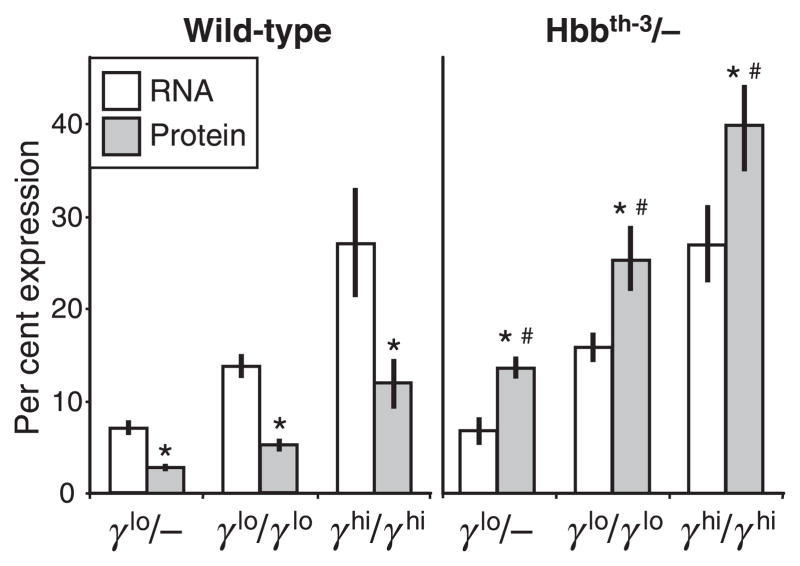
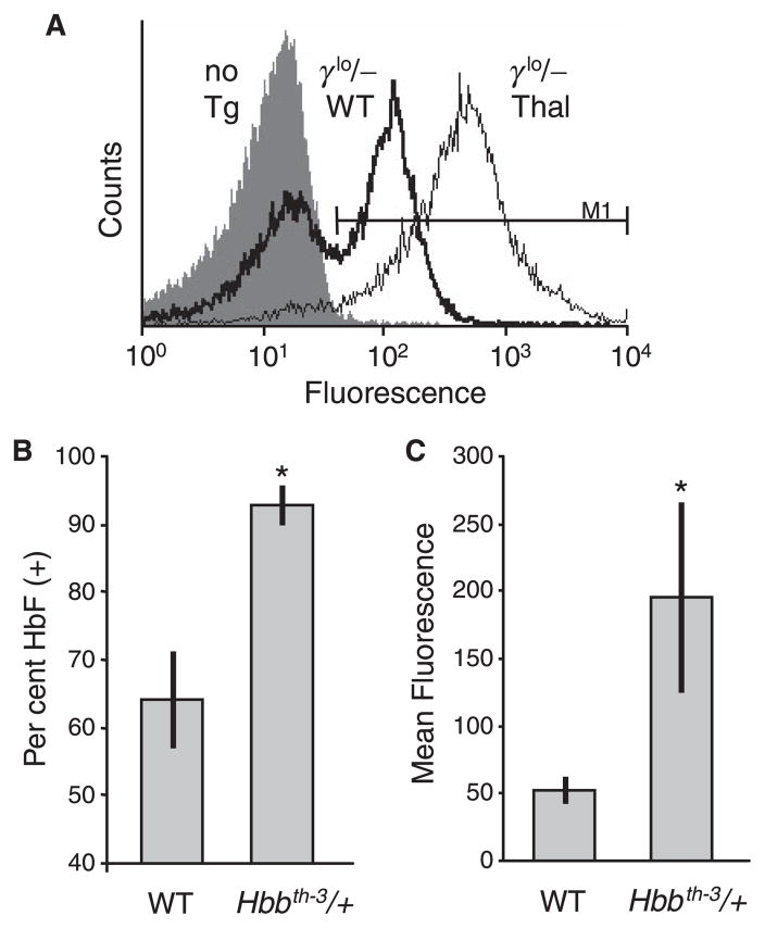
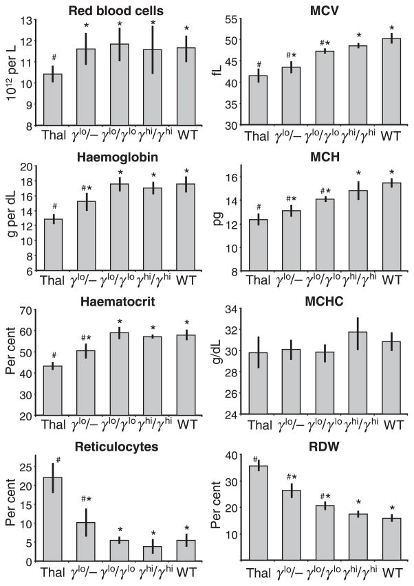
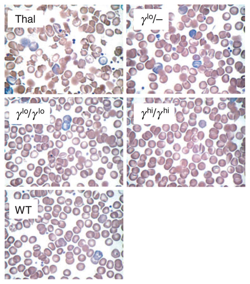
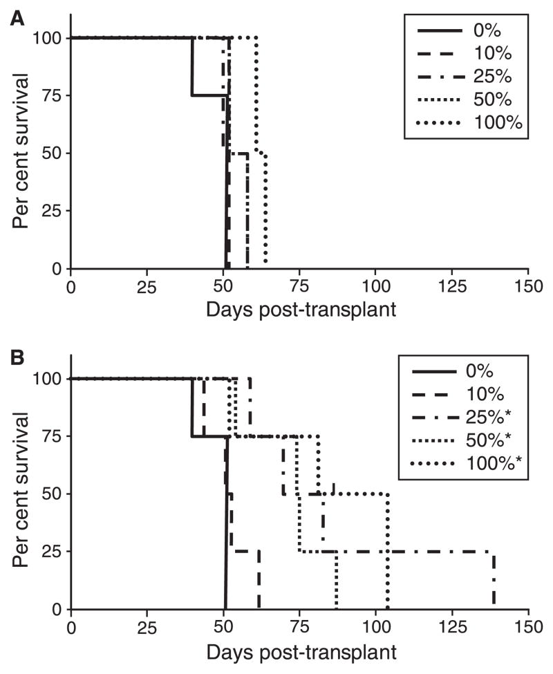
Murine models of beta-thalassaemia have been used to test therapeutic globin gene vectors. However, the level of gamma-globin expression necessary to achieve full phenotypic correction in these models is unclear. In order to address this issue, we carried out breeding and transplantation studies in murine models of beta-thalassaemia intermedia (Hbb(th-3)/+) and severe beta-thalassaemia major (Hbb(th-3)/Hbb(th-3)) using transgenic lines expressing various levels of human gamma-globin. Expression of gamma-globin RNA at a modest 7-14% of total alpha-globin RNA resulted in the selective survival of HbF(+) erythrocytes, a fivefold increase in total HbF, and a phenotypic improvement in the beta-thalassaemia intermedia model. Full normalisation of erythrocyte indices in this model required gamma-globin RNA expression at 27% of alpha-globin, resulting in an average 40% (6.8 g/dl) HbF. Studies using the homozygous Hbb(th-3) model of lethal beta-thalassaemia major demonstrated that even this high level of gamma-globin expression, for reasons related to the function of the hybrid globin tetramers, could only prolong, but not fully support, survival. Taken together, these results indicate that only the heterozygous Hbb(th-3) model of beta-thalassaemia intermedia can be reliably used for the pre-clinical assessment of gamma-globin gene therapy vectors, as well as other means of gamma-globin gene induction.


Figures

Fig 1
Fig 1
γ-Globin transgenic lines. The γ-globin expression cassettes in the transgenic mouse lines 1279 (Enver et al, 1989; Constantoulakis et al, 1991), and 135/4-2 (Stamatoyannopoulos et al, 1993), include a 2.5 kb locus control region (μLCR) containing DNase hypersensitive sites 1–4 from the human β-globin locus linked to a coding cassette for human Aγ-globin that includes either a −1348 bp 5′-promoter region (line 1279) or a −382 bp 5′-promoter region (line 135/4-2). The line 1279 allele (denoted γlo) contains three transgene copies in cis, while the line 135/4-2 allele (denoted γhi) contains nine copies in cis. Grey boxes, μLCR; filled and open boxes, Aγ-globin exons and introns respectively; heavy line: 5′- and 3′-regions flanking the Aγ-globin coding region.
Fig 2
Fig 2
γ-Globin expression in wild type and β-thalassaemia intermedia mice. Peripheral blood was collected at 10–20 weeks of age from wild type and Hbbth–3/+ individuals containing one or two copies of the γlo transgene alleles (γlo/− and γlolo respectively) or two copies of the γhi transgene allele (γhihi). Samples were analysed for the amount of γ-globin RNA (as a percentage of total mouse α-globin, open bars) by RNase protection, and for the amount of γ-globin protein (in the form of hybrid mouse/human HbF as a percentage of total Hb, grey bars) by high-performance liquid chromatography. Results are presented as mean ± SD for a total of five to 12 individuals for each group for the RNA analysis and seven to 29 individuals for each group for the protein analysis. *P < 0.001 vs. RNA; P < 0.001 vs. wild type.
Fig 3
Fig 3
In vivo selection of red blood cell (RBC) expression γ-globin. Peripheral blood was collected from non-transgenic wild-type (WT) mice, WT mice containing a single copy of the γlo transgene alleles (γlo/−), and Hbbth–3/+ thalassaemia mice containing a single copy of the γlo transgene allele, fixed and stained with a phycoerythrin-conjugated monoclonal antibody to HbF, and analysed on a flow cytometer. (A) Typical analysis showing the lack of staining in a non-transgenic control (no Tg; filled histogram), as well as the shift from a heterocellular pattern of γ-globin expression in a transgenic WT mouse (γlo/− WT; heavy line) to a pancellular and significantly elevated pattern of expression in a Hbbth–3/+ mouse (γlo/− Thal; light line). The marker used to define HbF(+) is indicated. (B) Percentage of HbF(+) RBC in WT versus Hbbth–3/+ mice containing a single copy of the γlo transgene allele showing a selective expansion of these cells in the thalassaemia background. Data represents mean ± SD for at least six individuals. (C) Mean fluorescence of HbF(+) RBC from panel (B), showing a concomitant increase in the intensity of staining in the thalassaemia background. *P < 0.01 vs. WT.
Fig 4
Fig 4
Effects of γ-globin expression on haemopoietic and red blood cell (RBC) indices. Peripheral blood was collected at 10–20 weeks of age from none-transgenic Hbbth–3/+ (Thal) and wild-type (WT) individuals, as well as from Hbbth–3/+ individuals containing one or two copies of the γlo transgene alleles (γlo/− and γlolo respectively) or two copies of the γhi transgene allele (γhihi). Samples were analysed for total RBC counts, total haemoglobin, haematocrit, the percentage of reticulocytes, as well as mean corpuscular volume, mean corpuscular haemoglobin, mean corpuscular haemoglobin concentration and red cell distribution width. Results represent mean ± SD for three to 11 individuals for each group. *P < 0.05 vs. the Thal controls; P < 0.05 vs. the WT control.
Fig 5
Fig 5
Effects of γ-globin expression on red blood cell histology. Wright–Giemsa stained peripheral blood smears from individual animals are presented, including wild-type C57BL/6J, non-transgenic β-thalassaemia intermedia Hbbth–3/+ (Thal) or Hbbth–3/+ individuals containing one copy of the γlo transgene allele (γlo/−), two copies of the γlo transgene allele (γlolo) or two copies of the γhi transgene allele (γhihi) (original magnification ×60).
Fig 6
Fig 6
Effects of γ-globin expression on survival of mice transplanted with Hbbth–3/Hbbth–3 fetal liver cells (FLCs). FLCs were collected from day 15.5 homozygous Hbbth–3/Hbbth–3 fetuses containing either no transgenes or two copies of either the γlo of γhi transgene alleles (γlolo or γhihi respectively). These cells were mixed in different ratios (shown as the percentage of transgenic FLCs), and transplanted into myeloablated congenic C57BL/6J recipients. Transplant recipients were monitored closely and killed when moribund or severely anemic with hematocrits <15%. (A) Survival of recipients transplanted with mixtures of γlolo FLCs. (B) Survival of recipients transplanted with mixtures of γhihi FLCs. *P ≤ 0.01 vs. 0% controls.

Similar articles

Cited by

References

    1. Antonchuck J, Hyland CD, Hilton DJ, Alexander WS. Synergistic effects of erythropoiesis, thrombopoiesis, and stem cell competitiveness in mice deficient in thrombopoietin and steel factor receptors. Blood. 2004;104:1306–1313. - PubMed
    1. Arcasoy MO, Romana M, Fabry ME, Skarpidi E, Nagel RL, Forget BG. High levels of human γ-globin gene expression in adult mice carrying a transgene of deletional-type hereditary persistence of fetal hemoglobin. Molecular and Cellular Biology. 1997;17:2076–2089. - PMC - PubMed
    1. Charache S, Clegg JB, Weatherall DJ. The negro variety of hereditary persistence of fetal haemoglobin is a mild form of thalassaemia. British Journal of Haematology. 1976;34:527–534. - PubMed
    1. Constantoulakis P, Josephson B, Mangahas L, Papayannopoulou T, Enver T, Costantini F, Stamatoyannopoulos G. Locus control region – a gamma transgenic mice: a new model for studying the induction of fetal hemoglobin in the adult. Blood. 1991;77:1326–1333. - PubMed
    1. Davis BH, Ornvold K, Bigelow NC. Flow cytometric reticulocyte maturity index: a useful laboratory parameter of erythropoietic activity in anemia. Cytometry. 1995;22:35–39. - PubMed

Publication types