. 2006 Jul 5;103(27):10420-10425.
doi: 10.1073/pnas.0604343103. Epub 2006 Jun 27.

The QseC sensor kinase: a bacterial adrenergic receptor

Affiliations

The QseC sensor kinase: a bacterial adrenergic receptor

Marcie B Clarke et al. Proc Natl Acad Sci U S A. .

Abstract

Quorum sensing is a cell-to-cell signaling mechanism in which bacteria respond to hormone-like molecules called autoinducers (AIs). The AI-3 quorum-sensing system is also involved in interkingdom signaling with the eukaryotic hormones epinephrine/norepinephrine. This signaling activates transcription of virulence genes in enterohemorrhagic Escherichia coli O157:H7. However, this signaling system has never been shown to be involved in virulence in vivo, and the bacterial receptor for these signals had not been identified. Here, we show that the QseC sensor kinase is a bacterial receptor for the host epinephrine/norepinephrine and the AI-3 produced by the gastrointestinal microbial flora. We also found that an alpha-adrenergic antagonist can specifically block the QseC response to these signals. Furthermore, we demonstrated that a qseC mutant is attenuated for virulence in a rabbit animal model, underscoring the importance of this signaling system in virulence in vivo. Finally, an in silico search found that the periplasmic sensing domain of QseC is conserved among several bacterial species. Thus, QseC is a bacterial adrenergic receptor that activates virulence genes in response to interkingdom cross-signaling. We anticipate that these studies will be a starting point in understanding bacterial-host hormone signaling at the biochemical level. Given the role that this system plays in bacterial virulence, further characterization of this unique signaling mechanism may be important for developing novel classes of antimicrobials.


Conflict of interest statement

Conflict of interest statement: No conflicts declared.

Figures

Fig. 1.
Fig. 1.
QseC increases autophosphorylation in response to epi. (A) Graphic depiction of the inside-out orientation of QseC in the liposome. QseC has two transmembrane domains and a HK domain, indicating that its membrane location may allow autophosphorylation upon sensing its specific environmental cue. QseC also contains an ATPase domain, which allows it to exhibit phosphatase activity toward QseB, and a conserved EAL domain that may confer cyclic diguanylate phosphodiesterase activity. (B) QseC weakly autophosphorylates in the liposome, but increases its autophosphorylation in response to epi.
Fig. 2.
Fig. 2.
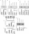
QseC specifically senses epi/NE/AI-3. Each graph represents results from three separate experiments. Student’s t test was performed to determine whether the results were statistically significant as compared with the control (no signal added). (A) DcuS increases autophosphorylation in response to fumarate but not to epi. (B) QseC autophosphorylation in the presence of 5 μM epi, 50 μM gastrin, 50 μM galanin, and 50 μM secretin. (C) QseC autophosphorylation in the presence of 5 μM epi, 50 μM PO, 50 μM PE, and 5 μM epi with 50 μM PE or PO. (D) QseC autophosphorylation in the presence of 5 and 10 μM [3H]NE and 5 μM NE with 50 μM PE. NE induces QseC autophosphorylation, and NE-induced autophosphorylation is also inhibited by PE. (E) Graphical representation of the amount of [3H]NE bound to QseC after incubation with 5 and 10 μM [3H]NE alone or in the presence of 50 μM PE. (F) Graphical representation of the amount of QseC autophosphorylation (cpm measurement of P32) after incubation with 5 and 10 μM [3H]NE, alone or in the presence of 50 μM PE. (G) QseC significantly increases autophosphorylation in response to AI-3 but not to AI-2 (negative control).
Fig. 3.
Fig. 3.
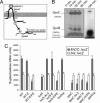
QseB/QseC is a functional two-component system. (A) Model of signaling begins with QseC responding to epi/NE and AI-3. QseC increases its autophosphorylation and transfers its phosphate to QseB. Phosphorylated QseB then activates both its own transcription and that of flhDC. (B) Autophosphorylation of QseC and phosphotransfer to QseB in liposomes loaded with 50 μM epi. QseC responds to epi and autophosphorylates after 10 min. At 30 min, QseC begins to transfer its phosphate to QseB, the response regulator. By 120 min, most of the phosphate has been transferred to QseB. (C) Transcriptional activation of flhDC::lacZ in WT, qseC, and luxS mutants and complemented strains (luxScp and qseCcp) in response to 100 nM AI-3, 5 μM epi (E), and 50 μM PE.
Fig. 4.
Fig. 4.
A qseC mutant is attenuated for virulence in rabbits. (A) Cumulative weight gain of rabbits inoculated with WT, qseC, or PBS by day 6. Development of diarrhea by rabbits inoculated with PBS (B), WT (C), and qseC (D). The WT caused diarrhea in six of seven rabbits by day 6 after inoculation. None of the PBS control animals presented diarrhea. The qseC mutant was attenuated in rabbits in comparison with WT, with two of eight rabbits presenting diarrhea.

Similar articles

Cited by

References

    1. Berg R. D. Trends Microbiol. 1996;4:430–435. - PubMed
    1. Hooper L. V., Gordon J. I. Science. 2001;292:1115–1118. - PubMed
    1. Sperandio V., Torres A. G., Jarvis B., Nataro J. P., Kaper J. B. Proc. Natl. Acad. Sci. USA. 2003;100:8951–8956. - PMC - PubMed
    1. Nataro J. P., Kaper J. B. Clin. Microbiol. Rev. 1998;11:142–201. - PMC - PubMed
    1. Grebe T. W., Stock J. B. Adv. Microb. Physiol. 1999;41:139–227. - PubMed

Publication types