. 2007 Jan;292(1):F330-9.
doi: 10.1152/ajprenal.00059.2006. Epub 2006 Jun 27.

Sustained renal interstitial macrophage infiltration following chronic angiotensin II infusions

Affiliations

Sustained renal interstitial macrophage infiltration following chronic angiotensin II infusions

Yuri Ozawa et al. Am J Physiol Renal Physiol. 2007 Jan.

Abstract

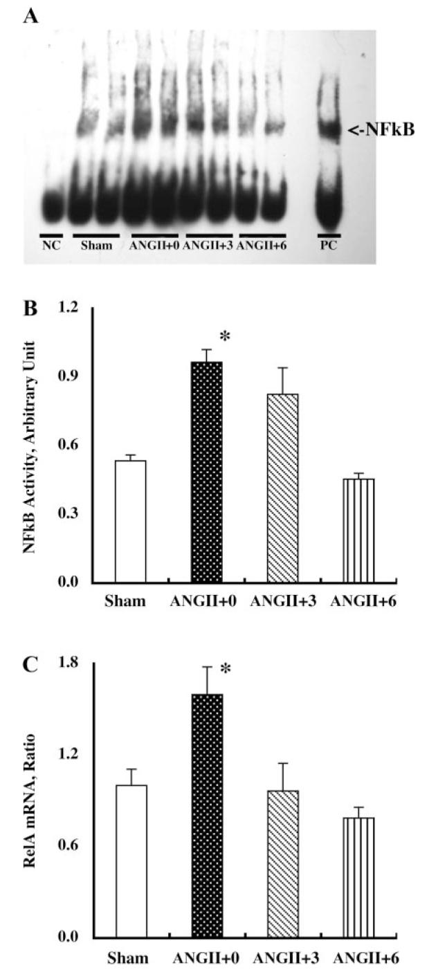
Chronic angiotensin (ANG) II infusions into rats lead to augmented intrarenal levels of ANG II and inflammatory factors, impaired renal function, and progressive hypertension. Residual effects persist after cessation of ANG II infusions, as manifested by a hypertensive response to high-salt intake. This study was performed to determine the residual cytokines and chemokines following the cessation of ANG II infusion. Male Sprague-Dawley rats, maintained on a normal diet, received either a sham operation or continuous ANG II infusion (120 ng/min) subcutaneously via minipumps. The ANG II-infused rats were further subdivided into three subgroups. Minipumps were removed on day 12 with subsequent harvesting of kidneys at 0, 3, and 6 days after cessation of ANG II infusion. After 12 days of ANG II infusion, systolic blood pressure, interstitial fibrosis, preglomerular hypertrophy, and interstitial macrophage infiltration were significantly enhanced compared with the shams. By 3 days following the cessation of ANG II infusion, systolic blood pressure was normalized; however, interstitial fibrosis and preglomerular hypertrophy were still present. Furthermore, increased interstitial macrophage infiltration was still present 6 days after cessation of ANG II infusion. Importantly, augmented mRNA levels of monocyte chemotactic protein (MCP)-1 (1.55 +/- 0.15 vs. 1.00 +/- 0.13, relative ratio) and transforming growth factor (TGF)-beta(1) (1.52 +/- 0.16 vs. 1.00 +/- 0.08) persisted 6 days after the withdrawal of ANG II infusion (1.60 +/- 0.20 for MCP-1 and 1.43 +/- 0.17 for TGF-beta(1)). Thus, the ANG II-induced activation of MCP-1 and TGF-beta(1) is sustained and may account for the persistent effect of chronic ANG II infusions on interstitial macrophage infiltration, suggesting a possible mechanism for the development of salt sensitivity in ANG II-dependent hypertension.


Figures

Fig. 1
Fig. 1
A: temporal profile of systolic blood pressure (BP) of each group. Systolic BP was similar among the groups before the treatments. Angiotensin (ANG) II infusion progressively increased systolic BP compared with the sham operation. Following cessation of ANG II infusion, systolic BP returned to normal range within 3 days. *P < 0.05 compared with the corresponding day 0 group and P < 0.05 compared with the corresponding sham group at that time period. B: urinary excretion rate of total protein in each group. After 12 days of ANG II infusion, urinary protein excretion rate was significantly enhanced compared with that of the sham-operated rats. Cessation of ANG II infusion normalized proteinuria within 3 days. *P < 0.05 compared with the sham group. N = 11, 14, 10, and 12 for sham, ANG II+0, ANG II+3, and ANG II+6, respectively.
Fig. 2
Fig. 2
Angiotensinogen (AGT) mRNA levels in renal cortex (A) and kidney ANG II contents (B) in each group. After 12 days of ANG II infusion, AGT mRNA levels and kidney ANG II contents were significantly increased compared with that of the sham-operated rats. Cessation of ANG II infusion normalized AGT mRNA levels and kidney ANG II contents within 3 days. *P < 0.05 compared with the sham group. N = 11, 14, 10, and 12 for sham, ANG II+0, ANG II+3, and ANG II+6, respectively.
Fig. 3
Fig. 3
A: representative electromobility shift assay (EMSA) of nuclear factor-κB (NF-κB) in each group. After 12 days of ANG II infusion, NF-κB activity (B) and RelA mRNA levels (C) were significantly enhanced compared with that of the sham-operated rats. Cessation of ANG II infusion normalized NF-κB activity and RelA mRNA levels within 3 days. NC, negative control sample; PC, positive control sample. *P ) 0.05 compared with the sham group. The detailed procedure of EMSA was described in materials and methods. N = 11, 14, 10, and 12 for sham, ANG II+0, ANG II+3, and ANG II+6, respectively.
Fig. 4
Fig. 4
A–E: representative Masson's trichromestained sections (A–D) from each group (magnification: ×40). Chronic ANG II infusions (B) significantly increased collagen-positive regions compared with the sham-operated rats (A). Evidence of interstitial fibrosis was still present at 3 days after cessation of ANG II infusion (C), but returned to near control levels by 6 days after (D). The detailed procedure of the semiautomatic image analysis of interstitial fibrosis was described in materials and methods. After 12 days of ANG II infusion, mRNA levels of matrix metalloproteinase (MMP)-9 (F) and tissue plasminogen activator (tPA) (G) were significantly enhanced compared with that of the sham-operated rats. Cessation of ANG II infusion normalized mRNA levels of MMP-9 and tPA within 3 days. ×P < 0.05 compared with the sham group. N = 11, 14, 10, and 12 for sham, ANG II+0, ANG II+3, and ANG II+6, respectively.
Fig. 5
Fig. 5
A–F: representative immunohistochemical slides (A–E) for α-smooth muscle isoform of actin from each group (magnification: ×40). Chronic ANG II infusions (B) significantly increased preglomerular arteriolar (AA) wall thickness compared with the sham-operated rats (A). Evidence of preglomerular hypertrophy was still present at 3 days after cessation of ANG II infusion (C), but returned to near control levels by 6 days after (D). E: a negative control slide where the primary antibody was omitted and was replaced with phosphate-buffered saline. The detailed procedure of the semiautomatic image analysis of preglomerular hypertrophy with the robotic system was described previously (28). G: to address the cellular proliferative activity in AA walls, the numbers of the AA cells expressing proliferating cell nuclear antigen (PCNA) were counted. The average numbers of positively stained cells were not altered among the groups. *P < 0.05 compared with the sham group. N = 11, 14, 10, and 12 for sham, ANG II+0, ANG II+3, and ANG II+6, respectively.
Fig. 6
Fig. 6
A–F: representative immunohistochemical slides (A–E) for CD68, a surface marker of macrophage/monocyte from each group (magnification: ×40). Chronic ANG II infusions (B) significantly increased interstitial inflammation compared with the sham-operated rats (A). Evidence of interstitial inflammation was still present at 3 days (C) and 6 days (D) after cessation of ANG II infusion. E: negative control slide where the primary antibody was omitted and was replaced with phosphate-buffered saline. The detailed procedure of the semiautomatic image analysis of interstitial inflammation with the robotic system was described previously (28). After 12 days of ANG II infusion, mRNA levels of monocyte chemotactic protein (MCP)-1 (G) and transforming growth factor (TGF)-β1 (H) were significantly enhanced compared with that of the sham-operated rats. The augmented mRNA levels of MCP-1 and TGF-β1 were persistent after 6 days of the withdrawal from continuous ANG II infusion. *P < 0.05 compared with the sham group. N = 11, 14, 10, and 12 for sham, ANG II+0, ANG II+3, and ANG II+6, respectively.

Similar articles

Cited by

References

    1. Alvarez V, Quiroz Y, Nava M, Pons H, Rodriguez-Iturbe B. Overload proteinuria is followed by salt-sensitive hypertension caused by renal infiltration of immune cells. Am J Physiol Renal Physiol. 2002;283:F1132–F1141. - PubMed
    1. Arthur MJ. Fibrogenesis II. Metalloproteinases and their inhibitors in liver fibrosis. Am J Physiol Gastrointest Liver Physiol. 2000;279:G245–G249. - PubMed
    1. Atkinson JJ, Senior RM. Matrix metalloproteinase-9 in lung remodeling. Am J Respir Cell Mol Biol. 2003;28:12–24. - PubMed
    1. Border WA, Noble N. Maximizing hemodynamic-independent effects of angiotensin II antagonists in fibrotic diseases. Semin Nephrol. 2001;21:563–572. - PubMed
    1. Border WA, Noble NA. TGF-beta in kidney fibrosis: a target for gene therapy. Kidney Int. 1997;51:1388–1396. - PubMed

Publication types

MeSH terms

LinkOut - more resources