Comparative Study
. 2006 Oct 15;108(8):2755-63.
doi: 10.1182/blood-2006-02-005488. Epub 2006 Jun 27.

Gene-expression profiling of Waldenstrom macroglobulinemia reveals a phenotype more similar to chronic lymphocytic leukemia than multiple myeloma

Affiliations
Comparative Study

Gene-expression profiling of Waldenstrom macroglobulinemia reveals a phenotype more similar to chronic lymphocytic leukemia than multiple myeloma

Wee J Chng et al. Blood. .

Abstract

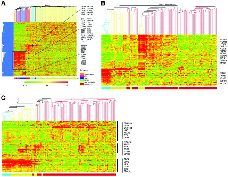
Waldenström macroglobulinemia (WM) is a B-cell malignancy characterized by the ability of the B-cell clone to differentiate into plasma cells. Although the clinical syndrome and the pathologic characteristics are well defined, little is known about its biology and controversy still exists regarding its cell of origin. In this gene-expression study, we compared the transcription profiles of WM with those of other malignant B cells including (chronic lymphocytic leukemia [CLL] and multiple myeloma [MM]) as well as normal cells (peripheral-blood B cells and bone marrow plasma cells). We found that WM has a homogenous gene expression regardless of 6q deletion status and clusters with CLL and normal B cells on unsupervised clustering with very similar expression profiles. Only a small gene set has expression profiles unique to WM compared to CLL and MM. The most significantly up-regulated gene is IL6 and the most significantly associated pathway for this set of genes is MAPK signaling. Thus, IL6 and its downstream signaling may be of biologic importance in WM. Further elucidation of the role of IL-6 in WM is warranted as this may offer a potential therapeutic avenue.


Figures

Figure 1.
Figure 1.
The GEP of WM is closer to CLL than MM. (A) Using 2162 gene probes that are variably expressed across the WM, CLL, and MM samples, unsupervised clustering of these samples together with normal B cells, PCs, MGUS, and SMM was performed. Selected genes that clustered together and were overexpressed in different sample clusters are highlighted. Restricting our analysis to only WM, CLL, and MM samples, unsupervised clustering was performed using published (B) CLL and (C) MM signatures. WM samples clustered predominantly with CLL samples and also exhibited closer approximation of the GEP using both signatures to CLL. Some genes of interests are highlighted. In both heatmaps, the colored bar at the bottom indicates the tumor type: blue, CLL; yellow, WM; and red, MM. The scale of the gene-expression data are similar to that in panel A.
Figure 2.
Figure 2.
Contributions of genes from contaminating cell populations. (A) Closer inspection of some of the genes within the gene cluster overexpressed in normal B cells, CLL, and WM revealed some interesting differences. In particular, the concurrent overexpression of NPTT (tdt), MME (CD10), and VPREB1 in the WM samples suggested likely contamination with pre-B cells. In addition, ZAP70 and CCNB1 (as a representative gene from the proliferation cluster overexpressed in a subset of patients with WM) overexpression is tightly associated with samples overexpressing NPTT (tdt), MME (CD10), and VPREB1. (B) To verify that tdt expression was originating from contaminating pre-B cells, immunostaining for tdt was performed on BM biopsies from patients with WM with and without the pre-B-cell contamination signature. This figure is representative for samples with the contamination signature. Malignant cells (CD20+) formed intramedullary clusters (marked by arrows; left panel, hematoxylin and eosin staining; middle panel, CD20 staining). In contrast, nuclear tdt staining was seen in scattered interstitial cells that represented pre-B cells (right panel). In samples without the contamination signature, no tdt+ cells were seen. All images were acquired using a 40×/0.8 numeric aperture objective lens. The microscope used is a Zeiss Axioskop (Carl Zeiss Microimaging, Thornwood, NY). Images were captured by the Olympus DP70 CCD camera using DP controller image capture software (Olympus, Center Valley, PA).
Figure 3.
Figure 3.
Gene-expression signature unique to WM. Seventy-three genes, 48 up-regulated and 25 down-regulated, constitute a gene-expression signature unique to WM. Here the samples are ordered according to diagnosis and genes according to fold difference in expression between WM and CLL and MM. Some interesting genes are highlighted. For a more complete list, see Table S1. The scale of the gene-expression data is similar to that in Figure 1A.

Similar articles

Cited by

References

    1. Owen RG, Treon SP, Al-Katib A, et al. Clinicopathological definition of Waldenström's macroglobulinemia: consensus panel recommendations from the Second International Workshop on Waldenström's macroglobulinemia. Semin Oncol. 2003;30: 110-115. - PubMed
    1. Kriangkum J, Taylor BJ, Strachan E, et al. Impaired class switch recombination (CSR) in Waldenstrom macroglobulinemia (WM) despite apparently normal CSR machinery. Blood. 2006; 107: 2920-2027. - PubMed
    1. Kriangkum J, Taylor BJ, Treon SP, Mant MJ, Belch AR, Pilarski LM. Clonotypic IgM V/D/J sequence analysis in Waldenstrom macroglobulinemia suggests an unusual B-cell origin and an expansion of polyclonal B cells in peripheral blood. Blood. 2004;104: 2134-2142. - PubMed
    1. Ackroyd S, O'Connor SJ, Owen RG. Rarity of IgH translocations in Waldenstrom macroglobulinemia. Cancer Genet Cytogenet. 2005;163: 77-80. - PubMed
    1. Schop RF, Kuehl WM, Van Wier SA, et al. Waldenstrom macroglobulinemia neoplastic cells lack immunoglobulin heavy chain locus translocations but have frequent 6q deletions. Blood. 2002;100: 2996-3001. - PubMed

Publication types

MeSH terms