Comparative Study
. 2006 Aug;12(8):1556-68.
doi: 10.1261/rna.106506. Epub 2006 Jun 27.

Regulation of poly(A) binding protein function in translation: Characterization of the Paip2 homolog, Paip2B

Affiliations
Comparative Study

Regulation of poly(A) binding protein function in translation: Characterization of the Paip2 homolog, Paip2B

Juan José Berlanga et al. RNA. 2006 Aug.

Abstract

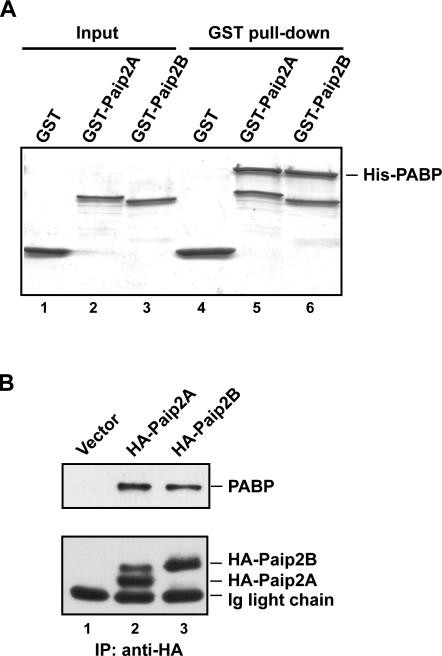
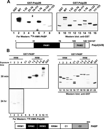
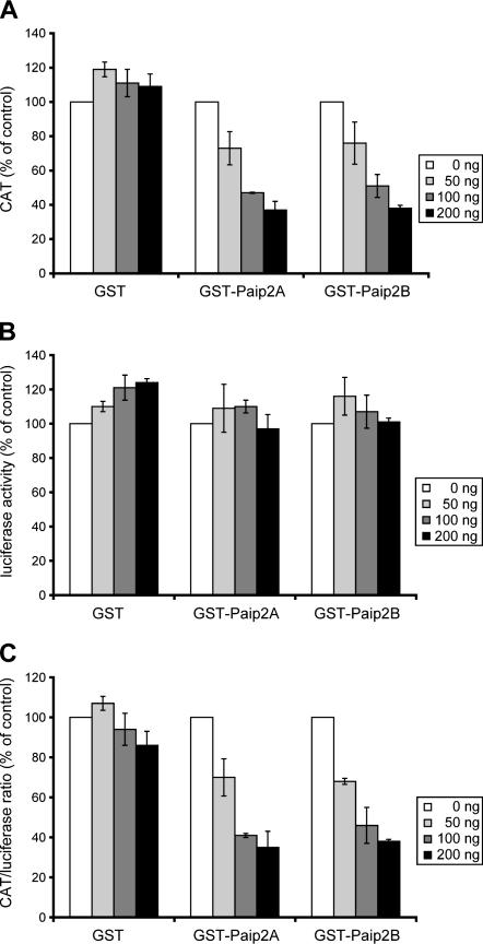
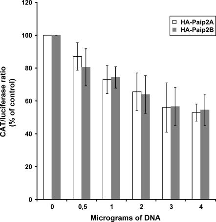
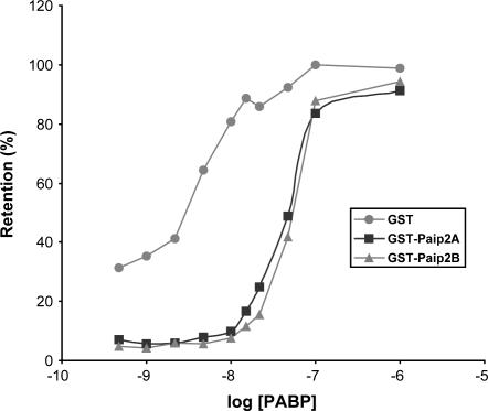
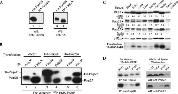
The 5' cap and 3' poly(A) tail of eukaryotic mRNAs act synergistically to enhance translation. This synergy is mediated via interactions between eIF4G (a component of the eIF4F cap binding complex) and poly(A) binding protein (PABP). Paip2 (PABP-interacting protein 2) binds PABP and inhibits translation both in vitro and in vivo by decreasing the affinity of PABP for polyadenylated RNA. Here, we describe the functional characteristics of Paip2B, a Paip2 homolog. A full-length brain cDNA of Paip2B encodes a protein that shares 59% identity and 80% similarity with Paip2 (Paip2A), with the highest conservation in the two PABP binding domains. Paip2B acts in a manner similar to Paip2A to inhibit translation of capped and polyadenylated mRNAs both in vitro and in vivo by displacing PABP from the poly(A) tail. Also, similar to Paip2A, Paip2B does not affect the translation mediated by the internal ribosome entry site (IRES) of hepatitis C virus (HCV). However, Paip2A and Paip2B differ with respect to both mRNA and protein distribution in different tissues and cell lines. Paip2A is more highly ubiquitinated than is Paip2B and is degraded more rapidly by the proteasome. Paip2 protein degradation may constitute a primary mechanism by which cells regulate PABP activity in translation.


Figures

FIGURE 1.
FIGURE 1.
Paip2B and Paip2A protein sequence alignment. (A) The amino acid sequences corresponding to human Paip2B and Paip2A proteins were aligned by using the CLUSTAL W (1.8) program. Identical residues are shown in black, similar residues are in gray, and sequence gaps are indicated by dashes. The proteins share 59% identity and 80% similarity. (B) Multiple sequence alignment of Paip2 proteins from different organisms using CLUSTAL W (1.8). Asterisks indicate residues that are completely conserved. Black shading indicates that the residues are present in at least six of the nine sequences. Dark gray shading indicates very conservative substitutions, and light gray shading indicates less conservative substitutions. (C) Neighbor-joining phylogenetic tree of Paip2 proteins using the two-parameter model of Kimura. The numbers at the nodes indicate bootstrap percentages after 1000 replications of bootstrap sampling. The bar indicates genetic distance.
FIGURE 2.
FIGURE 2.
Paip2B binds PABP in vitro and in vivo. (A) In GST pull-down experiments, complexes containing recombinant His-PABP and either GST, GST-Paip2A, or GST-Paip2B were immobilized by using glutathione-Sepharose beads, as described in the Materials and Methods. Bound proteins were eluted with Laemmli loading buffer and analyzed by SDS-PAGE and Coomassie blue staining. (B) 293T cells were transfected with plasmids containing either no insert or encoding HA-Paip2A or HA-Paip2B. Cell extracts were subjected to immunoprecipitation using anti-HA. The immune complexes were analyzed by Western blot using anti-PABP (upper panel) or anti-HA (lower panel).
FIGURE 3.
FIGURE 3.
Identification of respective binding sites within PABP and Paip2B. (A) Affinity-purified GST, GST-Paip2B (WT), and GST-Paip2B fragments were resolved by SDS-PAGE and assayed for PABP binding by far-Western blotting using a 32P-HMK-PABP probe (left panel) and by Western blotting using a polyclonal antibody to GST (for loading control, right panel). (B) Purified GST, GST-PABP (WT), and GST-PABP fragments (different combinations of RRM and C-terminal regions) were resolved by SDS-PAGE and analyzed for Paip2B binding by far-Western blotting using a 32P-HMK-Paip2B probe (left panel) and by Western blotting using a polyclonal antibody to GST (for loading control, right panel). Schematic representations of Paip2A/B and PABP shown below the respective panels identify the binding sites.
FIGURE 4.
FIGURE 4.
Effect of Paip2B on in vitro translation of capped and polyadenylated bicistronic CAT-HCV IRES-luciferase mRNA. Capped and polyadenylated bicistronic CAT-HCV IRES-luciferase mRNA was translated by using Krebs-2 cell-free translation reactions in the presence of increasing concentrations of GST (control), GST-Paip2B, or GST-Paip2A. (A) First cistron, cap-dependent, and poly(A)-dependent translation. (B) Second cistron, cap-independent, and poly(A)-independent translation. (C) First and second cistron translation ratio. CAT protein and luciferase activity values represent the average of at least three independent experiments. Error bars, SD of mean values.
FIGURE 5.
FIGURE 5.
Effect of Paip2B on in vivo translation of capped and polyadenylated bicistronic CAT-HCV IRES-luciferase mRNA. 293T cells were cotransfected with a fixed amount of plasmid encoding bicistronic CAT-HCV IRES-luciferase mRNA and increasing amounts of plasmids encoding HA-Paip2B or HA-Paip2A. At 24 h post-transfection, cells were lysed and CAT expression and luciferase activity were measured in the lysates. The results are presented as the ratio of the expression of CAT to that of luciferase. CAT protein and luciferase activity values represent the average of at least three independent experiments. Error bars, SD of mean values.
FIGURE 6.
FIGURE 6.
Paip2B, similar to Paip2A, displaces PAPB from poly(A) RNA. Filter binding assays were performed as described in the Materials and Methods using constant concentrations of 32P-poly(A) RNA (3000 cpm, 0.01 nM) and either GST-Paip2A, GST-Paip2B, or GST (100nM), and increasing concentrations of PABP, as indicated. Radioactivity in the spots was counted in a scintillation counter, and the results were plotted as the percentage of radioactive probe retained on the filter. The values represent the mean of three independent experiments that yielded very similar results.
Figure 7.
Figure 7.
Paip2B and Paip2A mRNA distribution. Membranes containing electrophoretically separated mRNAs from different human tissues (Human Multiple Tissue Northern blot membranes from Clontech), were hybridized by using labeled cDNA probes specific for Paip2B, Paip2A, and actin, as described in the Materials and Methods.
FIGURE 8.
FIGURE 8.
Characterization of an antibody specific for Paip2B: tissue-specific distribution of Paip2 proteins. A Paip2B-specific antibody was obtained by immunizing rabbits with GST-Paip2B (residues 1–21). (A) 293T cells were transfected with plasmids encoding HA-Paip2A or HA-Paip2B, and cell extracts were analyzed by Western blot using anti-Paip2B and anti-HA. (B) 293T cells were transfected with plasmids containing either no insert (negative control) or encoding either HA-Paip2A or HA-Paip2B. Cell extracts were subjected to immunoprecipitation using anti-Paip2A or anti-Paip2B, as indicated. The immune complexes were analyzed by far-Western blotting using 32P-HMK-PABP as a probe. The electrophoretic migration of endogenous and HA-tagged Paip2 proteins is indicated. (C) Equal amounts of total protein (150 μg) from mouse tissue extracts were subjected to SDS-PAGE and analyzed by Western blot using antibodies against Paip2B, Paip2A, PABP, or eIF2α (as a loading control), as indicated, and by far-Western blot using a 32P-HMK-PABP probe (bottom panel). The values under the Western blot panels represent the intensities of bands in each tissue normalized with respect to the corresponding eIF2alpha bands. For comparison, the value obtained for the small intestine sample was set as one. (D) 293T, HeLa, COS-7, and NIH 3T3 cell extracts were prepared, and equal amounts of protein were subjected either to immunoprecipitation using anti-Paip2B or anti-Paip2A antibodies or to SDS-PAGE (50 μg of whole-cell extract) and Western blotting using the same antibodies. The immune complexes were analyzed by far-Western blotting using a 32P-HMK-PABP probe.
FIGURE 9.
FIGURE 9.
Differential regulation of Paip2A and Paip2B protein levels. (A) HEK 293T cells were transiently transfected by using increasing amounts of plasmids encoding HA-Paip2A or HA-Paip2B, as indicated. At 24 h post-transfection, cells were lysed and lysates were analyzed by Western blot using anti-HA. Samples from two independent experiments were loaded. (B) HEK 293T cells were transfected with plasmids encoding HA-tagged proteins (PABP, Paip2A, or Paip2B) and metabolically labeled for 2 h with [35S]methionine and chased with cold methionine for 5 h in the absence or presence of 50 μM LLnL (proteasome inhibitor). Cell lysates were subjected to immunoprecipitation with anti-HA antibody, and immune complexes were washed and resolved by SDS-PAGE gels, dried, and exposed to X-ray film. (C) The same procedure as in B, but cells were metabolically labeled for 1 h with [35S]methionine in the absence or presence of 10 μM MG132 (proteasome inhibitor) and were chased or not chased with cold methionine for 5 h, in the same conditions. After immunoprecipitation with anti-HA antibody, immune complexes were first washed and proteins resolved by SDS-PAGE gels and then transferred to nitrocellulose membranes and exposed to X-ray film (upper panel). After autoradiography membranes were analyzed by Western blot using anti-HA and anti-PABP antibodies (lower panels) as indicated. Numbers under the upper autoradiography panel represent the intensity of bands in each lane. For comparison, the values obtained at chase time 0 in the absence of MG132 were taken as 100%. The numbers between brackets were obtained when the values at chase time 0 in the presence of MG132 were taken as 100%. (D) Cells were cotransfected with plasmids encoding either HA-Paip2A or HA-Paip2B and a plasmid encoding His6-ubiquitin. At the indicated times before lysis, some of the cells were treated with 50 μM LLnL. Cells were lysed in guanidine-HCl denaturing buffer, and His6-tagged complexes were purified by using a metal affinity resin as described in the Materials and Methods. Purified proteins were eluted from the resin with 1× Laemmli sample buffer, and the presence of HA-Paip2A or HA-Paip2B was revealed by Western blotting with anti-HA (upper panel). Aliquots of cell lysates were also analyzed in the same way for the presence of HA-Paip2A and HA-Paip2B. (E) The same procedure as in D (upper panel), but cells were also transfected with an empty plasmid or with plasmids encoding either untagged Paip2A or Paip2B in combination with the His6-ubiquitin plasmid. Half of the cells were treated with LLnL for 6 h before lysis. Proteins were visualized by Western blot using appropriate specific antisera for Paip2A and Paip2B proteins.

References

    1. Altschul S.F., Gish W., Miller W., Myers E.W., Lipman D.J. Basic local alignment search tool. J. Mol. Biol. 1990;215:403–410. - PubMed
    1. Blanar M.A., Rutter W.J. Interaction cloning: Identification of a helix-loop-helix zipper protein that interacts with c-Fos. Science. 1992;256:1014–1018. - PubMed
    1. Bravo J., Aguilar-Henonin L., Olmedo G., Guzman P. Four distinct classes of proteins as interaction partners of the PABC domain of Arabidopsis thaliana Poly(A)-binding proteins. Mol. Genet. Genomics. 2005;272:651–665. - PubMed
    1. Callaghan M.J., Russell A.J., Woollatt E., Sutherland G.R., Sutherland R.L., Watts C.K. Identification of a human HECT family protein with homology to the Drosophila tumor suppressor gene hyperplastic discs. Oncogene. 1998;17:3479–3491. - PubMed
    1. Combet C., Blanchet C., Geourjon C., Deleage G. NPS@: Network protein sequence analysis. Trends Biochem. Sci. 2000;25:147–150. - PubMed

Publication types

MeSH terms