. 2004 May;1(2):165-76.
doi: 10.1017/s1740925x04000237.

Downregulation of uPA, uPAR and MMP-9 using small, interfering, hairpin RNA (siRNA) inhibits glioma cell invasion, angiogenesis and tumor growth

Affiliations

Downregulation of uPA, uPAR and MMP-9 using small, interfering, hairpin RNA (siRNA) inhibits glioma cell invasion, angiogenesis and tumor growth

Christopher S Gondi et al. Neuron Glia Biol. 2004 May.

Abstract

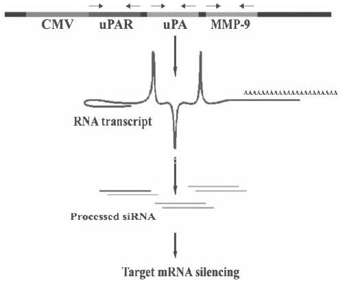
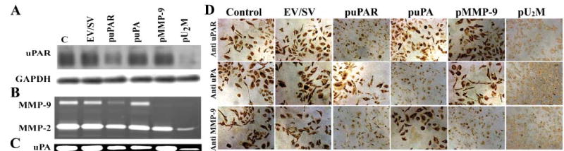
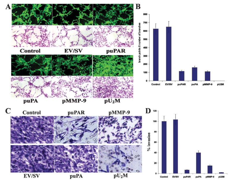
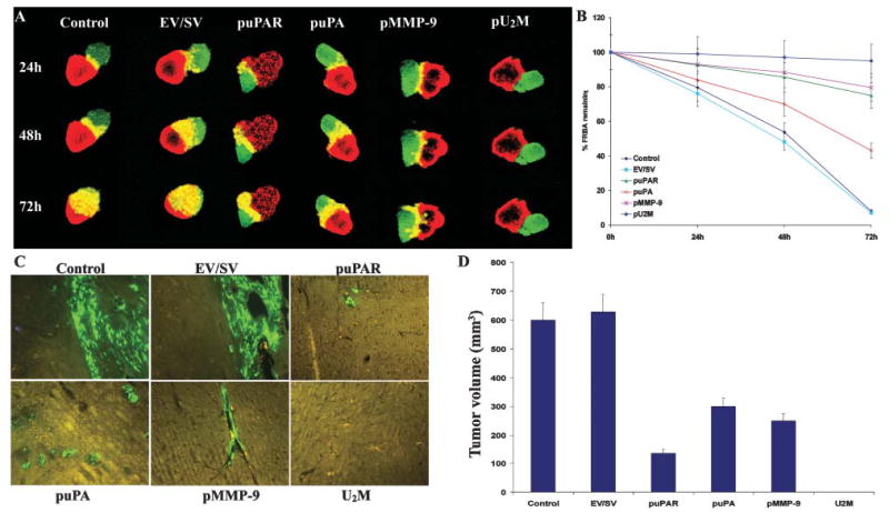
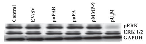
The diffuse, extensive infiltration of malignant gliomas into the surrounding normal brain is believed to rely on modification of the proteolysis of extracellular matrix components. Our previous results clearly demonstrate that uPA, uPAR and MMP-9 concentrations increase significantly during tumor progression and that tumor growth can be inhibited with antisense stable clones of these molecules. Because antisense-mediated gene silencing does not completely inhibit the translation of target mRNA and high concentrations of antisense molecules are required to achieve gene silencing, we used the RNAi approach to silence uPA, uPAR and MMP-9 in this study. We examined a cytomegalovirus (CMV) promoter-driven DNA-template approach to induce hairpin RNA (hpRNA)-triggered RNAi to inhibit uPA, uPAR and MMP-9 gene expression with a single construct. uPAR protein levels and enzymatic activity of uPA and MMP-9 were found to significantly decrease in cells transfected with a plasmid expressing hairpin siRNA for uPAR, uPA and MMP-9. pU(2)M-transfected SNB19 cells significantly decreased uPA, uPAR and MMP-9 expression compared to mock and EV/SV-transfected cells, determined by immunohistochemical analysis. Furthermore, the effect of the single constructs for these molecules was a specific inhibition of their respective protein levels, as demonstrated by immunohistochemical analysis. After transfection with a plasmid vector expressing dsRNA for uPA, uPAR and MMP-9, glioma-cell invasion was retarded compared with mock and EV/SV-treated groups, demonstrated by Matrigel-invasion assay and spheroid-invasion assay. Downregulation of uPA, uPAR and MMP-9 using RNAi inhibited angiogenesis in an in vitro (co-culture) model. Direct intratumoral injections of plasmid DNA expressing hpRNA for uPA, uPAR and MMP-9 significantly regressed pre-established intracranial tumors in nude mice. In addition, cells treated with RNAi for uPAR, uPA and MMP-9 showed reduced pERK levels compared with parental and EV/SV-treated SNB19 cells. Our results support the therapeutic potential of RNAi as a method for gene therapy in treating gliomas.


Figures

Fig. 1
Fig. 1. Schematic of the possible formation of hpRNA molecules from a single, CMV-driven, tri-inverted repeat construct for uPAR, uPA and MMP-9
The powerful CMV viral promoter drives the formation of an RNA molecule that possesses self-complementary inverted repeats for uPA, uPAR and MMP-9.
Fig. 2
Fig. 2. Western blotting, fibrin/gelatin zymography and immunohistochemical analysis of uPA and MMP-9
SNB19 cells were either mock transfected or transfected with an empty vector/scrambled vector (EV/SV) and vectors encoding siRNA uPAR (puPAR), uPA (puPA), MMP-9 (pMMP-9) and a combination of the three together (pU2M). After a 3-day-incubation period, total cell lysates were prepared in extraction buffer and 50 μg of protein from these samples were separated by 12% non-reducing SDS-PAGE and immunoblotted with anti-uPAR antibody (A). GAPDH was immunoprobed simultaneously as a loading control. Conditioned medium was collected from these samples (20 μg) and gelatin and fibrin zymography performed to detect MMP-9 (B) and uPA activity (C). (D) SNB19 cells (1×10⁴) were seeded onto Lab-Tek II chamber slides and either mock transfected or transfected with EV/SV and vectors encoding siRNA puPA, puPAR and pMMP-9 either singly or together (pU2M). After 72 hours, cells were fixed, washed for 1 hour with blocking buffer and stained for uPAR, uPA and MMP-9 expression using specific antibodies for uPA, uPAR and MMP-9.
Fig. 3
Fig. 3. Inhibition of glioma angiogenesis and invasion by siRNA constructs
SNB19 cells (2×10⁴) were seeded onto 8-well-chamber slides and transfected with EV/SV and vectors encoding siRNA uPAR (puPAR), uPA (puPA), MMP-9 (pMMP-9) and a combination of three together (pU2M). After a 24-hour-incubation period, the medium was removed, cells were co-cultured with either 4×10⁴ human endothelial cells or 4×10⁴ endothelial cells alone and were grown in the presence of conditioned media. After 72 hours endothelial cells were stained for factor VIII antigen in the co-cultures (green florescence). Cells grown in the preserved conditioned media were H&E stained and examined under either a florescent microscope or a bright field microscope (A). (B) Quantification of angiogenesis in co-cultures infected with EV/SV, puPAR, puPA, pMMP-9 and pU2M vectors. Values are mean±SD of four experiments. SNB19 cells were trypsinized 72 hours after transfection with EV/SV, puPAR, puPA, pMMP-9 and pU2M, washed with PBS and resuspended in serum-free medium. (C) Invasion assays were carried out in a 12-well transwell unit on polycarbonate filters with 8-μm pores coated with matrigel (0.7 mg ml−1). After a 24-hour-incubation period, the cells that had passed through the filter into the lower wells were stained, counted and photographed under a bright-field microscope. (D) The invasion was quantified as described Methods (D). Values are mean±SD of four experiments.
Fig. 4
Fig. 4. Inhibition and regression of invasiveness and tumor growth by siRNA in spheroid and intracranial assays
(A) Invasiveness of glioma spheroids was measured by co-culturing glioma spheroids with fetal rat brain aggregates. Spheroids of SNB19 cells were transfected with EV/SV, puPA, puPAR, pMMP-9 and pU2M and stained with DiI and co-cultured with DiO-stained fetal rat brain aggregates. Serial, 1-μm-thick sections were obtained from the surface through the center of the co-cultures with a confocal laser scanning microscope at the indicated time points. (B) The remaining volume of the rat brain aggregate transfected with EV/SV, puPA, puPAR, pMMP-9 and pU2M was measured as described in Methods. The values are mean ±SD of three experiments. (C,D) RNA-mediated regression of pre-established tumor growth. SNB19 GFP cells in suspension (2×10⁶ in 10 μl serum-free medium) were injected intracranially. One week later, the mice were injected with either EV/SV or siRNA-expressing vectors (puPAR, puPA, pMMP-9 and pU2M) using an Alzet mini osmotic pump (constructs diluted to 1.5 μg ml−1 in PBS and injected at 0.25 μg hour−1, with six mice in each group). After a 5-week follow-up period, mice were sacrificed, their brains removed, paraffin embedded and sectioned. Sections were observed under fluorescence microscopy for GFP-expressing cells. (D) Semi-quantification of tumor volume in control, EV/SV, puPAR, puPA, pMMP-9 and pU2M-treated groups was assessed after 5 weeks. Data are shown mean±SD of six animals from each group.
Fig. 5
Fig. 5. RNAi-mediated downregulation of uPAR, uPA and MMP-9 reduces phosphorylation of ERK
Western blot analysis of total and phosphorylated ERK (pERK) protein after transfection of glioblastoma cells with EV/SV, puPAR, puPA, pMMP-9 and pU2M constructs. GAPDH levels served as loading control.

References

    1. Alemany R, Gomez-Manzano C, Balague C, Yung WK, Curiel DT, Kyritsis AP, Fueyo J. Gene therapy for gliomas: molecular targets, adenoviral vectors, and oncolytic adenoviruses. Experimental Cell Research. 1999;252:1–12. - PubMed
    1. Alonso DF, Farias EF, Ladeda V, Davel L, Puricelli L, Bal de Kier JE. Effects of synthetic urokinase inhibitors on local invasion and metastasis in a murine mammary tumor model. Breast Cancer Research Treatment. 1996;40:209–223. - PubMed
    1. Alonso DF, Tejera AM, Farias EF, Bal de Kier JE, Gomez DE. Inhibition of mammary tumor cell adhesion, migration, and invasion by the selective synthetic urokinase inhibitor B428. Anticancer Research. 1998;18:4499–4504. - PubMed
    1. Andreasen PA, Kjoller L, Christensen L, Duffy MJ. The urokinase-type plasminogen activator system in cancer metastasis: a review. International Journal of Cancer. 1997;72:1–22. - PubMed
    1. Behzadian MA, Windsor LJ, Ghaly N, Liou G, Tsai NT, Caldwell RB. VEGF-induced paracellular permeability in cultured endothelial cells involves urokinase and its receptor. FASEB Journal. 2003;17:752–754. - PubMed

LinkOut - more resources