

Transcranial motor-evoked potentials monitoring can detect spinal cord ischemia more rapidly than spinal cord-evoked potentials monitoring during aortic occlusion in rats
- PMID: 16804674
- PMCID: PMC2200716
- DOI: 10.1007/s00586-006-0165-1
Transcranial motor-evoked potentials monitoring can detect spinal cord ischemia more rapidly than spinal cord-evoked potentials monitoring during aortic occlusion in rats
Abstract
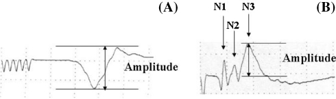
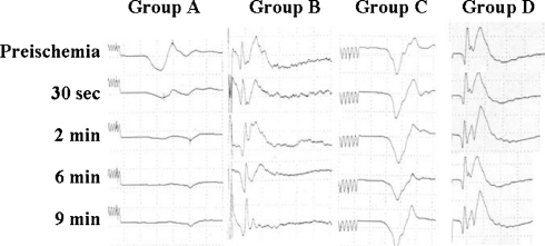
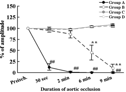
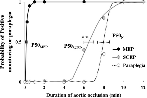
In this study, we evaluated the efficacy of transcranial motor-evoked potentials (tc-MEPs), compared with segmental spinal cord-evoked potentials (SCEPs), for detecting spinal cord ischemia (SCI) and assessed the relationship between neurological outcome and tc-MEPs or SCEPs in the rat aortic occlusion model. In the rats, SCI was induced by aortic occlusion for 10 min with a balloon catheter. At first, tc-MEPs (Group A: n = 6) or segmental SCEPs (Group B: n = 6) was recorded during SCI. Second, in using the quantal bioassay for the relationship between an interval of aortic occlusion and the probability of positive response in tc-MEPs or segmental SCEPs, the P50(MEP) and P50(SCEP) which represent the interval of aortic occlusion associated with 50% probability of assessment of ischemic spinal cord dysfunction by tc-MEP and SCEP were analyzed. The amplitude of tc-MEPs decreased significantly at 30 s and disappeared completely at 2 min after aortic occlusion. In Group B, it took about 6 min after aortic occlusion to diminish SCEP signal amplitude by approximately 50%. P50(MEP) obtained in the quantal analysis was 0.3 +/- 0.1 min. P50(SCEP) was calculated as 6.2 +/- 0.5 min that was significantly (P < 0.01) longer than P50(MEP). Our data indicated that tc-MEP monitoring could detect the onset of SCI so rapidly in comparison with segmental SCEP monitoring, which could provide therapeutic windows in a surgical approach that includes spinal cord protection.
Figures
Similar articles
-
Myogenic transcranial motor evoked potentials monitoring cannot always predict neurologic outcome after spinal cord ischemia in rats.J Thorac Cardiovasc Surg. 2005 Jan;129(1):46-52. doi: 10.1016/j.jtcvs.2004.05.007. J Thorac Cardiovasc Surg. 2005. PMID: 15632824
-
Transcranial myogenic motor-evoked potentials after transient spinal cord ischemia predicts neurologic outcome in rabbits.J Vasc Surg. 2004 Jan;39(1):207-13. doi: 10.1016/s0741-5214(03)01050-4. J Vasc Surg. 2004. PMID: 14718841
-
[Changes of somatosensory and transcranial magnetic stimulation motor evoked potentials in experimental spinal cord injury].Zhonghua Yi Xue Za Zhi. 2008 Mar 18;88(11):773-7. Zhonghua Yi Xue Za Zhi. 2008. PMID: 18683688 Chinese.
-
[Evoked potentials monitoring in aortic surgery].Kyobu Geka. 2014 Jul;67(8):630-5. Kyobu Geka. 2014. PMID: 25138930 Review. Japanese.
-
Intraoperative neurophysiological monitoring of the spinal cord during spinal cord and spine surgery: a review focus on the corticospinal tracts.Clin Neurophysiol. 2008 Feb;119(2):248-64. doi: 10.1016/j.clinph.2007.09.135. Epub 2007 Nov 28. Clin Neurophysiol. 2008. PMID: 18053764 Review.
Cited by
-
Magnetic resonance imaging and motor-evoked potentials in spinal cord infarction: report of two cases.Neurol Sci. 2010 Aug;31(4):505-9. doi: 10.1007/s10072-010-0263-z. Epub 2010 May 5. Neurol Sci. 2010. PMID: 20443040
-
In Vivo Neuroprotective Effect of Histidine-Tryptophan-Ketoglutarate Solution in an Ischemia/Reperfusion Spinal Cord Injury Animal Model.Korean J Thorac Cardiovasc Surg. 2016 Aug;49(4):232-41. doi: 10.5090/kjtcs.2016.49.4.232. Epub 2016 Aug 5. Korean J Thorac Cardiovasc Surg. 2016. PMID: 27525231 Free PMC article.
-
Heat shock proteins as biomarkers for the rapid detection of brain and spinal cord ischemia: a review and comparison to other methods of detection in thoracic aneurysm repair.Cell Stress Chaperones. 2011 Mar;16(2):119-31. doi: 10.1007/s12192-010-0224-8. Epub 2010 Aug 30. Cell Stress Chaperones. 2011. PMID: 20803353 Free PMC article. Review.
-
Attenuation of Cortically Evoked Motor-Neuron Potential in Streptozotocin-Induced Diabetic Rats: A Study about the Effect of Diabetes upon Cortical-Initiated Movement.Biomed Res Int. 2020 Feb 25;2020:1942534. doi: 10.1155/2020/1942534. eCollection 2020. Biomed Res Int. 2020. PMID: 32185194 Free PMC article.
-
Identification of injury type using somatosensory and motor evoked potentials in a rat spinal cord injury model.Neural Regen Res. 2023 Feb;18(2):422-427. doi: 10.4103/1673-5374.346458. Neural Regen Res. 2023. PMID: 35900440 Free PMC article.
References
-
- Crawford ES, Crawford JL, Safi HJ, Coselli JS, Hess KR, Brooks B, Norton HJ, Glaeser DH. Thoracoabdominal aortic aneurysms: preoperative and intraoperative factors determining immediate- and long-term results of operations in 605 patients. J Vasc Surg. 1986;3:389–404. doi: 10.1067/mva.1986.avs0030389. - DOI - PubMed
Publication types
MeSH terms
LinkOut - more resources
Full Text Sources
Research Materials
