Comparative Study
. 2006 Jun;22(6):562-74.
doi: 10.3928/1081-597X-20060601-08.

Effect of prophylactic and therapeutic mitomycin C on corneal apoptosis, cellular proliferation, haze, and long-term keratocyte density in rabbits

Affiliations
Comparative Study

Effect of prophylactic and therapeutic mitomycin C on corneal apoptosis, cellular proliferation, haze, and long-term keratocyte density in rabbits

Marcelo V Netto et al. J Refract Surg. 2006 Jun.

Abstract

Purpose: To determine the mechanism through which topical mitomycin C prevents and treats corneal haze after photorefractive keratectomy (PRK) and to examine the effects of dosage and duration of exposure.

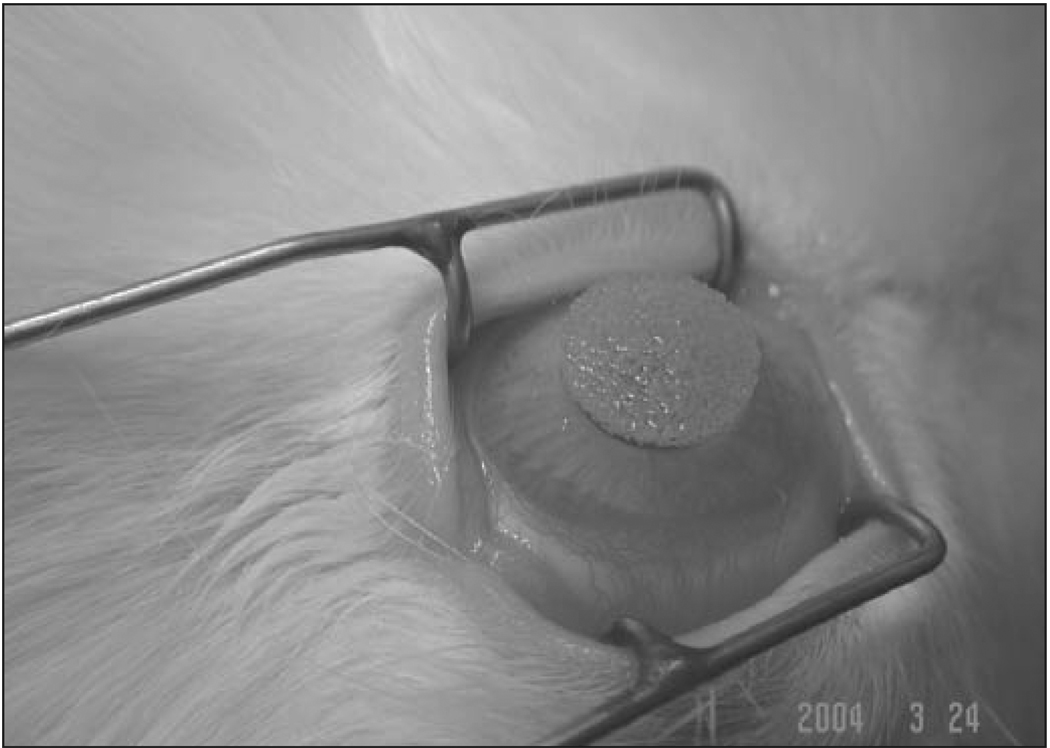
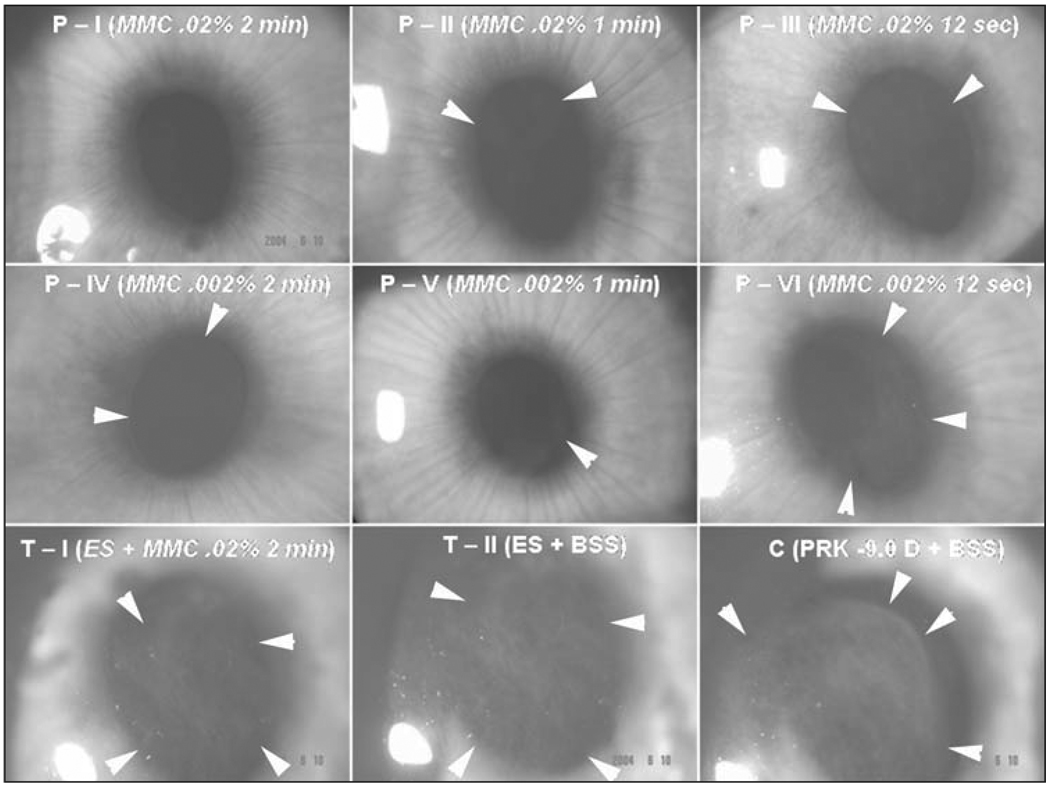
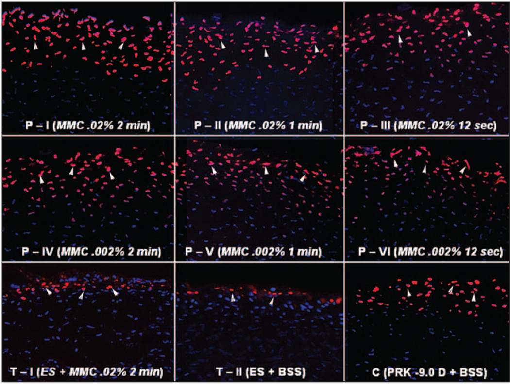
Methods: In 224 New Zealand rabbits, -9.0 diopter PRK with mitomycin C or balanced salt solution was performed. Haze level was graded at the slit-lamp. Rabbits were sacrificed at 4 hours, 24 hours, 4 weeks, or 6 months after surgery and immunohistochemistry was performed with TUNEL assay, Ki67, and alpha-SMA.

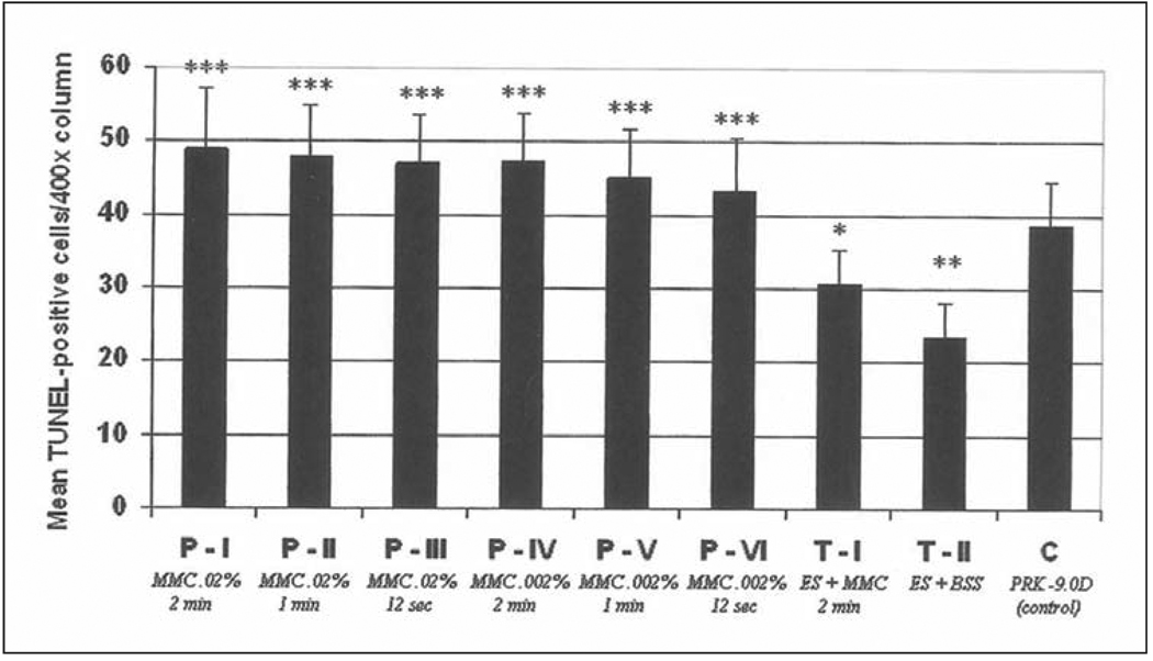
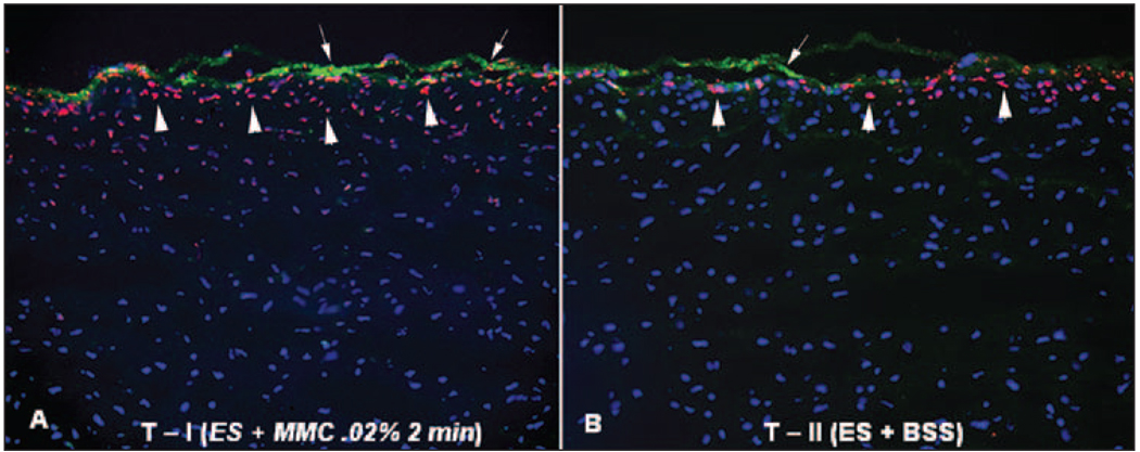
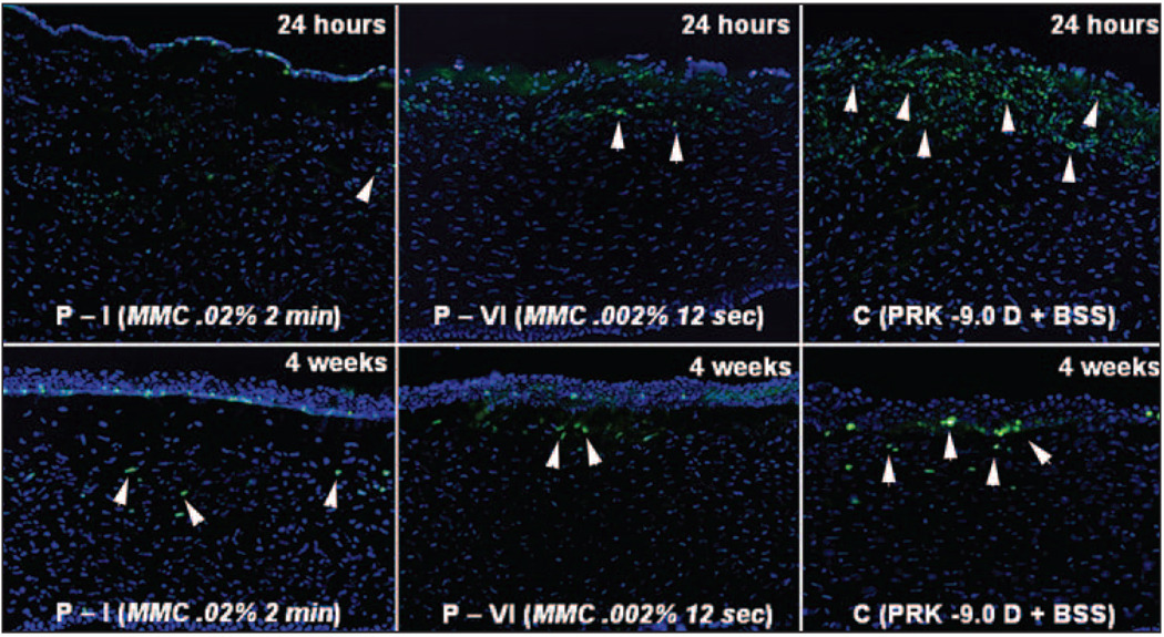
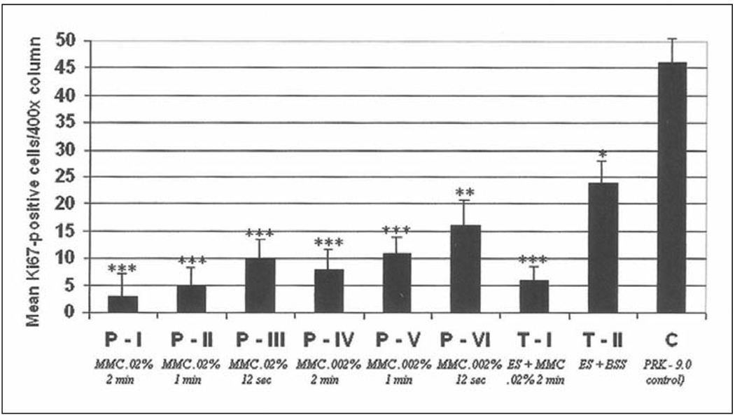
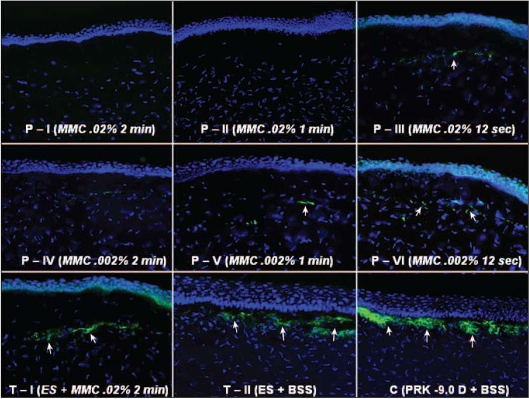
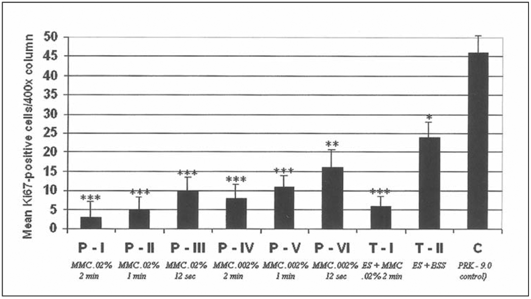
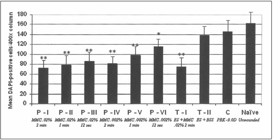
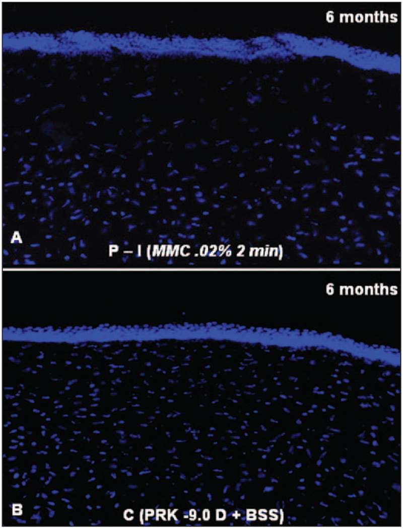
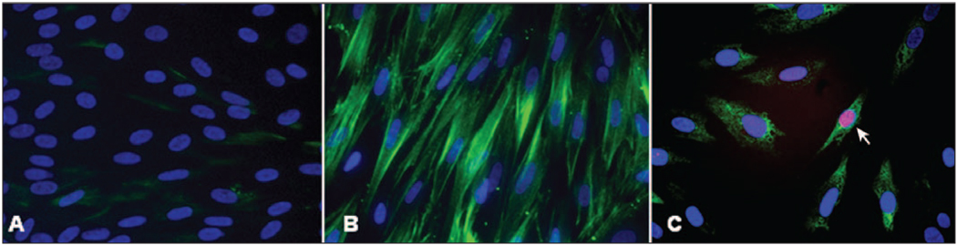
Results: TUNEL-positive apoptotic cells marginally increased in all mitomycin C groups whereas Ki67-positive mitotic cells decreased significantly following mitomycin C application. A greater decrease in myofibroblasts was noted with prophylactic mitomycin C treatment than therapeutic mitomycin C treatment. There was, however, an anterior stromal acellular zone (approximately 20% of the total stroma) in eyes treated with mitomycin C, which persisted to the maximum follow-up of 6 months.

Conclusions: Mitomycin C treatment induces apoptosis of keratocytes and myofibroblasts, but the predominate effect in inhibiting or treating haze appears to be at the level of blocked replication of keratocytes or other progenitor cells of myofibroblasts. Treatment with 0.002% mitomycin C for 12 seconds to 1 minute appears to be just as effective as higher concentrations for longer duration in the rabbit model. However, a persistent decrease in keratocyte density in the anterior stroma could be a warning sign for future complications and treatment should be reserved for patients with significant risk of developing haze after PRK.


Figures

Figure 1
Figure 1
Topical application of mitomycin C on the rabbit cornea using a soaked circular sponge.
Figure 2
Figure 2
Representative slit-lamp photographs showing subepithelial haze patterns and locations restricted to the area of corneal ablation. Faint 0.5 to 1.5 subepithelial corneal haze can be seen in the prophylactic mitomycin C groups P-I to P-VI with differing concentrations of mitomycin C and exposure times noted. Grade II subepithelial haze is seen in one specimen (T-1) that was not very effectively treated with therapeutic 0.02% mitomycin C for 2 minutes. Moderate to severe subepithelial corneal haze (grade III) can be noted in the therapeutic control group treated with BSS (T-II) and the prophylactic control group (C). Note that the subepithelial haze tends to terminate exactly at the margin of the excimer laser ablation (arrows in C) (original magnification ×10).
Figure 3
Figure 3
TUNEL assay for apoptotic cells 4 hours after surgery of the central cornea from rabbits that had −9.0 D PRK for myopia followed by mitomycin C or BSS application. Different concentrations and exposure times of mitomycin C are indicated. These are representative results for each group. Cell nuclei are stained blue with DAPI and TUNEL-positive cells are stained red (arrows) (original magnification ×200).
Figure 4
Figure 4
Quantitation of TUNEL-positive apoptotic cells in the anterior stroma (per 400× column) at 4 hours after laser surgery in all prophylactic and therapeutic mitomycin C groups and the control (BSS) group. *, **, *** are used to indicate groups that are significantly different from the control group. In addition, * is significantly different from **, * is significantly different from ***, and ** is significantly different from ***.
Figure 5
Figure 5
TUNEL assay and α-smooth muscle actin double-staining of the central rabbit corneas at 4 hours after epithelial scrape followed by mitomycin C or BSS application in eyes that had −9.0 D PRK 4 weeks earlier with development of significant subepithelial corneal haze (therapeutic treatment with mitomycin C or BSS). All cell nuclei are stained blue with DAPI. TUNEL-positive cells are stained red (arrows) and α-smooth muscle actin-positive cells are stained green (arrowheads). Overall, significantly more TUNEL-positive cells were noted in A) group T-I than in B) group T-II (original magnification ×200).
Figure 6
Figure 6
Immunohistochemical Ki67 staining to detect cell mitosis in the central cornea in eyes that had −9.0 D PRK for myopia followed by mitomycin C or BSS application at 24 hours or 4 weeks after treatment. There is marked cell proliferation (arrowheads) in the anterior stroma in the BSS group (C) at 24 hours, with some still detectable 4 weeks after treatment. Minimal proliferation cells in the anterior stroma are noted in corneas treated with 0.02% mitomycin C for 2 minutes (P-I) at 24 hours after treatment. In corneas treated with 0.002% mitomycin C for 12 seconds (P-VI), there are more anterior stromal cells undergoing mitosis at 24 hours after treatment than the higher concentration mitomycin C group, but still far fewer than in the control group (original magnification ×200).
Figure 7
Figure 7
Quantitation of Ki67-positive cells in the anterior stroma (per 400× column) at 24 hours after laser surgery in prophylactic and therapeutic mitomycin C-treated groups and the BSS-treated control group. *, **, *** are used to indicate groups that are significantly different from the control group. In addition, * is significantly different from **, * is significantly different from ***, and ** is significantly different from ***.
Figure 8
Figure 8
Immunohistochemical staining for α-smooth muscle actin in the central cornea of representative sections of the different groups at 4 weeks after surgery. Cells nuclei are stained blue with DAPI and α-smooth muscle actin-positive myofibroblasts are stained green (arrows). Note that in mitomycin C-treated corneas myofibroblasts lie beneath an acellular to pauci-cellular zone of anterior stroma whereas in BSS-treated corneas (either therapeutic control group T-II or PRK control) the myofibroblasts are localized immediately beneath the epithelium. Also note that more DAPI stained nuclei are present in the anterior stroma of all of the prophylactic group corneas treated with 0.002% mitomycin C (P-IV, P-V, and P-VI) compared to the corneas treated with 0.02% mitomycin C (P-I, P-II, and P-III) (original magnification ×400).
Figure 9
Figure 9
Quantitation of α-smooth muscle actin-positive cells in the anterior stroma (per 400× column) at 4 weeks after laser surgery in prophylactic and therapeutic mitomycin C groups and the BSS-treated controls. *, **, *** are used to indicate groups that are significantly different from the control group. In addition, * is significantly different from **, * is significantly different from ***, and ** is significantly different from ***.
Figure 10
Figure 10
Quantitation of DAPI-positive cells in the anterior stroma (per 400× column) at 4 weeks after laser surgery in prophylactic and therapeutic mitomycin C-treated groups and BSS-treated control groups. *, **, *** are used to indicate groups that are significantly different from the control group. In addition, * is significantly different from **, * is significantly different from ***, and ** is significantly different from ***.
Figure 11
Figure 11
DAPI staining of cell nuclei in the central corneas from rabbits at 6 months after −9.0 D PRK and prophylactic treatment with A) 0.02% mitomycin C or B) BSS for 2 minutes. Note the markedly diminished keratocyte density in the anterior stroma of the cornea after mitomycin C treatment (original magnification ×400).
Figure 12
Figure 12
Mitomycin C-induced apoptosis of myofibroblasts in vitro. A) Keratocytes cultured in the absence of TGFβ1. Nuclei stain with DAPI, with no detectible α-smooth muscle actin-positive cells. B) Myofibroblasts generated in the presence of TGFβ1 stain green for α-smooth muscle actin, with nuclei staining with DAPI. C) After treatment of myofibroblasts with 0.02% mitomycin C for 2 minutes, cells undergo triple staining—immunohistochemistry for α-smooth muscle actin, TUNEL assay, and DAPI staining. Cells are shrunken and the nucleus of one cell (arrow) is stained with the TUNEL assay.

References

    1. Rajan MS, Jaycock P, O’Brart D, Nystrom HH, Marshall J. A long-term study of photorefractive keratectomy; 12-year followup. Ophthalmology. 2004;111:1813–1824. - PubMed
    1. El-Maghraby A, Salah T, Waring GO, 3rd, Klyce SD, Ibrahim O. Randomized bilateral comparison of excimer laser in situ keratomileusis and photorefractive keratectomy for 2.50 to 8.00 diopters of myopia. Ophthalmology. 1999;106:447–457. - PubMed
    1. Hersh PS, Stulting RD, Steinert RF, Waring GO, III, Thompson KP, O’Connell M, Doney K, Schein OD. Results of phase III excimer laser photorefractive keratectomy for myopia. The Summit PRK Study Group. Ophthalmology. 1997;104:1535–1553. - PubMed
    1. Mohan RR, Hutcheon AE, Choi R, Hong J, Lee J, Mohan RR, Ambrosio R, Jr, Zieske JD, Wilson SE. Apoptosis, necrosis, proliferation, and myofibroblast generation in the stroma following LASIK and PRK. Exp Eye Res. 2003;76:71–87. - PubMed
    1. Moller-Pedersen T, Cavanagh HD, Petroll WM, Jester JV. Corneal haze development after PRK is regulated by volume of stromal tissue removal. Cornea. 1998;17:627–639. - PubMed

Publication types

MeSH terms

Substances