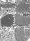
Endoplasmic reticulum architecture: structures in flux
- PMID: 16806883
- PMCID: PMC4264046
- DOI: 10.1016/j.ceb.2006.06.008
Endoplasmic reticulum architecture: structures in flux
Abstract
The endoplasmic reticulum (ER) is a dynamic pleiomorphic organelle containing continuous but distinct subdomains. The diversity of ER structures parallels its many functions, including secretory protein biogenesis, lipid synthesis, drug metabolism and Ca2+ signaling. Recent studies are revealing how elaborate ER structures arise in response to subtle changes in protein levels, dynamics, and interactions as well as in response to alterations in cytosolic ion concentrations. Subdomain formation appears to be governed by principles of self-organization. Once formed, ER subdomains remain malleable and can be rapidly transformed into alternative structures in response to altered conditions. The mechanisms that modulate ER structure are likely to be important for the generation of the characteristic shapes of other organelles.
Figures

References
-
- Baumann O, Walz B. Endoplasmic reticulum of animal cells and its organization into structural and functional domains. Int Rev Cytol. 2001;205:149–214. - PubMed
-
- Snapp EL. Endoplasmic reticulum biogenesis: proliferation and differentiation. In: Mullins C, editor. The biogenesis of cellular organelles Molecular Biology Intelligence Unit. C. Landes Bioscence, Georgetown TX and Kluwer Academic/Plenum Publishers; New York: 2004. pp. 63–95.
-
- Kepes F, Rambourg A, Satiat-Jeunemaitre B. Morphodynamics of the secretory pathway. Int Rev Cytol. 2005;242:55–120. - PubMed
Publication types
MeSH terms
Grants and funding
LinkOut - more resources
Full Text Sources
Miscellaneous

