. 2006 Jun 28;26(26):6958-67.
doi: 10.1523/JNEUROSCI.0613-06.2006.

Brief bursts of parallel fiber activity trigger calcium signals in bergmann glia

Affiliations

Brief bursts of parallel fiber activity trigger calcium signals in bergmann glia

Michael Beierlein et al. J Neurosci. .

Abstract

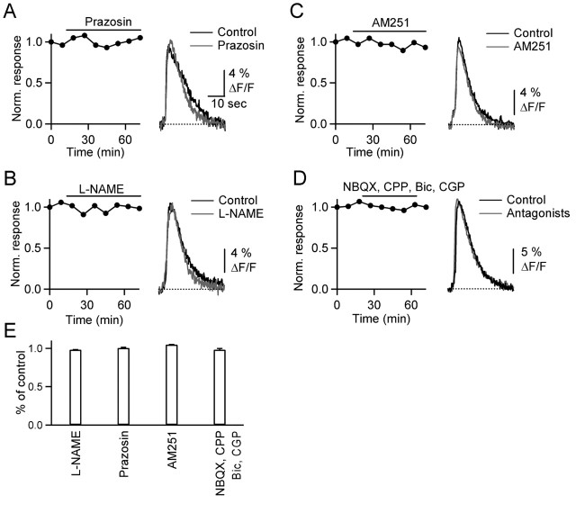
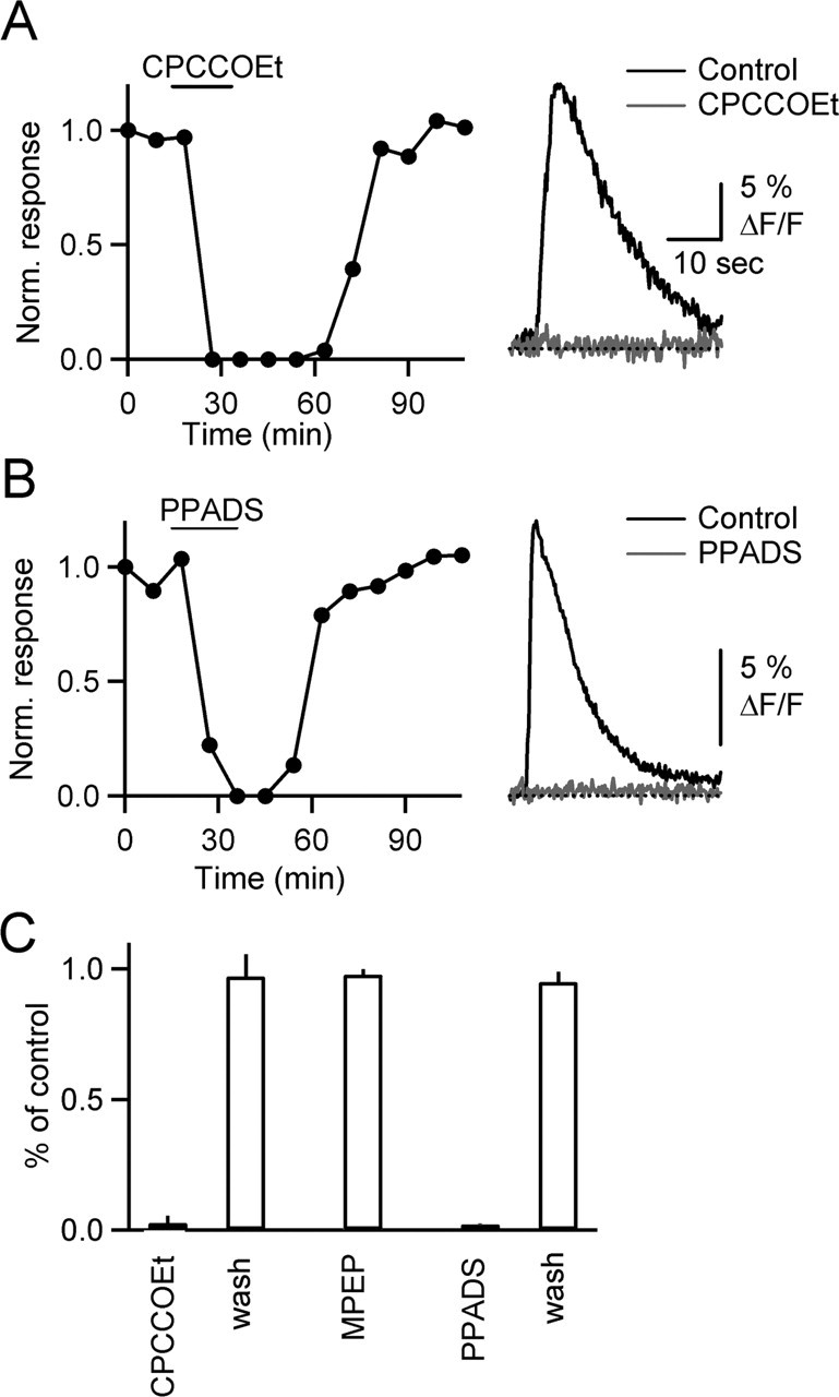
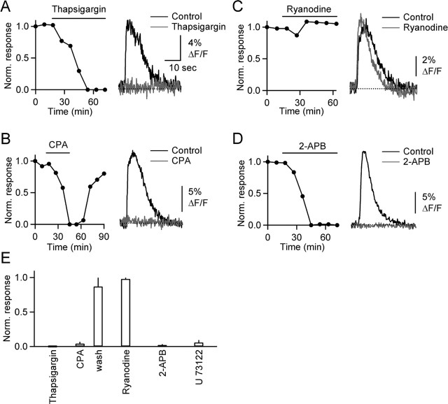
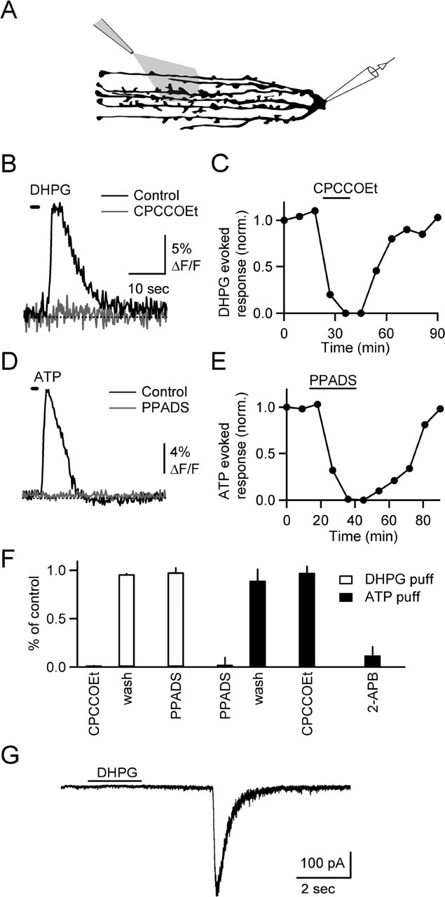
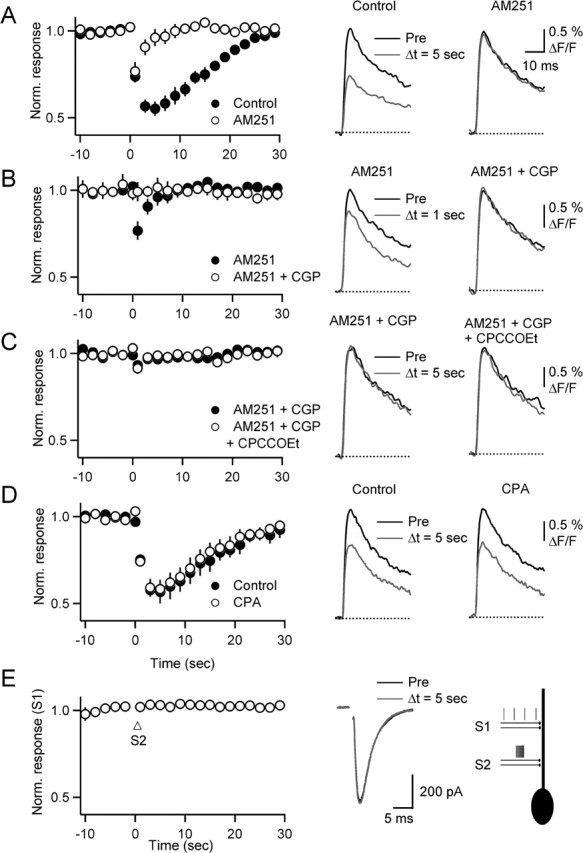
Changes in synaptic strength during ongoing activity are often mediated by neuromodulators. At the synapse between cerebellar granule cell parallel fibers (PFs) and Purkinje cells (PCs), brief bursts of stimuli can evoke endocannabinoid release from PCs and GABA release from interneurons that both inhibit transmission by activating presynaptic G-protein-coupled receptors. Studies in several brain regions suggest that synaptic activity can also evoke calcium signals in astrocytes, thereby causing them to release a transmitter, which acts presynaptically to regulate neurotransmitter release. In the cerebellum, Bergmann glia cells (BGs) are intimately associated with PF synapses. However, the mechanisms leading to calcium signals in BGs under physiological conditions and the role of BGs in regulating ongoing synaptic transmission are poorly understood. We found that brief bursts of PF activity evoke calcium signals in BGs that are triggered by the activation of metabotropic glutamate receptor 1 and purinergic receptors and mediated by calcium release from IP3-sensitive internal stores. We found no evidence for modulation of release from PFs mediated by BGs, even when endocannabinoid- and GABA-mediated presynaptic modulation was prominent. Thus, despite the fact that PF activation can reliably evoke calcium transients within BGs, it appears that BGs do not regulate synaptic transmission on the time scale of seconds to tens of seconds. Instead, endocannabinoid release from PCs and GABA release from molecular layer interneurons provide the primary means of feedback that dynamically regulate release from PF synapses.


Figures

Figure 1.
Figure 1.
Brief bursts of PF stimulation evoke calcium responses in BGs. Data shown here and in Figure 2 were obtained in sagittal slices. A, 2PLSM image of BGs filled with Alexa-594. The area outlined by the box indicates the region selected for calcium imaging. B, Close-up of BG process, as visualized with either 2PLSM (top) or CCD optics (bottom). The white box outlines the area used for the fluorescence measurement in C. The dot indicates site of stimulus (stim) electrode. C, Calcium response (top) and current (bottom) evoked by train of stimuli (10 pulses, 50 Hz). The inset shows the current recorded during stimulation on an expanded time scale.
Figure 2.
Figure 2.
Characteristics of BG calcium signals. A–C, A representative experiment is shown that characterizes the spatiotemporal spread of calcium. A, 2PLSM (left) and CCD image (right) of BG process. B, Calcium response evoked by PF stimulus burst (10 pulses, 50 Hz), in region indicated by red line in A. The arrow indicates onset of stimulus train. C, Spatial spread of calcium wave. The sequence of fluorescence CCD images (50 ms frame duration) shows initiation and spread of calcium response (top row), followed by a more uniform decay (bottom row). Times indicated are relative to the onset of the stimulus train. The color bar indicates fluorescence change (% ΔF/F). D–G, In another experiment, the effect of different numbers of stimuli on BG calcium transients was assessed. D, 2PLSM (left) and CCD images (right) of a BG process. E, CCD images show peak fluorescence changes in glial process evoked by 50 Hz stimulus trains of either 1, 3, or 10 stimuli. Times indicated are relative to stimulus onset. F, Calcium transients evoked by trains of 3 (left) or 10 stimuli (right), in region proximal (black) and distal (gray) to initiation site. Regions of measurements are outlined in D. G, Stimulus dependence of peak fluorescence change at proximal (black) and distal (gray) site outlined in D, evoked by 50 Hz trains. stim, Stimulus location.
Figure 3.
Figure 3.
PF-evoked calcium transients are not triggered by activation of α1-adrenergic receptors, nitric oxide release, CB1 receptors, or AMPA, NMDA, or GABA receptors. Data shown here and in the following figures, except for Figure 6, were obtained in transverse slices. A–D, Graphs on the left plot normalized peak fluorescence transients in BG processes evoked by PF stimulation (10 stimuli, 50 Hz) every 9 min, during bath application of the α1-adrenergic receptor antagonist prazosin (A) (100 μm), the nitric oxide synthase blocker L-NAME (B) (100 μm), the CB1 receptor antagonist AM251 (C) (2 μm), and NBQX (2,3-dihydroxy-6-nitro-7-sulfamoyl-benzo[f]quinoxaline) (D) (5 μm), CPP 3-[(R)-2-carboxypiperazin-4-yl]-propyl-1-phosphonic acid (5 μm), bicuculline (20 μm), and CGP 55845A (2 μm). Traces (right) show responses from same experiments under control conditions (black) and after bath application of antagonists (gray). E, Summary of effects of antagonists on the amplitude of fluorescence transients (n = 5 cells for each antagonist). Bic, Bicuculline; CGP, CGP 55845A.
Figure 4.
Figure 4.
Calcium signals in BGs require mGluR1 and P2R activation. A, B, Graphs on the left plot normalized peak fluorescence transients in BG process, evoked by PF stimulation (10 stimuli, 50 Hz), during bath application of the mGluR1 antagonist CPCCOEt (A) (100 μm) and the P2R antagonist PPADS (B) (50 μm). Traces (right) show responses under control conditions (black) and after bath application of antagonists (gray). C, Summary of effects of antagonists on the amplitude of fluorescence transients [n = 10 cells for CPCCOEt and PPADS, n = 5 for 2-methyl-6-(phenylethynyl)-pyridine (MPEP)].
Figure 5.
Figure 5.
Calcium signals are mediated by release from IP3-sensitive internal stores. A–D, Graphs plot normalized peak fluorescence transients in BG process, evoked by PF stimulus trains (10 stimuli, 50 Hz), during bath application of thapsigargin (A) (10 μm), CPA (B) (20 μm), ryanodine (C) (50 μm), and 2-APB (D) (50 μm). Traces show responses under control conditions (black) and after bath application of antagonists (gray). E, Summary of effects of antagonists on the amplitude of fluorescence transients (n = 5 cells for each antagonist).
Figure 6.
Figure 6.
Agonist-induced calcium transients in BGs. A, Experimental setup. DHPG (100 μm) or ATP (100 μm) were puffed onto slice for 1–2 s. Bath contained 500 nm TTX. B, DHPG-evoked calcium transient in BG processes (black) is blocked by bath application of CPCCOEt (100 μm; gray). The time of DHPG application is indicated by horizontal bar. C, Time course of experiment shown in B. D, ATP-evoked calcium transient (black) is blocked by PPADS (gray). E, Time course of experiment shown in D. F, Summary of effects of antagonists (n = 5 for each condition) on calcium signals evoked by DHPG (open bars) and ATP (filled bars). G, DHPG evoked slow EPSC recorded in a voltage-clamped stellate cell. Bath contained 0 mm MgCl2 and 500 nm TTX.
Figure 7.
Figure 7.
BGs do not control neurotransmitter release from PFs. Parallel fibers were loaded with Magnesium Green AM, and stimulus-evoked calcium transients were recorded with a photodiode. Summary graphs (left column) show normalized calcium transients evoked by single stimuli (0.5 Hz) before and after a burst (10 stimuli, 50 Hz) delivered at t = 0 s. Representative traces (right columns) show calcium transients from single experiments preceding (Pre; black) and after (gray) burst stimulation, under control condition (left column) and after drug application (right column). A, Reduction in presynaptic calcium influx after burst is partially blocked by AM251. B, Remaining reduction is completely blocked by CGP 55845A. Data in A and B are from same set of experiments. C, In the presence of AM251 and CGP 55845A, the addition of CPCCOEt has no effect on presynaptic calcium transients. D, CPA has no effect on the transient reduction of transmitter release that is mediated by GABAB and CB1 receptors. E, Calcium transients in BGs do not lead to modulation of PF EPSC in PCs. PCs were recorded in voltage clamp, and EPSCs were evoked by stimulating two independent PF pathways (S1 and S2). Graph plots normalized responses (n = 5 PCs) evoked by low-frequency (0.5 Hz) stimulation of S1. Arrow indicates onset of burst stimulation (10 stimuli, 50 Hz) of S2. Traces (middle) show sample EPSCs evoked in S1 before (black) and 5 s after (gray) burst stimulation of S2. Right, Experimental setup.

References

    1. Angulo MC, Kozlov AS, Charpak S, Audinat E (2004). Glutamate released from glial cells synchronizes neuronal activity in the hippocampus. J Neurosci 24:6920–6927. - PMC - PubMed
    1. Atluri PP, Regehr WG (1996). Determinants of the time course of facilitation at the granule cell to Purkinje cell synapse. J Neurosci 16:5661–5671. - PMC - PubMed
    1. Auld DS, Robitaille R (2003). Glial cells and neurotransmission: an inclusive view of synaptic function. Neuron 40:389–400. - PubMed
    1. Bergles DE, Dzubay JA, Jahr CE (1997). Glutamate transporter currents in bergmann glial cells follow the time course of extrasynaptic glutamate. Proc Natl Acad Sci USA 94:14821–14825. - PMC - PubMed
    1. Brasnjo G, Otis TS (2001). Neuronal glutamate transporters control activation of postsynaptic metabotropic glutamate receptors and influence cerebellar long-term depression. Neuron 31:607–616. - PubMed

Publication types

Substances

LinkOut - more resources