. 2006 Jun 28;26(26):6968-78.
doi: 10.1523/JNEUROSCI.1013-06.2006.

N-cadherin transsynaptically regulates short-term plasticity at glutamatergic synapses in embryonic stem cell-derived neurons

Affiliations

N-cadherin transsynaptically regulates short-term plasticity at glutamatergic synapses in embryonic stem cell-derived neurons

Kay Jüngling et al. J Neurosci. .

Abstract

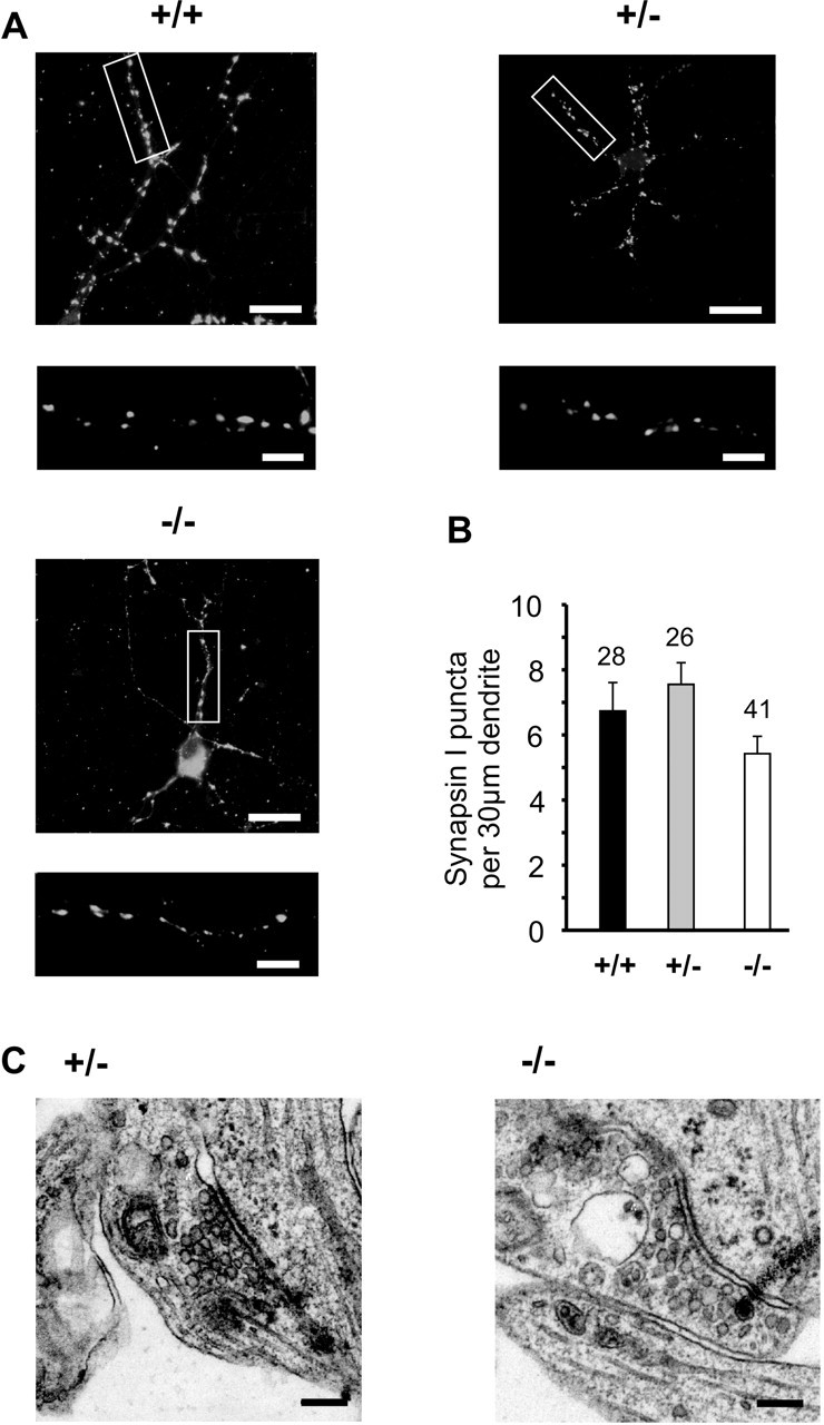
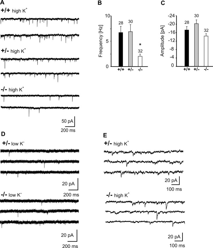
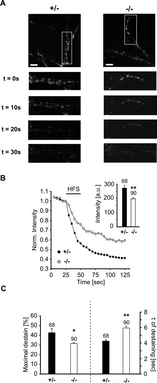
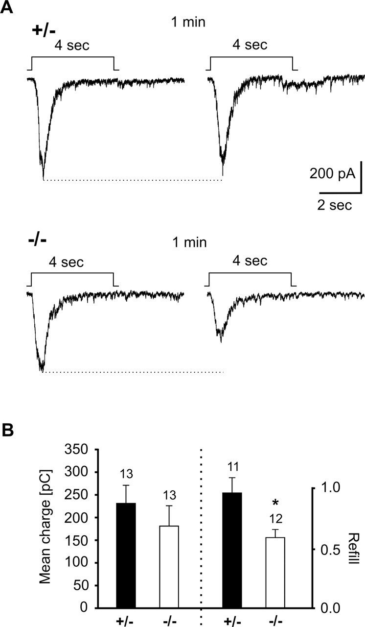
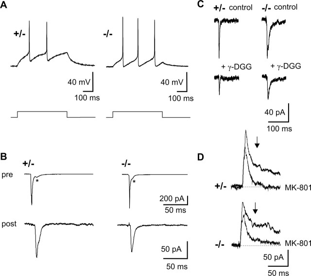
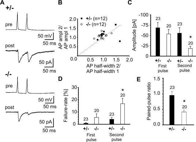
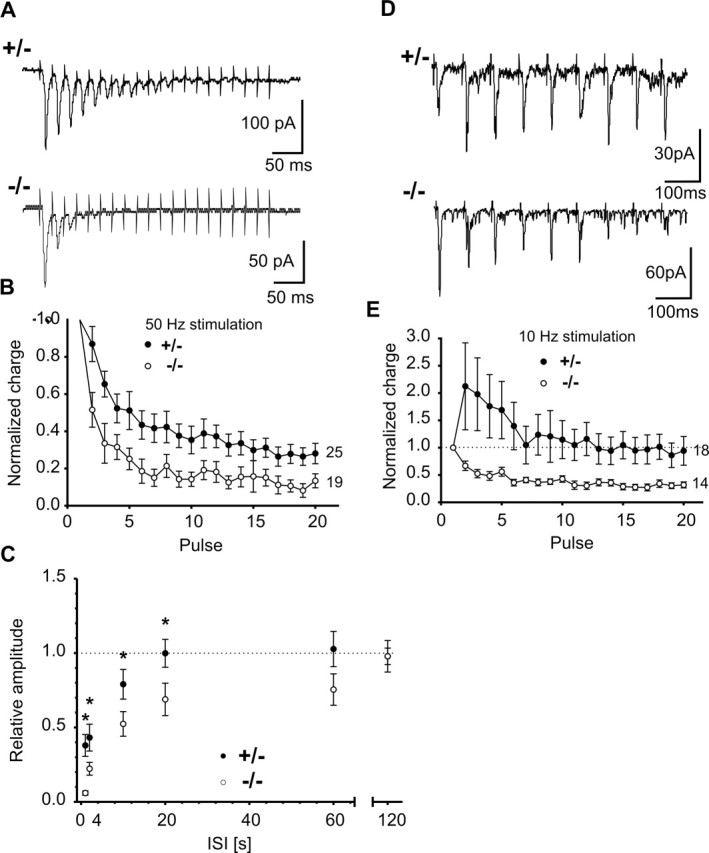
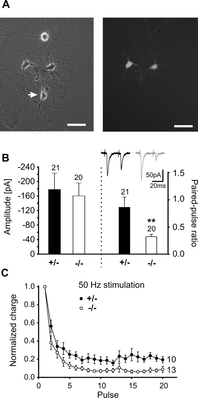
The cell adhesion molecule N-cadherin has been proposed to regulate synapse formation in mammalian central neurons. This is based on its synaptic localization enabling alignment of presynaptic and postsynaptic specializations by an adhesion mechanism. However, a potential role of N-cadherin in regulating synaptic transmission has remained elusive. In this paper, a functional analysis of N-cadherin knock-out synapses was enabled by in vitro neuronal differentiation of mouse embryonic stem cells circumventing the early embryonic lethality of mice genetically null for N-cadherin. In our in vitro system, initial synapse formation was not altered in the absence of N-cadherin, which might be attributable to compensatory mechanisms. Here, we demonstrate that N-cadherin is required for regulating presynaptic function at glutamatergic synapses. An impairment in the availability of vesicles for exocytosis became apparent selectively during high activity. Short-term plasticity was strongly altered with synaptic depression enhanced in the absence of N-cadherin. Most intriguingly, facilitation was converted to depression under specific stimulation conditions. This indicates an important role of N-cadherin in the control of short-term plasticity. To analyze, whether N-cadherin regulates presynaptic function by a transsynaptic mechanism, we studied chimeric cultures consisting of wild-type neocortical neurons and ES cell-derived neurons. With N-cadherin absent only postsynaptically, we observed a similar increase in short-term synaptic depression as found in its complete absence. This indicates a retrograde control of short-term plasticity by N-cadherin. In summary, our results revealed an unexpected involvement of a synaptic adhesion molecule in the regulation of short-term plasticity at glutamatergic synapses.


Figures

Figure 1.
Figure 1.
In vitro neuronal differentiation of N-cadherin knock-out, ES cell-derived neurons. A, Scheme of ES cell in vitro differentiation and purification of ES cell-derived neurons. EB, Embryoid body. B, Western blot analysis of N-cadherin expression in wild-type (+/+), heterozygous (+/−), and homozygous N-cadherin knock-out (−/−) ES cells. C, ES cell-derived neurons cultured on glial microislands at 10 DIV. Scale bars, 16 μm.
Figure 2.
Figure 2.
Initial synapse formation is not altered in N-cadherin knock-out, ES cell-derived neurons. A, Synapsin I immunostaining of presynaptic boutons at 10–12 DIV. The box in the top panel is shown enlarged in the bottom panel. Scale bars: top, 12 μm; bottom, 3 μm. B, Quantitative comparison of the dendritic density of synapsin I puncta. n, dendritic segments. C, Typical asymmetric synaptic profiles in cultures of N-Cad+/− and N-Cad−/− neurons. Scale bar, 100 nm. Ultrastructural characteristics were similar to those described for primary cultured cortical neurons. In this and all subsequent figures, genotypes are indicated by +/+, +/−, −/−; error bars represent SEM; and significant differences are indicated by asterisks (∗p < 0.05; ∗∗p < 0.01).
Figure 3.
Figure 3.
AMPA receptor-mediated miniature EPSCs revealed an impairment of presynaptic function during high activity. A, Example traces of AMPA mEPSCs evoked by elevated extracellular K+ (30 mm). B, C, Comparison of mean frequencies (B) and amplitudes (C) of AMPA mEPSCs at 30 mm K+. n (cells) is indicated. Note the significantly reduced frequency, indicating a presynaptic defect. D, Example traces of AMPA mEPSCs at 5 mm extracellular K+. E, Example traces of GABAA mPSCs evoked by elevated extracellular K+ (30 mm).
Figure 4.
Figure 4.
Reduced high-frequency stimulation-evoked vesicle exocytosis as revealed by FM4-64 staining/destaining of presynaptic release sites. A, Examples of high-frequency stimulation (HFS)-induced destaining of FM4-64-labeled presynaptic release sites. Dendritic segments in boxed areas in the top panels are shown enlarged below. Time after onset of HFS is indicated. Scale bars, 10 μm. B, Typical destaining kinetics of individual FM4-64 puncta in N-Cad+/− (filled dots) and N-Cad−/− (open dots) neurons. HFS is indicated by black bar. Inset, Initial mean fluorescence intensity without normalization. C, Quantitative analysis revealed a reduced amount of destaining (left part) and an increased mean time constant of destaining (right part) in N-Cad−/− neurons, indicating reduced vesicle exocytosis. Destaining kinetics were fitted monoexponentially. n indicates the number of FM4-64 puncta analyzed.
Figure 5.
Figure 5.
Impaired refill of the readily releasable vesicle pool in N-cadherin knock-out synapses. A, Example traces of AMPA EPSC bursts elicited by two successive hypertonic sucrose applications in N-Cad+/− and N-Cad−/− neurons. The application protocol is indicated above the EPSC traces. B, Quantitative comparison of the mean charge influx during the first sucrose application (left part) and the mean refill (ratio of charge influx during second and first sucrose response) of the readily releasable pool (right part). Note the substantially reduced refill in N-cadherin knock-out synapses.
Figure 6.
Figure 6.
Properties of glutamatergic synaptic transmission evoked by a single stimulus are not altered in N-cadherin knock-out synapses. A, Typical action potentials evoked by a depolarizing current pulse (bottom) in N-Cad+/− and N-Cad−/− neurons. B, AMPA EPSCs (post) elicited by action currents in the presynaptic cell soma (pre) in a paired recording. Asterisks indicate autaptic PSCs. ampl, Amplitude. C, Partial block of evoked AMPA EPSCs by 1 mm γ-DGG. D, MK-801 (40 μm) block of evoked NMDA PSC component at +40 mV holding potential (measured 40 ms after onset of PSC, as indicated by arrows in D). The control response before and the fourth response after addition of MK-801 are superimposed.
Figure 7.
Figure 7.
Increased synaptic depression in N-cadherin knock-out synapses as revealed by paired-pulse analysis. A, Typical paired-pulse behavior of action potential-evoked AMPA EPSCs in paired recordings in N-Cad+/− and N-Cad−/− neurons. Presynaptic action potentials elicited by a 5 ms depolarizing twin pulse (pre) and the corresponding AMPA EPSCs (post) are shown. B, Mean amplitudes and mean half-widths of action potentials did not differ between N-Cad+/− and N-Cad−/− neurons. The ratio of the AP amplitudes and the ratio of the AP half widths of APs elicited by the second and the first pulse in individual presynaptic cells are shown. Filled circles: N-Cad+/−; open circles: N-Cad−/−. C, D, Mean amplitudes (including failures) (C) and mean failure rates (D) of AMPA EPSCs. Responses to the first action potential were not different, whereas responses to the second action potential showed a reduced amplitude and an increased failure rate in N-Cad−/− zneurons. E, Quantitative comparison of paired-pulse ratio of AMPA EPSCs. Note the markedly enhanced paired-pulse depression in N-cadherin knock-out synapses. n (cells) is indicated.
Figure 8.
Figure 8.
Altered short-term plasticity in N-cadherin knock-out synapses as revealed by tetanic stimulation. A, Typical responses to high-frequency stimulation in N-Cad+/− and N-Cad−/− neurons. AMPA EPSCs were elicited by 20 action potentials at 50 Hz. B, Quantitative comparison of responses to high-frequency stimulation in N-Cad+/− (filled circles) and N-Cad−/− (open circles) synapses revealed strongly increased synaptic depression. C, Recovery from depression elicited by 20 action potentials at 50 Hz. The normalized AMPA EPSC amplitude evoked by a test pulse (N-Cad+/−, n = 8; N-Cad−/−, n = 11) at different time intervals [interstimulus interval (ISI): 1, 2, 10, 20, 60, and 120 s] after the end of the conditioning high-frequency pulse train is shown. Note the significantly slower recovery in N-Cad−/− synapses. D, Typical AMPA EPSCs evoked by 10 Hz stimulation in N-Cad+/− and N-Cad−/− neurons. E, Quantitative comparison of responses to 10 Hz stimulation in N-Cad+/− and N-Cad−/− synapses revealed a switch from facilitation to depression. n (cells) is indicated.
Figure 9.
Figure 9.
Transsynaptic retrograde regulation of short-term plasticity by N-cadherin. A, Chimeric microisland culture (left) consisting of EGFP-expressing, wild-type neocortical neurons (right) and unlabeled ES cell-derived neurons (arrow) at 12 DIV. Paired recordings were obtained from a presynaptic wild-type neuron and a postsynaptic N-Cad+/− or N-Cad−/− neuron. B, Right, Mean amplitudes of AMPA EPSCs elicited by the first stimulus of a paired-pulse protocol. Left, Paired-pulse analysis of evoked AMPA EPSCs revealed an increased paired-pulse depression in the postsynaptic absence of N-cadherin. Insets, Original recordings. C, High-frequency stimulation of synapses between presynaptic wild-type neurons and postsynaptic N-Cad+/− or N-Cad−/− neurons revealed an increased depression of AMPA EPSCs in the postsynaptic absence of N-cadherin. n (cells) is indicated.

References

    1. Abe K, Chisaka O, van Roy F, Takeichi M (2004). Stability of dendritic spines and synaptic contacts is controlled by αN-catenin. Nat Neurosci 7:357–363. - PubMed
    1. Angst BD, Marcozzi C, Magee AI (2001). The cadherin superfamily: diversity in form and function. J Cell Sci 114:629–641. - PubMed
    1. Bamji SX, Shimazu K, Kimes N, Huelsken J, Birchmeier W, Lu B, Reichardt LF (2003). Role of β-catenin in synaptic vesicle localization and presynaptic assembly. Neuron 40:719–731. - PMC - PubMed
    1. Benson DL, Tanaka H (1998). N-cadherin redistribution during synaptogenesis in hippocampal neurons. J Neurosci 18:6892–6904. - PMC - PubMed
    1. Bozdagi O, Shan W, Tanaka H, Benson DL, Huntley GW (2000). Increasing numbers of synaptic puncta during late-phase LTP: N-cadherin is synthesized, recruited to synaptic sites, and required for potentiation. Neuron 28:245–259. - PubMed

Publication types

MeSH terms