. 2006 Jun 28;26(26):7014-21.
doi: 10.1523/JNEUROSCI.0573-06.2006.

Identification of an endoplasmic reticulum-retention motif in an intracellular loop of the kainate receptor subunit KA2

Affiliations

Identification of an endoplasmic reticulum-retention motif in an intracellular loop of the kainate receptor subunit KA2

Yukiko Nasu-Nishimura et al. J Neurosci. .

Erratum in

  • J Neurosci. 2006 Jul 12;26(28):7541

Abstract

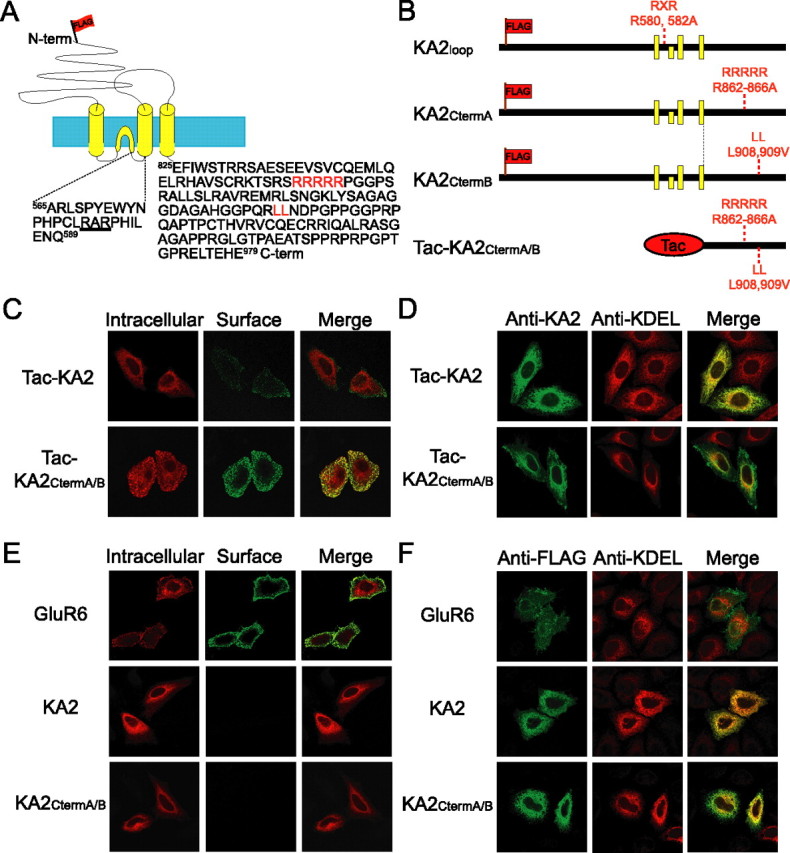
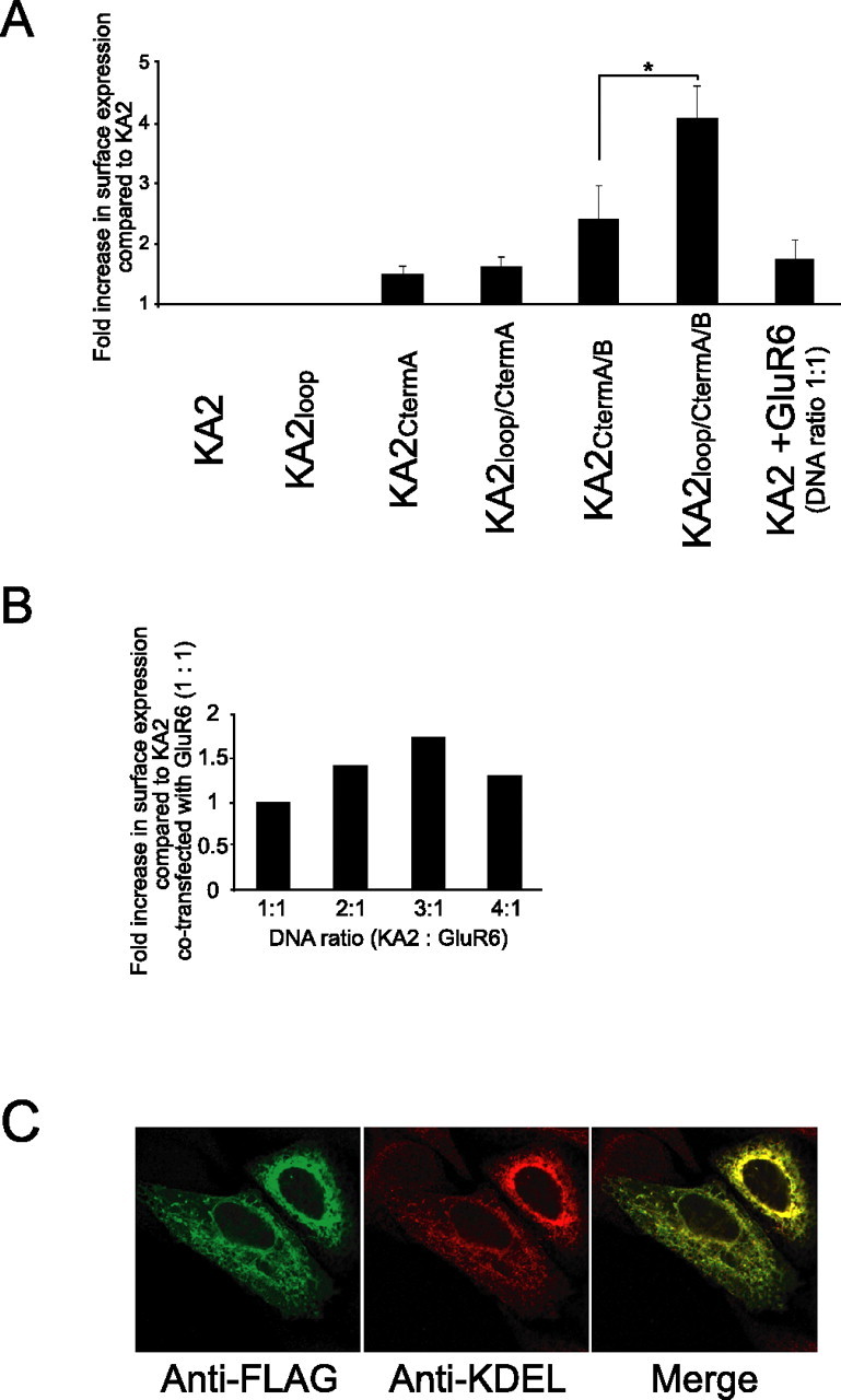
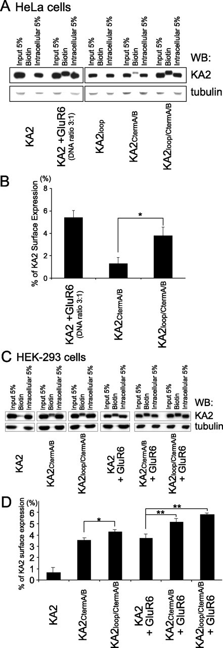
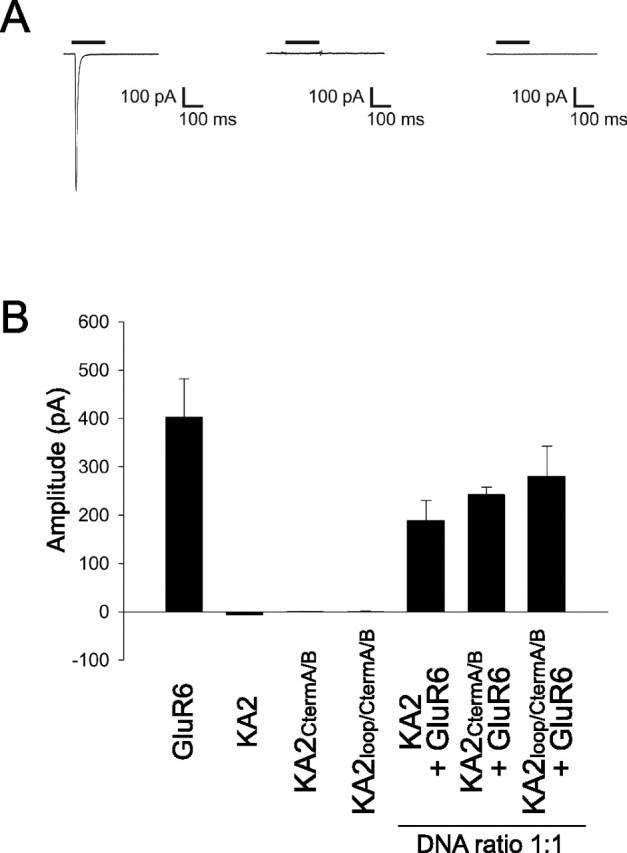
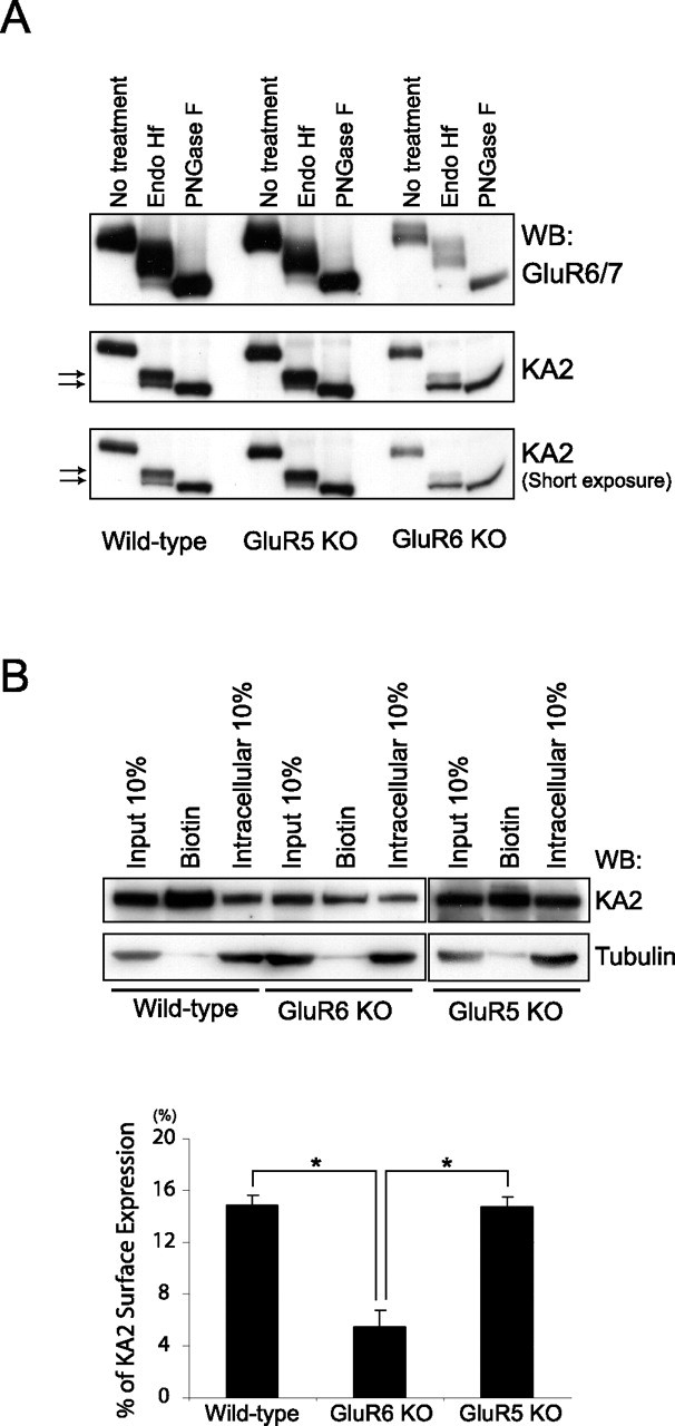
Neuronal kainate receptors are typically heteromeric complexes composed of GluR5-7 and KA1-2 subunits. Although GluR5-7 can exist as functional homomeric channels, the KA subunits cannot. KA2 is widely expressed in the CNS, and KA2/GluR6 heteromers are the most prevalent subunit composition in brain. Previous work has identified endoplasmic reticulum (ER)-retention motifs in the C terminus of KA2, which prevent surface expression of KA2 homomers. However, we find that, when these motifs are mutated, only a small fraction of KA2 is surface expressed. We now identify an additional ER retention motif in the intracellular loop region of KA2, which, when mutated together with the C-terminal motifs, significantly increases the level of KA2 surface expression. However, electrophysiological analysis of surface-expressed KA2 homomers indicates that they do not form functional ion channels. In heterologous cells, a large fraction of KA2 remains intracellular even when the trafficking motifs are mutated or when GluR6 is coexpressed. Therefore, we analyzed the trafficking of endogenous KA2 in vivo. We find that native KA2 surface expression is dramatically reduced in GluR6 knock-out mice compared with wild-type mice. In contrast, KA2 trafficking was unaffected in the GluR5 knock-out. Thus, our study demonstrates that trafficking motifs in both the intracellular loop and C terminus regulate KA2 surface expression; however, in neurons, GluR6 oligomerization is required for egress of KA2 from the ER and transport to the cell surface. The combination of these mechanisms likely prevents surface expression of nonfunctional KA2 homomers and ensures a high level of GluR6/KA2 heteromeric kainate receptors.


Figures

Figure 1.
Figure 1.
Mutations of C-terminal trafficking motifs do not allow robust surface expression of full-length KA2. A, Schematic diagram depicting the topology of KA2. The KA2 C terminus has 155 aa and contains several trafficking motifs that have been described previously: an arginine-rich motif (RRRRR) and a dileucine sequence (LL) are depicted in red. A consensus arginine-based endocytic motif (RxR) within the intracellular loop is underlined. The FLAG epitope is depicted as a flag inserted just after the signal sequence in the N terminus of KA2. The yellow and blue boxes represent membrane domains and lipid bilayer, respectively. B, Diagram illustrating mutations made in KA2. Tac-KA2 is a chimeric protein consisting of the plasma membrane protein Tac with the C terminus of KA2. The yellow boxes represent membrane domains. Mutations in one or more of the trafficking motifs are indicated with a subscript representing the region of the mutation. C, Tac-KA2CtermA/B is efficiently expressed on the plasma membrane. Live HeLa cells transfected with Tac-KA2 or Tac-KA2CtermA/B were labeled with anti-Tac antibody (surface) and Alexa 488-conjugated secondary antibody (green) on ice. The cells were fixed, permeabilized, and labeled with anti-Tac antibody (intracellular) and Alexa 568-conjugated secondary antibody (red). D, Tac-KA2CtermA/B is localized at the plasma membrane as well as the ER. HeLa cells transfected with Tac-KA2 or Tac-KA2CtermA/B were fixed, permeabilized, and stained with antibodies against the C terminus of KA2 (green) and the ER marker KDEL (red). E, KA2CtermA/B is not expressed on the plasma membrane. Live HeLa cells transfected with FLAG-GluR6, FLAG-KA2, or FLAG-KA2CtermA/B were labeled with anti-FLAG antibody (surface) and Alexa 488-conjugated secondary antibody (green) on ice. The cells were fixed, permeabilized, and labeled with anti-FLAG antibody (intracellular) and Alexa 568-conjugated secondary antibody (red). FLAG-GluR6 was used as a positive control for surface-expressed receptor. F, Full-length KA2 is retained in the ER when expressed in HeLa cells, even with mutations in C-terminal ER-retention motifs. HeLa cells transfected with FLAG-GluR6, FLAG-KA2, or FLAG-KA2CtermA/B were fixed, permeabilized, and stained with antibodies against FLAG (green) and the ER marker KDEL (red). The merged images show the extensive colocalization of KA2CtermA/B with KDEL.
Figure 2.
Figure 2.
Identification of an arginine-based ER-retention motif (RxR) in an intracellular loop of KA2. A, FACS analysis of the surface expression of full-length KA2 mutants expressed in HeLa cells. HeLa cells were transfected with FLAG-KA2 wild-type and FLAG-KA2 that contains mutations of putative trafficking motifs within the intracellular loop and the C terminus. Surface expression was analyzed by FACS as described in Materials and Methods. When arginines 580-RxR-582 were mutated to alanines in combination with C-terminal A/B mutations, this KA2loop/CtermA/B mutant showed a fourfold increase in surface expression compared with wild-type KA2. Data represent means ± SEM of the fold increase in surface expression compared with KA2 based on a percentage of the cells with surface-expressed KA2 in transfected cells [∗p < 0.05 (n=7) relative to the surface expression of KA2CtermA/B mutant; Student's t test]. B, Expression of GluR6 affects the surface expression of KA2. GluR6 and FLAG-tagged KA2 were transfected into HeLa cells at different ratios, and the surface expression of KA2 was evaluated by FACS analysis as described in Materials and Methods. C, KA2 is largely ER retained when coexpressed with GluR6 in HeLa cells. FLAG-tagged KA2 and GluR6 were transfected into HeLa cells at a 3:1 ratio. The cells were fixed, permeabilized, and stained with antibodies against FLAG (green) and the ER marker KDEL (red). The merged image shows the extensive colocalization of KA2 with KDEL.
Figure 3.
Figure 3.
Surface expression of KA2 is most robust when the RxR loop mutation is combined with mutations of the C-terminal trafficking motifs as revealed using a biotinylation assay. A, C, HeLa cells (A) or HEK-293 cells (C) expressing combinations of wild-type KA2 or mutant KA2 with or without GluR6 were analyzed using the biotinylation assay. The biotinylated surface protein fractions of the whole-cell extracts were separated from the intracellular fractions by conjugation with streptavidin beads and visualized by Western blotting (WB). When arginines 580-RxR-582 within the KA2 intracellular loop were mutated to alanines in combination with C-terminal A/B mutations, this KA2loop/CtermA/B mutant was more heavily biotinylated than KA2 containing only the C-terminal A/B mutations (KA2CtermA/B). B, D, Surface expression of wild-type or mutant KA2 alone or cotransfected with GluR6 expressed in HeLa cells (B) and HEK-293 cells (D) was quantitated by measuring the band intensity of the biotinylated fraction compared with the intensity of the band representing 5% of total input using NIH Image software. Error bars indicate SEM. ∗p < 0.04 (n = 4 for B; n = 3 for D); ∗∗p < 0.01 (n = 3), Student's t test.
Figure 4.
Figure 4.
Homomeric wild-type KA2 or KA2 containing C-terminal and loop ER-retention motif mutations do not exhibit kainate-evoked currents in HEK-293 cells. A, Averaged current during whole-cell patch-clamp recordings from HEK-293 cells in response to kainate (300 μm; black bar) for cells expressing GluR6 (left), wild-type KA2 (center), or KA2loop/CtermA/B (right) (traces are average of the response to 5–8 consecutive applications of kainate). B, Summary data bar graph of mean kainate-evoked current amplitude in HEK-293 cells expressing GluR6 (n = 13), KA2 (n = 5), KA2CtermA/B (n = 12), KA2loop/CtermA/B (n = 9), GluR6 and KA2 (n = 12), GluR6 and KA2CtermA/B (n = 6), and GluR6 and KA2loop/CtermA/B (n = 6). Error bars indicate SEM.
Figure 5.
Figure 5.
KA2 does not traffic to the cell surface efficiently in neurons lacking GluR6. A, The majority of KA2 in GluR6 KO brain is ER retained. The whole-brain lysate (50 μg) from wild-type, GluR5 KO, and GluR6 KO mouse was treated with endo H or PNGase F, and immunoblots were probed with GluR6/7 or KA2 antibodies. Mature forms of both GluR6 and KA2 display partial endo H sensitivity. B, Less KA2 is expressed on the cell surface in primary cultured GluR6 KO cortical neurons than in wild-type or GluR5 KO cortical neurons. Primary cultures of cortical neurons were obtained from P0–P1 pups. The neurons were maintained for 2 weeks and surface-biotinylated with sulfo-NHS-LC-biotin. The biotinylated surface protein fractions of the whole-cell extracts were separated from the intracellular fractions by conjugation with streptavidin beads, resolved by SDS-PAGE, and probed with KA2 or tubulin antibodies. Surface expression of KA2 was quantitated by measuring the band intensity of biotinylated fraction compared with the intensity of the band representing 10% of total input using NIH Image software. Error bars indicate SEM. ∗p < 0.01 (n = 5 for wild-type and GluR6 KO; n = 3 for GluR5 KO), Student's t test. WB, Western blot.

References

    1. Bahn S, Volk B, Wisden W (1994). Kainate receptor gene expression in the developing rat brain. J Neurosci 14:5525–5547. - PMC - PubMed
    1. Christensen JK, Paternain AV, Selak S, Ahring PK, Lerma J (2004). A mosaic of functional kainate receptors in hippocampal interneurons. J Neurosci 24:8986–8993. - PMC - PubMed
    1. Coussen F, Normand E, Marchal C, Costet P, Choquet D, Lambert M, Mege RM, Mulle C (2002). Recruitment of the kainate receptor subunit glutamate receptor 6 by cadherin/catenin complexes. J Neurosci 22:6426–6436. - PMC - PubMed
    1. Coussen F, Perrais D, Jaskolski F, Sachidhanandam S, Normand E, Bockaert J, Marin P, Mulle C (2005). Co-assembly of two GluR6 kainate receptor splice variants within a functional protein complex. Neuron 47:555–566. - PubMed
    1. Ellgaard L, Helenius A (2003). Quality control in the endoplasmic reticulum. Nat Rev Mol Cell Biol 4:181–191. - PubMed

Publication types

MeSH terms

LinkOut - more resources