Comparative Study
. 2006 Jul-Aug;26(4-6):943-57.
doi: 10.1007/s10571-006-9096-y. Epub 2006 Jun 29.

Hippocampal neuropathology of diabetes mellitus is relieved by estrogen treatment

Affiliations
Comparative Study

Hippocampal neuropathology of diabetes mellitus is relieved by estrogen treatment

Flavia E Saravia et al. Cell Mol Neurobiol. 2006 Jul-Aug.

Abstract

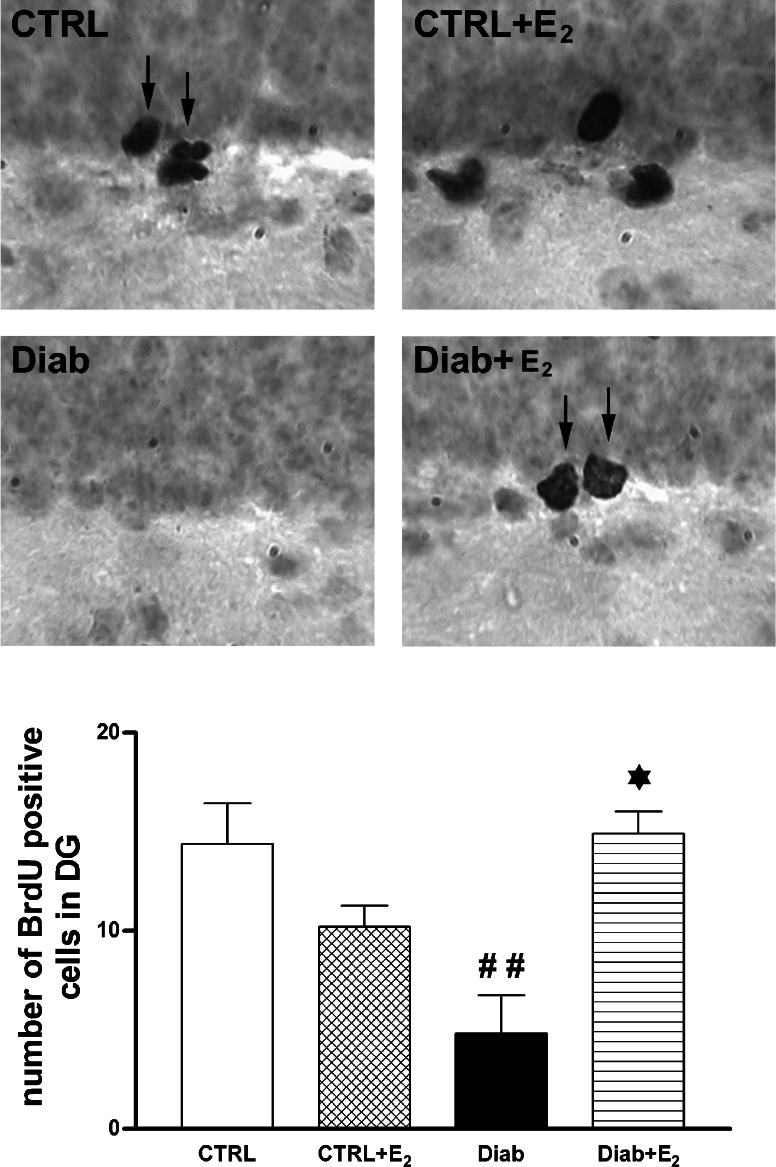
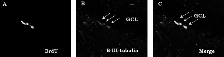
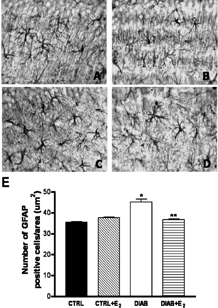
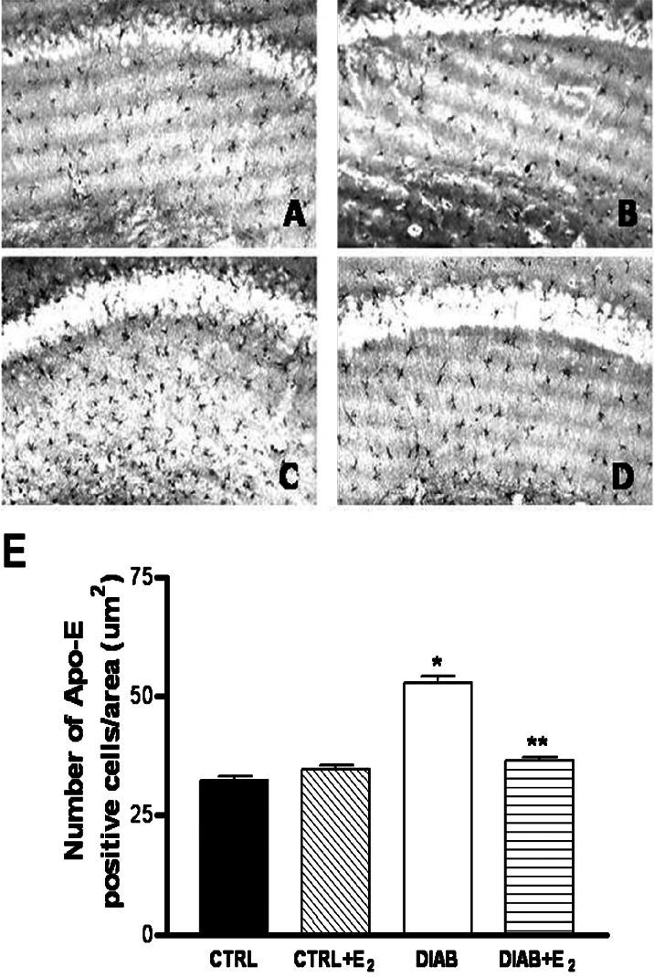
1. A recently recognized complication of uncontrolled diabetes mellitus is the encephalopathy involving, among other regions, the hippocampus. Since estrogens bring neuroprotection in cases of brain injury and degenerative diseases, we have studied if estradiol (E2) administration counteracts some hippocampal abnormalities of streptozotocin (STZ)-diabetic adult mice. 2. We first report the ability of E2 to modulate neurogenesis in the dentate gyrus (DG) and subventricular zone (SVZ) of diabetic mice. Using bromodeoxyuridine (BrdU) to label newly generated cells, a strong reduction in cell proliferation was obtained in DG and SVZ of mice sacrificed 20 days after STZ administration. The reduction was completely relieved by 10 days of E2 pellet implantation, which increased 30-fold the circulating E2 levels. 3. Diabetic mice also showed abnormal expression of astrocyte markers in hippocampus. Thus, increased number of GFAP(+) cells, indicative of astrogliosis, and increased number of apolipoprotein-E (Apo-E)(+) astrocytes, a marker of ongoing neuronal dysfunction, was found in stratum radiatum below the CA1 hippocampal subfield of diabetic mice. Both parameters were reverted to normal by the E2 regime that upregulated cell proliferation. 4. The studies demonstrated that hippocampal neuropathology of uncontrolled diabetes is a reversible condition and sensitive to estrogen treatment. Studies in animal models may open up new venues for understanding the beneficial role of steroid hormones in diabetic encephalopathy.


Figures

Fig. 1.
Fig. 1.
Effects of diabetes and estradiol treatment on cell proliferation in the dentate gyrus (DG). Control and diabetic mice were implanted with a pellet containing cholesterol (CTRL) (DIAB) or cholesterol plus 200 μg 17-β; estradiol (CTRL + E2, DIAB+E2). Upper photomicrograp: Representative examples of BrdU+ cells in a CTRL mouse and a CTRL + E2 mouse, the pronounced decrease in a diabetic mouse and the recovery of BrdU+ cells in a diabetic mouse receiving E2 (DIAB+E2). Lower graph: Computerized image analysis of the number of BrdU-immunopositive cells in DG. (##) Significantly smaller than all other groups (p < 0.01 by ANOVA followed by Bonferroni's posthoc test). (Modified from Saravia et al., 2004).
Fig. 2.
Fig. 2.
Representative confocal laser scanning microscopic images corresponding to BrdU-labelled cells in the dentate gyrus from a control mouse (A) and to cells exhibiting an immature neuronal phenotype (β-III-tubulin) (B). Merging showed colocaIization of some BrdU-labelled cells with β-III-tubulin (C) indicated by arrows in B and C. Scale bar: 10 μm, GCL: Granular cell layer.
Fig. 3.
Fig. 3.
Photomicrographs showing glial fibrillary acidic protein-positive (GFAP) cells in hippocampal stratum radiatum from a control C57BL/6 mouse(A), control mouse receiving an estradiol pellet (B), a diabetic mouse (C), and an estrogen-treated diabetic mouse (D). Note the GFAP-immunopositive astroglial reaction in (C) and the returned-to-normal level in (D). Magnification: 400×. (E) Quantitative analysis of GFAP+ cells per area (65×103 μm2) in control (CTRL), control plus estradiol (CTRL+E2), diabetic (DIAB), and diabetic mice treated with estradiol (DIAB+E2). Statistical analysis was carried out by one-way ANOVA followed by Bonferroni's test (n=5 animals per group). *DIAB vs. CTRL and CTRL+E2: p < 0.05; ** DIAB+E2 vs. DIAB: p < 0.05.
Fig. 4.
Fig. 4.
Photomicrographs showing Apolipoprotein E (Apo-E)+ cells in hippocampal stratum radiatum from control C57BL/6 mouse (A), control mouse receiving an estradiol pellet (B), a diabetic mouse (C), and an estrogen-treated diabetic mouse (D). Note the numerous positive cells in (C) and the returned-to-normal level in (D). Magnification: 200×. (E) Quantitative analysis of Apo-E+ cells per area (65×103 μm2) in control (CTRL), control plus estradiol (CTRL+E2), diabetic (DIAB), and diabetic mice treated with estradiol (DIAB+E2). Statistical analysis was carried out by one-way ANOVA followed by Bonferroni's test (n=5 animals per group). *DIAB vs. CTRL and CTRL+E2: p < 0.05; ** DIAB+E2 vs. DIAB: p < 0.05.

Similar articles

Cited by

References

    1. Adeghate, E., and Ponery, A. S. (2001). The effect of 17 beta-estradiol on weight, blood glucose and plasma insulin levels in diabetic rats. Gynecol. Endocrinol.15:433–438. - PubMed
    1. Anderson, M. F., Aberg, M. A. I., Nilsson, M., and Eriksson, P. S. (2002). Insulin-like growth factor-I and neurogenesis in the adult mammalian brain. Dev. Brain Res.4:115–122. - PubMed
    1. Artola, A., Kamal, A., Ramakers, G. M., Gardoni, F., DiLuca, M., Biessels, G. J., Cattabeni, F., and Gispen, W. H. (2002). Synaptic plasticity in the diabetic brain: Advanced aging? Prog. Brain Res. 138:305–314. - PubMed
    1. Behl, C. (2002). Estrogen as a neuroprotective hormone. Nat. Rev. Neurosci. 3:433–442. - PubMed
    1. Bestetti, G., and Rossi, G. L. (1980). Hypothalamic lesions in rats with long-term streptozotocin-induced diabetes mellitus. Acta Neurophatol.52:119–127. - PubMed

Publication types

LinkOut - more resources