Review
. 2006 Jun 30;47(3):293-306.
doi: 10.3349/ymj.2006.47.3.293.

An update of the defensive barrier function of skin

Affiliations
Review

An update of the defensive barrier function of skin

Seung Hun Lee et al. Yonsei Med J. .

Abstract

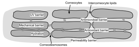
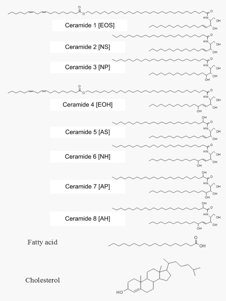
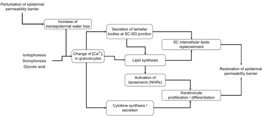
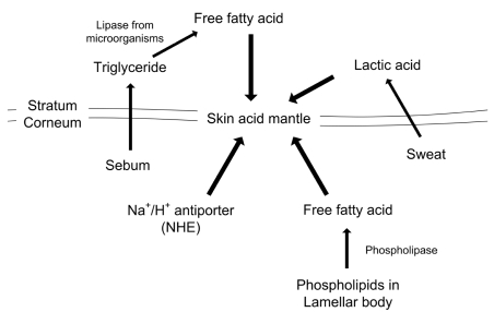
Skin, as the outermost organ in the human body, continuously confronts the external environment and serves as a primary defense system. The protective functions of skin include UV-protection, anti-oxidant and antimicrobial functions. In addition to these protections, skin also acts as a sensory organ and the primary regulator of body temperature. Within these important functions, the epidermal permeability barrier, which controls the transcutaneous movement of water and other electrolytes, is probably the most important. This permeability barrier resides in the stratum corneum, a resilient layer composed of corneocytes and stratum corneum intercellular lipids. Since the first realization of the structural and biochemical diversities involved in the stratum corneum, a tremendous amount of work has been performed to elucidate its roles and functions in the skin, and in humans in general. The perturbation of the epidermal permeability barrier, previously speculated to be just a symptom involved in skin diseases, is currently considered to be a primary pathophysiologic factor for many skin diseases. In addition, much of the evidence provides support for the idea that various protective functions in the skin are closely related or even co-regulated. In this review, the recent achievements of skin researchers focusing on the functions of the epidermal permeability barrier and their importance in skin disease, such as atopic dermatitis and psoriasis, are introduced.


Figures

Fig. 1
Fig. 1
The localization of various protective functions in the stratum corneum. While the corneocytes (bricks) serve as UV and mechanical barriers as well as playing a hydrating role in the stratum corneum, the intercellular lipids perform the functions of antimicrobial barrier, anti-oxidant barrier and permeability barrier. It should be noted, however, that regulation of these protective functions are closely related and modulation of one function can affect other functions.
Fig. 2
Fig. 2
The structure of the major components of the stratum corneum intercellular lipids. Numbers 1 to 8 are ceramides and represent a thin layer with chromatographic mobility, with ceramide 1 being least polar and ceramide 8 the most polar. The letters in square brackets are the structural classifications of the ceramide as suggested by Motta et al. (Biochim Biophys Acta 1993;1182:145-51).
Fig. 3
Fig. 3
The epidermal permeability barrier homeostasis. When barrier function is perturbed, immediate secretion of preformed lamellar bodies occurs, followed by an increased synthesis of lamellar bodies and lipid precursors. Increased expression of inflammatory cytokines is observed after barrier disruption. Keratinocyte differentiation and proliferation are also increased after barrier disruption. The epidermal permeability barrier is restored through the above described homeostatic responses. Iontophoresis, sonophoresis and topical glycolic acid can also induce the down-stream homeostatic responses without changing the transepidermal water loss.
Fig. 4
Fig. 4
The endogenous and exogenous factors affecting stratum corneum acidification. Exogenous, free fatty acids are either derived from sweat glands or catalyzed from sebaceous gland-originated triglycerol by microorganism-secreted lipases. Endogenous free fatty acids are derived from phospholipids by phospholipase A2 (PLA2), both of which are secreted by lamellar bodies at the stratum corneum-stratum granulosum junction. The Na+/H+ antiporter is also involved in maintaining the skin acid mantle.
Fig. 5
Fig. 5
The activation of corneodesmosome degrading protease in the stratum corneum. SCCE/KLK7 and KLK17 are activated by the tryptic cleavage of a short amino-terminal domain of Pro-SCCE and pro-KLK14 by SCTE/KLK5. Pro-SCTE/KLK7 is autoactivated to yield active SCTE/KLK5. The cathepsin activator is unknown. The proteolytic activities of each protease are regulated by their chemical and biochemical environment, including the pH and the protease inhibitors in the stratum corneum. SCTE, stratum corneum tryptic enzyme; SCCE, stratum corneum chymotryptic enzyme; LEKTI, lymphepithelial Kazal-type 5 serine protease inhibitor; SKALP, skin-derived-antileukoprotease; ALP, antileukoproteinase; SLPI, secretory leukocyte protease inhibitor.

References

    1. Albanesi C, Scarponi C, Giustizieri ML, Girolomoni G. Keratinocytes in inflammatory skin diseases. Curr Drug Targets Inflamm Allergy. 2005;4:329–334. - PubMed
    1. Hearing VJ. Biogenensis of pigment granules: a sensitive way to regulate melanocyte function. J Dermatol Sci. 2005;37:3–14. - PubMed
    1. Lehmann B, Querings K, Reichrath J. Vitamin D and skin: new aspects for dermatology. Exp Dermatol. 2004;13(Suppl 4):11–15. - PubMed
    1. Garssen J, Vandebriel RJ, van Loveren H. Molecular aspects of UVB-induced immunosuppression. Arch Toxicol Suppl. 1997;19:97–109. - PubMed
    1. Ghoreishi M. Heat shock proteins in the pathogenesis of inflammatory skin diseases. J Med Dent Sci. 2000;47:143–150. - PubMed