

The role of hydration in the distribution of free radical trapping in directly ionized DNA
- PMID: 16808596
- PMCID: PMC1847792
- DOI: 10.1667/RR3585.1
The role of hydration in the distribution of free radical trapping in directly ionized DNA
Abstract
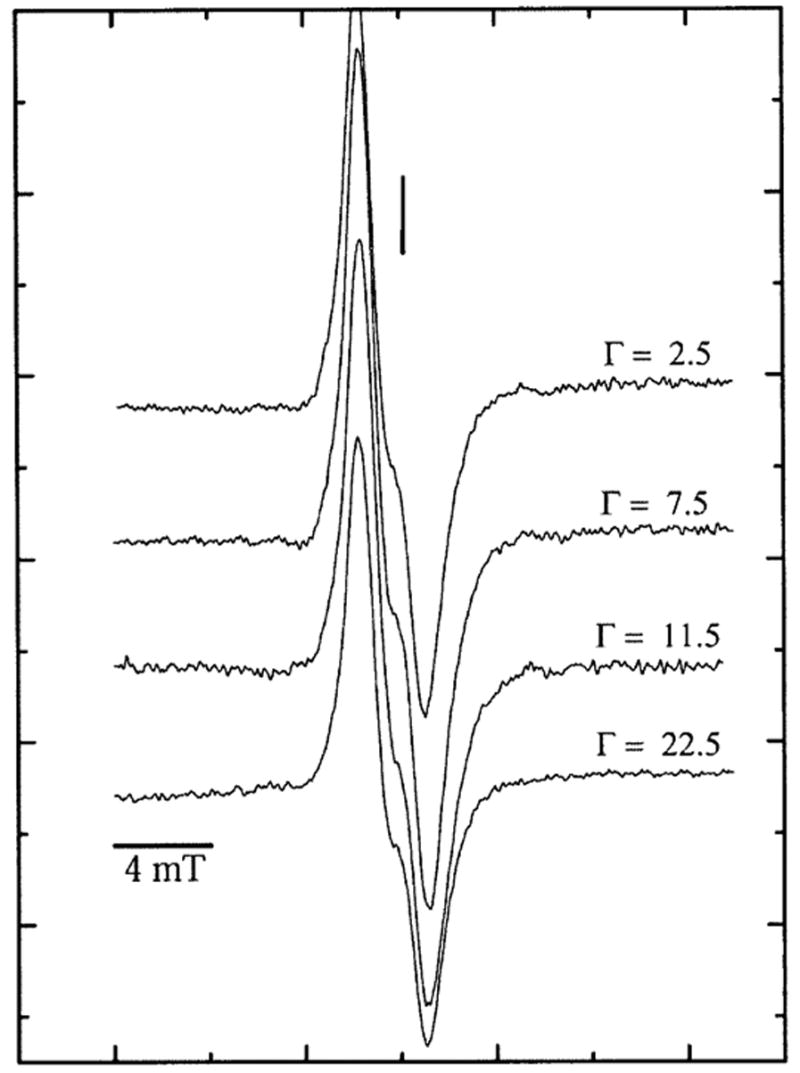
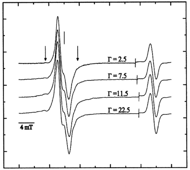
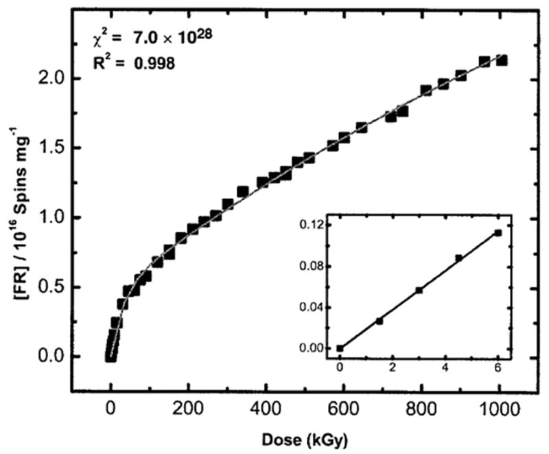
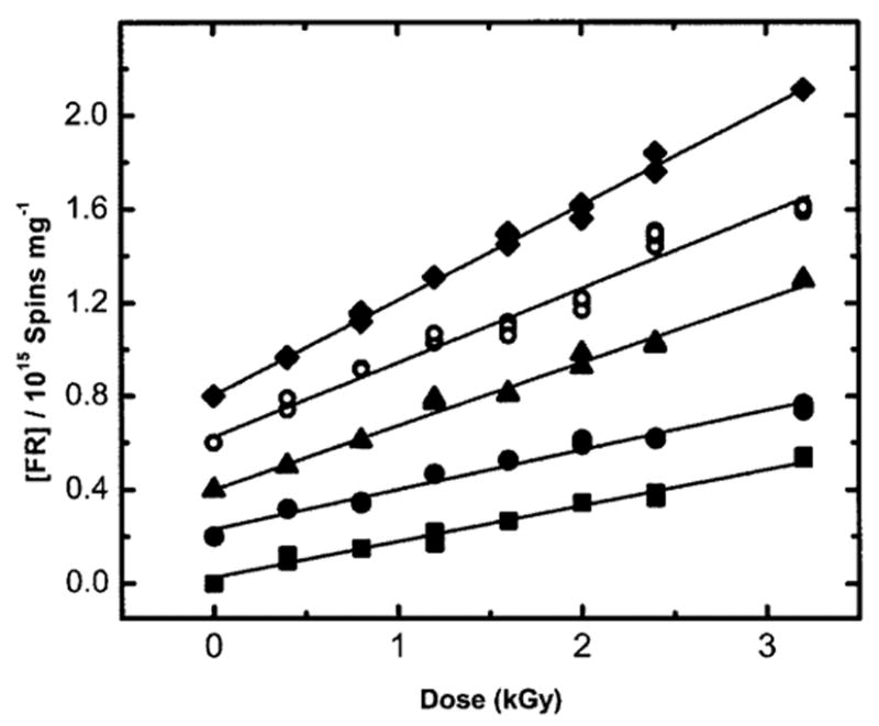
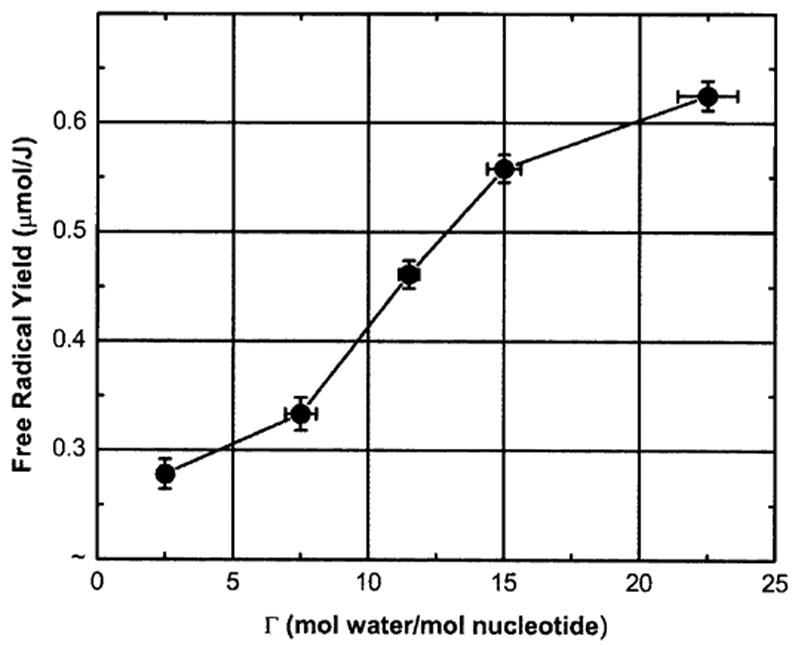
The purpose of this study was to elucidate the role of hydration (Gamma) in the distribution of free radical trapping in directly ionized DNA. Solid-state films of pUC18 (2686 bp) plasmids were hydrated to Gamma in the range 2.5 < or = Gamma < or = 22.5 mol water/mol nucleotide. Free radical yields, G(Sigmafr), measured by EPR at 4 K are seen to increase from 0.28 +/- 0.01 micromol/J at Gamma = 2.5 to 0.63 +/- 0.01 micromol/J at Gamma= 22.5, respectively. Based on a semi-empirical model of the free radical trapping events that follow the initial ionizations of the DNA components, we conclude that two-thirds of the holes formed on the inner solvation shell (Gamma < 10) transfer to the sugar-phosphate backbone. Likewise, of the holes produced by direct ionization of the sugar-phosphate, about one-third are trapped by deprotonation as neutral sugar-phosphate radical species, while the remaining two-thirds are found to transfer to the bases. This analysis provides the best measure to date for the probability of hole transfer (approximately 67%) into the base stack. It can thus be predicted that the distribution of holes formed in fully hydrated DNA at 4 K will be 78% on the bases and 22% on the sugar-phosphate. Adding the radicals due to electron attachment (confined to the pyrimidine bases), the distribution of all trapped radicals will be 89% on the bases and 11% on the sugar-phosphate backbone. This prediction is supported by partitioning results obtained from the high dose-response curves fitted to the two-component model. These results not only add to our understanding of how the holes redistribute after ionization but are also central to predicting the yield and location of strand breaks in DNA exposed to the direct effects of ionizing radiation.
Figures
Similar articles
-
Correlation of free radical yields with strand break yields produced in plasmid DNA by the direct effect of ionizing radiation.J Phys Chem B. 2005 Sep 8;109(35):16967-73. doi: 10.1021/jp0518409. J Phys Chem B. 2005. PMID: 16853159 Free PMC article.
-
An investigation into the mechanisms of DNA strand breakage by direct ionization of variably hydrated plasmid DNA.J Phys Chem B. 2006 Dec 28;110(51):26286-91. doi: 10.1021/jp065489i. J Phys Chem B. 2006. PMID: 17181287 Free PMC article.
-
The influence of hydration on the absolute yields of primary ionic free radicals in gamma-irradiated DNA at 77 K. I. Total radical yields.Radiat Res. 1993 Aug;135(2):146-54. Radiat Res. 1993. PMID: 8396268
-
Combination is the dominant free radical process initiated in DNA by ionizing radiation: an overview based on solid-state EPR studies.Int J Radiat Biol. 1994 Nov;66(5):491-7. doi: 10.1080/09553009414551511. Int J Radiat Biol. 1994. PMID: 7983436 Review.
-
One-electron oxidation reactions of purine and pyrimidine bases in cellular DNA.Int J Radiat Biol. 2014 Jun;90(6):423-32. doi: 10.3109/09553002.2013.877176. Epub 2014 Apr 3. Int J Radiat Biol. 2014. PMID: 24369822 Free PMC article. Review.
Cited by
-
Radioresistance of GGG sequences to prompt strand break formation from direct-type radiation damage.Radiat Environ Biophys. 2016 Nov;55(4):411-422. doi: 10.1007/s00411-016-0660-7. Epub 2016 Jun 27. Radiat Environ Biophys. 2016. PMID: 27349757 Free PMC article.
-
Unaltered free base release from d(CGCGCG)2 produced by the direct effect of ionizing radiation at 4 K and room temperature.Radiat Res. 2007 May;167(5):501-7. doi: 10.1667/RR0847.1. Radiat Res. 2007. PMID: 17474798 Free PMC article.
-
Proton-coupled electron transfer in DNA on formation of radiation-produced ion radicals.Chem Rev. 2010 Dec 8;110(12):7002-23. doi: 10.1021/cr100023g. Epub 2010 May 5. Chem Rev. 2010. PMID: 20443634 Free PMC article. Review. No abstract available.
-
On the chemical yield of base lesions, strand breaks, and clustered damage generated in plasmid DNA by the direct effect of X rays.Radiat Res. 2007 Sep;168(3):357-66. doi: 10.1667/RR0964.1. Radiat Res. 2007. PMID: 17705639 Free PMC article.
-
Structure reactivity relationship in the reaction of DNA guanyl radicals with hydroxybenzoates.Radiat Phys Chem Oxf Engl 1993. 2010 Nov 1;79(1):1144-1148. doi: 10.1016/j.radphyschem.2010.06.006. Radiat Phys Chem Oxf Engl 1993. 2010. PMID: 21966099 Free PMC article.
References
-
- Gregoli S, Olast M, Bertinchamps A. Radiolytic pathways in gamma-irradiated DNA: Influence of chemical and conformational factors. Radiat Res. 1982;89:238–254. - PubMed
-
- Hüttermann J, Röhrig M, Köhnlein W. Free radicals from irradiated lyophilized DNA: Influence of water of hydration. Int J Radiat Biol. 1992;61:299–313. - PubMed
-
- Swarts SG, Sevilla MD, Becker D, Tokar CJ, Wheeler KT. Radiation-induced DNA damage as a function of hydration. I. Release of unaltered bases. Radiat Res. 1992;129:333–344. - PubMed
-
- Mroczka N, Bernhard WA. Hydration effects on free radical yields in DNA X-irradiated at 4 K. Radiat Res. 1993;135:155–159. - PubMed
-
- Wang W, Becker D, Sevilla MD. The influence of hydration on the absolute yields of primary ionic free radicals in gamma-irradiated DNA at 77 K. I. Total radical yields. Radiat Res. 1993;135:146–154. - PubMed
Publication types
MeSH terms
Substances
Grants and funding
LinkOut - more resources
Full Text Sources
