Comparative Study
. 2006 Jul;80(14):6993-7008.
doi: 10.1128/JVI.00365-06.

Evaluating replication-defective vesicular stomatitis virus as a vaccine vehicle

Affiliations
Comparative Study

Evaluating replication-defective vesicular stomatitis virus as a vaccine vehicle

Ayaz M Majid et al. J Virol. 2006 Jul.

Abstract

We have generated replication-competent (VSV-C/E1/E2) and nonpropagating (VSVDeltaG-C/E1/E2) vesicular stomatitis virus (VSV) contiguously expressing the structural proteins of hepatitis C virus (HCV; core [C] and glycoproteins E1 and E2) and report on their immunogenicity in murine models. VSV-C/E1/E2 and VSVDeltaG-C/E1/E2 expressed high levels of HCV C, E1, and E2, which were authentically posttranslationally processed. Both VSV-expressed HCV E1-E2 glycoproteins were found to form noncovalently linked heterodimers and appeared to be correctly folded, as confirmed by coimmunoprecipitation analysis using conformationally sensitive anti-HCV-E2 monoclonal antibodies (MAbs). Intravenous or intraperitoneal immunization of BALB/c mice with VSV-C/E1/E2 or VSVDeltaG-C/E1/E2 resulted in significant and surprisingly comparable HCV core or E2 antibody responses compared to those of control mice. In addition, both virus types generated HCV C-, E1-, or E2-specific gamma interferon (IFN-gamma)-producing CD8(+) T cells, as determined by enzyme-linked immunospot (ELISPOT) analysis. Mice immunized with VSVDeltaG-C/E1/E2 were also protected against the formation of tumors expressing HCV E2 (CT26-hghE2t) and exhibited CT26-hghE2t-specific IFN-gamma-producing and E2-specific CD8(+) T-cell activity. Finally, recombinant vaccinia virus (vvHCV.S) expressing the HCV structural proteins replicated at significantly lower levels when inoculated into mice immunized with VSV-C/E1/E2 or VSVDeltaG-C/E1/E2, but not with control viruses. Our data therefore illustrate that potentially safer replication-defective VSV can be successfully engineered to express high levels of antigenically authentic HCV glycoproteins. In addition, this strategy may therefore serve in effective vaccine and immunotherapy-based approaches to the treatment of HCV-related disease.


Figures

FIG. 1.
FIG. 1.
Development and characterization of a nonpropagating VSV vector expressing hepatitis C virus structural proteins (VSVΔG-C/E1/E2). (A) Construction of nonpropagating rVSV. The complete VSV glycoprotein (G) was deleted from the parent pVSV-XN2 plasmid and replaced with a multiple cloning site linker to create the pVSVΔG vector. The contiguous HCV C/E1/E2 genes were amplified by PCR and inserted into the multiple cloning site in pVSVΔG between the matrix (M) and polymerase (L) genes of VSV-XN2. The VSVΔG or VSVΔG-C/E1/E2 viruses were made by transfecting a pVSVΔG plasmid encoding VSV G into BHK cells. Vaccinia virus encoding the T7 polymerase (vTF7-3) was used to infect these “helper” BHK cells 24 h later (MOI of 10) followed by transfection with pVSVΔG or pVSVΔG-C/E1/E2 pBl-N, -P, and -L. The cells expressing VSV G mediate incorporation of G protein into VSVΔG or VSVΔG-C/E1/E2 virions that can subsequently infect cells. However, since VSV G is no longer encoded in the genome, the virus will be unable to produce infectious virions in cells not expressing the G protein and is therefore replication defective. (B) Growth curve analysis of VSVΔG-C/E1/E1 and VSVΔG. A one-step growth curve analysis was performed in VSV G-expressing “helper” BHK cells. (C) Expression of VSV and HCV proteins in nonpropagating rVSV. BHK cells were infected with wild-type VSV (MOI of 10 for 15 h) or nonpropagating VSVΔG or VSVΔG-C/E1/E2 (MOI of 10 for 24 h), and cell lysates were subjected to Western blot (WB) analysis using mouse VSV-antiserum and the anti-HCV antibodies shown. poly Ab, polyclonal antibody.
FIG. 2.
FIG. 2.
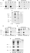
Characterization of E1 and E2 glycoproteins expressed by VSV using mouse or human conformation-dependent MAbs. Huh-7 cells were infected (MOI of 10 for 12 h) with VSV-C/E1/E2 or VSV-GFP, and the cell lysates were subjected to IP using anti-E2 H33 conformation-dependent or anti-E2 H52 conformation-independent MAb (as detailed in Materials and Methods). The immunoprecipitated protein G complexes were then subjected to reducing SDS-PAGE (A) analysis or nonreducing SDS-PAGE conditions (B) and detected using an anti-E2 polyclonal antibody following the Western blotting procedure. (C) Radiolabeled noncovalently linked E1/E2 heterodimers. Huh-7 cells were infected (MOI of 10) for 5 h with VSV-C/E1/E2 or VSV-GFP. Cells were depleted of Met and Cys before addition of 600 μCi [35S]Met/Cys. Cells were harvested, and lysates were subjected to IP (detailed in Materials and Methods) using the anti-E2 H33 conformationally sensitive MAb. The immune complexes were subjected to nonreducing SDS-PAGE and subsequently to autoradiography. E1/E2 heterodimers bind human conformation-dependent antibodies, as shown under reducing (D) and nonreducing (E) SDS-PAGE conditions. Huh-7 cells were infected as described above, and lysates were investigated using an IP procedure with CBH-7 and CBH-8 hMAbs followed by Western detection with anti-E2 polyclonal antibody (upper panels). The blots were then stripped and reprobed with anti-E1 A4 MAb (lower panels). Huh-7 cells were also infected with VSVΔG-C/E1/E2 or VSVΔG (MOI of 10 for 12 h), and the lysates were subjected to IP as described above using CBH-7 or CBH-8 hMAB followed by SDS-PAGE and Western detection with anti-E2 polyclonal antibody (F, upper panels). The blots were stripped and reprobed with anti-E1 A4 MAb (lower panels).
FIG. 3.
FIG. 3.
Comparison of levels of HCV antigen expression between VSVΔG-C/E1/E2 and VSV-C/E1/E2 and antibody responses to HCV core in immunized mice. BHK cells were infected with nonpropagating (VSVΔG or VSVΔG-C/E1/E2) or replication-competent VSV-C/E1/E2 at an MOI of 10 overnight. Harvested cell lysates were subjected to Western analysis with anti-HCV core (A), anti-HCV E1 A4 (B), or anti-E2 polyclonal antibody (polyAb) (C). The same concentration of total protein (20 μg) was used for the Western procedure. Six-week-old BALB/c mice (three mice per group) were vaccinated (day 1 and day 14) with 6 × 10⁶ PFU of VSV-C/E1/E2 or VSV-GFP (or 0.1 ml of PBS only) in parallel experiments with VSVΔG-C/E1/E2 or VSVΔG by the i.p. route (D and E) or i.v. injection (F and G). Serum from all mice was extracted on day 0 and day 21 and tested (in duplicate for each mouse) for anti-HCV core antibody using an ELISA (described in Materials and Methods). The results are presented as mean serum anti-HCV core optical density (O.D.) at 450 nm for each group, and error bars represent standard deviations within each group. Student's t test analysis was also performed for comparisons between the virus groups shown, and significant data are illustrated as P = <0.05.
FIG. 4.
FIG. 4.
Comparison of anti-HCV E2 antibody responses stimulated by intraperitoneal or intravenous injection of VSV-C/E1/E2 or VSVΔG-C/E1/E2. Sera from mice tested for anti-HCV core reactivity were used to investigate anti-HCV E2 antibody responses using an HCV E2-specific ELISA (Materials and Methods). Antibody responses to E2 were compared between replication-competent and nonpropagating viruses following i.p. injection (A and B, respectively) and i.v. administration (C and D). Responses are shown as means ± standard deviation of three mice tested in duplicate per group. Student's t test analysis was also performed on the data between virus groups, and significant results are shown (P = <0.05). O.D., optical density.
FIG. 5.
FIG. 5.
Analysis of T-helper-cell responses by examining IgG class-switching profiles by B cells. HCV core antibody-positive sera (day 21) from three mice vaccinated with VSVΔG-C/E1/E2 were tested (in duplicate) in the core HCV ELISA and subjected to the isotype-specific anti-mouse secondary antibodies shown. Results are presented as mean anti-HCV core optical density (O.D.) at 450 nm ± standard deviation for each isotype-specific antibody.
FIG. 6.
FIG. 6.
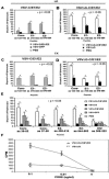
HCV-specific T-cell responses to core, E1, or E2. Splenectomies were performed on BALB/c mice sacrificed (day 21) following vaccination as described for antibody responses with VSV-C/E1/E2 or VSV-GFP and VSVΔG-C/E1/E2 or VSVΔG by the i.p. route (A and B, respectively) or i.v. injection (C and D). PBS controls were also included. Splenocytes were harvested from three mice per group, pooled, and plated in a precoated anti-mouse IFN-γ MAb 96-well format at dilutions of 1 × 10⁶ to 0.25 × 10⁶ (in triplicate) in RPMI medium containing IL-2 and HCV peptide as shown. Unstimulated (mock) splenocytes were also plated for each group. After 36 h at 37°C, ELISPOT analysis was performed (described in Materials and Methods). The results are presented as mean ELISPOTS per million splenocytes − any background ELISPOTS from unpulsed mock controls ± standard deviation from the means. P values represent Student's t test analysis of comparisons of the ELISPOTS between virus groups. Splenocytes were isolated from BALB/c mice immunized intraperitoneally (five per group) as described above and tested against a panel of additional CTL epitopes in HCV C or E2 (E). A control HIV peptide was also included. Lymphoproliferative responses in VSVΔG-C/E1/E2- or VSVΔG-immunized mice were also measured (as described in text) against HCV core 1a antigen (F). The results represent cpm as means of triplicate values at each cell concentration (error bars represent standard deviation).
FIG. 7.
FIG. 7.
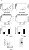
VSVΔG-C/E1/E2 vaccination protects against HCV E2-expressing CT26-hghE2t tumor. BALB/c mice (eight per group) were vaccinated with three injections 6 × 10⁶ PFU of VSVΔG or VSVΔG-C/E1/E2 intravenously (days 1, 14, and 28). Two weeks following the second boost, mice were challenged with 2 × 10⁶ wild-type CT26 cancer cells injected subcutaneously into mice immunized with VSVΔG or VSVΔG-C/E1/E2 (A and B, respectively). VSVΔG- or VSVΔG-C/E1/E2-immunized mice were also challenged with HCV-E2-expressing (CT26hghE2t) cancer cells (C and D, respectively). The tumors were observed at 3-day intervals, and the results are presented as tumor volumes (in cubic millimeters) for each mouse. The median CT26 and CT26-hghE2t tumor diameters (E and F, respectively) are shown for the first and last measurements for each challenged group. (G) IFN-γ ELISPOT analysis in CT26-hghE2t-challenged mice. Splenectomies were performed on VSVΔG-C/E1/E2 (or VSVΔG)-vaccinated mice (two mice from each group) at day 26 post-tumor challenge. Responder splenocytes were stimulated with target CT26-hghE2t cells or negative control HeLa cells at a ratio of 10:1 (dilutions in triplicate) for 1 week at 37°C prior to ELISPOT analysis as described in Materials and Methods. The results are presented as mean ELISPOTS (subtracted from mock unpulsed controls per group) ± standard deviation from the mean. (H) Western blot (WB) analysis of CT26 and CT26-hghE2t cell lines was performed using anti-E2 polyclonal antibody (poly Ab) prior to injection into mice. The blots were reprobed with anti-β-actin MAb.
FIG. 8.
FIG. 8.
VSVΔG-C/E1/E2 immunization of BALB/c mice inhibits viral replication of recombinant vaccinia virus (vvHCV.S) expressing HCV structural antigens. (A) Mice (five per group) were vaccinated intravenously (days 1, 14, and 28) with 6 × 10⁶ PFU/ml of VSVΔG or VSVΔG-C/E1/E2 and 2 weeks later challenged with 1 × 10⁷ PFU/ml of vvHCV.S administered by intraperitoneal injection. A nonimmunized mouse group also received the same dose of vvHCV.S. Ovaries were harvested from all mice on day 5 (peak viral titers), and following homogenization and freeze-thaw, viral titers were evaluated by plaque assay. The mean viral titers (error bars represent standard deviations) for each group are given. (B) Viral challenge (1 ×10⁷ PFU/ml) with control vaccinia virus Western Reserve strain (vv-WR). The experiment was performed in parallel and as described above. (C) VSV-C/E1/E2- or VSV-GFP-immunized animals (as above) were also challenged with 1 × 10⁷ PFU/ml of vvHCV.S administered by intraperitoneal injection. The ovaries were harvested on day 5, measured (mg), and processed as described above followed by plaque assay to evaluate viral titers. The results represent mean viral titers expressed as a ratio of residual virus and size of ovary. P values represent Student's t test analysis of comparisons of viral titers between groups.

Similar articles

Cited by

References

    1. Adams, D. O., T. Hall, Z. Steplewski, and H. Koprowski. 1984. Tumors undergoing rejection induced by monoclonal antibodies of the IgG2a isotype contain increased numbers of macrophages activated for a distinctive form of antibody-dependent cytolysis. Proc. Natl. Acad. Sci. USA 81:3506-3510. - PMC - PubMed
    1. Afdhal, N. H. 2004. The natural history of hepatitis C. Semin. Liver Dis. 24(Suppl. 2):3-8. - PubMed
    1. Agnello, V., A. Gyorgy, M. Elfahal, G. B. Knight, and Q.-X. Zhang. 1999. Hepatitis C virus and other Flaviviridae viruses enter cells via low density lipoprotein receptor. Proc. Natl. Acad. Sci. USA 96:12766-12771. - PMC - PubMed
    1. Balachandran, S., and G. N. Barber. 2000. Vesicular stomatitis virus (VSV) therapy of tumors. IUBMB Life 50:135-138. - PubMed
    1. Balachandran, S., M. Porosnicu, and G. N. Barber. 2001. Oncolytic activity of vesicular stomatitis virus is effective against tumors exhibiting aberrant p53, Ras, or Myc function and involves induction of apoptosis. J. Virol. 75:3474-3479. - PMC - PubMed

Publication types

MeSH terms