Comparative Study
. 2006 Jul;80(14):7009-19.
doi: 10.1128/JVI.00489-06.

PKR and RNase L contribute to protection against lethal West Nile Virus infection by controlling early viral spread in the periphery and replication in neurons

Affiliations
Comparative Study

PKR and RNase L contribute to protection against lethal West Nile Virus infection by controlling early viral spread in the periphery and replication in neurons

Melanie A Samuel et al. J Virol. 2006 Jul.

Abstract

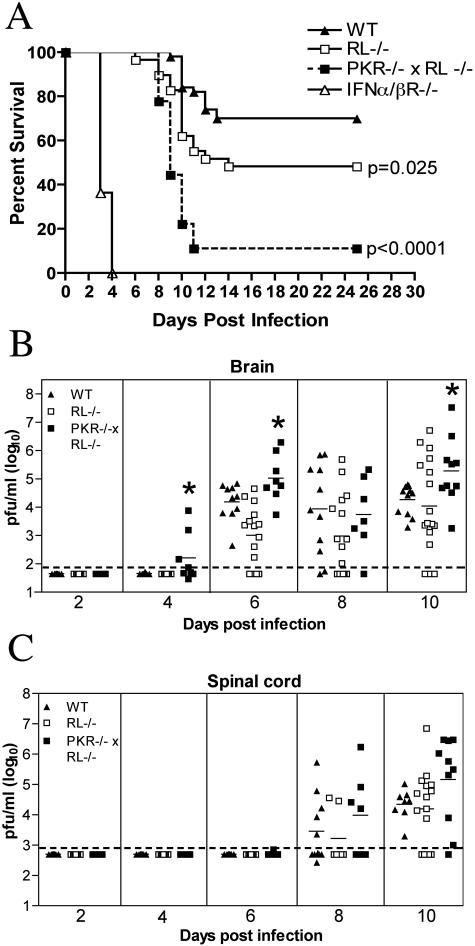
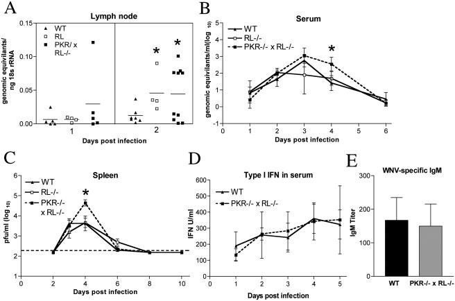
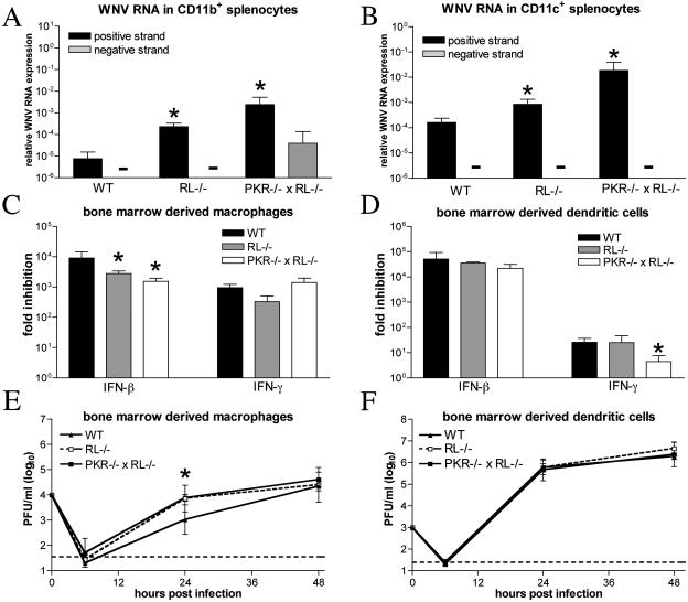
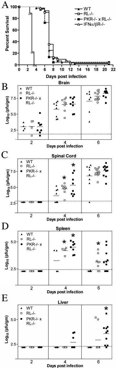
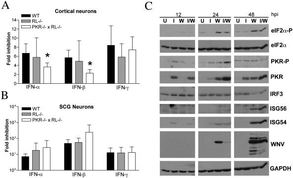
West Nile virus (WNV) is a neurotropic, mosquito-borne flavivirus that can cause lethal meningoencephalitis. Type I interferon (IFN) plays a critical role in controlling WNV replication, spread, and tropism. In this study, we begin to examine the effector mechanisms by which type I IFN inhibits WNV infection. Mice lacking both the interferon-induced, double-stranded-RNA-activated protein kinase (PKR) and the endoribonuclease of the 2',5'-oligoadenylate synthetase-RNase L system (PKR(-/-) x RL(-/-)) were highly susceptible to subcutaneous WNV infection, with a 90% mortality rate compared to the 30% mortality rate observed in congenic wild-type mice. PKR(-/-) x RL(-/-) mice had increased viral loads in their draining lymph nodes, sera, and spleens, which led to early viral entry into the central nervous system (CNS) and higher viral burden in neuronal tissues. Although mice lacking RNase L showed a higher CNS viral burden and an increased mortality, they were less susceptible than the PKR(-/-) x RL(-/-) mice; thus, we also infer an antiviral role for PKR in the control of WNV infection. Notably, a deficiency in both PKR and RNase L resulted in a decreased ability of type I IFN to inhibit WNV in primary macrophages and cortical neurons. In contrast, the peripheral neurons of the superior cervical ganglia of PKR(-/-) x RL(-/-) mice showed no deficiency in the IFN-mediated inhibition of WNV. Our data suggest that PKR and RNase L contribute to IFN-mediated protection in a cell-restricted manner and control WNV infection in peripheral tissues and some neuronal subtypes.


Figures

FIG. 1.
FIG. 1.
Survival and virologic analysis for wild-type (WT), RL−/−, PKR−/− × RL−/−, and IFN-α/βR−/− C57BL/6 mice. (A) Eight- to 10-week-old mice were inoculated with 102 PFU of WNV by footpad injection and followed for mortality for 25 days. The number of mice in these experiments was as follows: 50 wild-type, 29 RL−/−, 18 PKR−/− × RL−/−, and 11 IFN-α/βR−/− mice. Survival differences between wild-type and all immunodeficient mice were statistically significant (P ≤ 0.03). (B and C) Viral burdens in the CNS after WNV infection. Viral loads in the brain (B) and spinal cord (C) were determined from samples harvested on days 2, 4, 6, 8, and 10 using a viral plaque assay on BHK21 cells. Data are shown as the PFU per gram of tissue for 5 to 17 mice per time point. For all viral load data, the solid line represents the mean PFU per gram at the indicated time point, and the dotted line represents the limit of sensitivity of the assay. Asterisks indicate values that are statistically significant (P < 0.05) compared to those for wild-type mice.
FIG. 2.
FIG. 2.
PKR−/− × RL−/− mice show increased viral replication in the periphery. (A to C) Viral burdens in the lymph nodes, sera, and spleens of wild-type (WT), RL−/−, and PKR−/− × RL−/− mice following footpad infection with 102 PFU of WNV. (A) Levels of viral RNA were measured from inguinal lymph nodes ipsilateral to the site of infection by quantitative RT-PCR. Data are shown as the genomic equivalents of WNV RNA per ng of 18S rRNA. (B) Viral RNA levels in sera were analyzed via quantitative RT-PCR. Data shown are genomic equivalents of WNV RNA per milliliter of serum. (C) WNV burden in the spleen was measured by plaque assay using samples harvested on the indicated days and is expressed as PFU per gram of tissue. The dotted line represents the limit of sensitivity of the assay. (D) Type I IFN concentrations were determined from sera collected on days 1 to 5 after infection by using an EMCV bioassay in L929 cells. Data reflect the averages from serum samples harvested from three to five mice per time point and are shown as units of IFN/ml of serum. (E) WNV-specific IgM levels were determined from sera collected on day 4 postinfection by enzyme-linked immunosorbent assay against purified recombinant WNV E protein. Data represent the averages from samples harvested from eight mice per group and are expressed as units of optical density after subtraction of the background. Error bars indicate the standard deviations, and asterisks indicate values that are statistically significant (P < 0.05) compared to those for wild-type mice in all panels.
FIG. 3.
FIG. 3.
PKR and RNase L modulate viral infection in bone marrow-derived cells. (A and B) Total RNA from CD11b+ (A) and CD11c+ (B) splenocytes isolated on day 4 postinfection was analyzed for the levels of WNV positive and negative strands with strand-specific real-time RT-PCR. Data are expressed as the amounts of positive- and negative-strand RNA relative to 18S rRNA levels. Average values are from four to eight mice per group. BM-Mφ (C) and BM-DCs (D) were generated from wild-type, RL−/−, and PKR−/− × RL−/− mice and treated for 24 h with 1,000 IU/ml of mouse IFN-β or -γ prior to infection with an MOI of 0.1. The production of infectious virus at 24 h postinfection was determined by plaque assay. Average values represent results from quadruplicate samples harvested from three independent experiments. BM-Mφ (E) and BM-DCs (F) generated from wild-type, RL−/−, and PKR−/− × RL−/− mice were infected at an MOI of 0.01, and viral production was evaluated at the indicated times postinfection by plaque assay. Values are averages of results from quadruplicate samples generated from two independent experiments, and asterisks indicate differences that are statistically significant relative to results for wild-type mice (P < 0.05).
FIG. 4.
FIG. 4.
Survival and viral burden data for wild-type (WT), RL−/−, PKR−/− × RL−/−, and IFN-α/βR−/− C57BL/6 mice after intracranial inoculation with 101 PFU of WNV. (A) Data from two to three independent experiments were used to construct the survival curves, for which 44 wild-type, 31 RL−/−, 22 PKR−/− × RL−/−, and 9 IFN-α/ βR−/− mice were used. The mean times to death for wild-type, RL−/−, and PKR−/− × RL−/− mice were not statistically different, whereas the mean time to death for IFN-α/βR−/− mice was significantly decreased (P < 0.0001). (B through E) Viral burden in tissues after intracranial WNV infection. Viral loads in brain (B), spinal cord (C), spleen (D), and liver (E) were measured by plaque assay from samples harvested on days 2, 4, and 6 after infection and reflect results for four to eight mice per group. The dotted line represents the limit of sensitivity of the assay, and asterisks indicate values that are statistically significant (P < 0.05) compared to those for wild-type mice.
FIG. 5.
FIG. 5.
Effects of PKR and RNase L on IFN-mediated inhibition of WNV infection in neurons. (A and B) Primary cultures of cortical and SCG neurons were prepared from wild-type, RL−/−, and PKR−/−RL−/− mice. Cortical neurons (A) or SCG neurons (B) were treated 24 h prior to infection with 100 IU/ml of the indicated mouse IFN. Neurons were infected with WNV and evaluated for the production of infectious virus at 24 h by a plaque assay. Data represent averages of results from three to five independent experiments, and asterisks indicate values that are statistically significant (P < 0.05) compared to those for treated wild-type cells. (C) Whole-cell lysates were generated at the indicated times (hours postinfection [hpi]) from cortical neurons that were uninfected (lanes U), infected with WNV at an MOI of 0.1 (lanes W), treated with IFN-β at 100 IU/ml (lanes I), or infected with WNV and treated with IFN at the time of infection (lanes I/W). Protein levels of phosphorylated PKR (PKR-P), phosphorylated eIF2-α (eIF2α-P), total PKR, total eIF2-α, mouse IRF-3, ISG56, ISG54, WNV, and GAPDH were examined by immunoblot analysis.

References

    1. Alexopoulou, L., A. C. Holt, R. Medzhitov, and R. A. Flavell. 2001. Recognition of double-stranded RNA and activation of NF-kappaB by Toll-like receptor 3. Nature 413:732-738. - PubMed
    1. Al-khatib, K., B. R. Williams, R. H. Silverman, W. Halford, and D. J. Carr. 2003. The murine double-stranded RNA-dependent protein kinase PKR and the murine 2′,5′-oligoadenylate synthetase-dependent RNase L are required for IFN-beta-mediated resistance against herpes simplex virus type 1 in primary trigeminal ganglion culture. Virology 313:126-135. - PubMed
    1. Asnis, D. S., R. Conetta, A. A. Teixeira, G. Waldman, and B. A. Sampson. 2000. The West Nile virus outbreak of 1999 in New York: the Flushing Hospital experience. Clin. Infect. Dis. 30:413-418. - PubMed
    1. Austin, B. A., C. James, R. H. Silverman, and D. J. Carr. 2005. Critical role for the oligoadenylate synthetase/RNase L pathway in response to IFN-beta during acute ocular herpes simplex virus type 1 infection. J. Immunol. 175:1100-1106. - PubMed
    1. Balachandran, S., P. C. Roberts, L. E. Brown, H. Truong, A. K. Pattnaik, D. R. Archer, and G. N. Barber. 2000. Essential role for the dsRNA-dependent protein kinase PKR in innate immunity to viral infection. Immunity 13:129-141. - PubMed

Publication types

MeSH terms