Comparative Study
. 2006 Jul;80(14):7136-45.
doi: 10.1128/JVI.00509-06.

Comparative analysis of 22 coronavirus HKU1 genomes reveals a novel genotype and evidence of natural recombination in coronavirus HKU1

Affiliations
Comparative Study

Comparative analysis of 22 coronavirus HKU1 genomes reveals a novel genotype and evidence of natural recombination in coronavirus HKU1

Patrick C Y Woo et al. J Virol. 2006 Jul.

Abstract

We sequenced and compared the complete genomes of 22 strains of coronavirus HKU1 (CoV HKU1) obtained from nasopharyngeal aspirates of patients with respiratory tract infections over a 2-year period. Phylogenetic analysis of 24 putative proteins and polypeptides showed that the 22 CoV HKU1 strains fell into three clusters (genotype A, 13 strains; genotype B, 3 strains and genotype C, 6 strains). However, different phylogenetic relationships among the three clusters were observed in different regions of their genomes. From nsp4 to nsp6, the genotype A strains were clustered with the genotype B strains. For nsp7 and nsp8 and from nsp10 to nsp16, the genotype A strains were clustered with the genotype C strains. From hemagglutinin esterase (HE) to nucleocapsid (N), the genotype B strains were clustered closely with the genotype C strains. Bootscan analysis showed possible recombination between genotypes B and C from nucleotide positions 11,500 to 13,000, corresponding to the nsp6-nsp7 junction, giving rise to genotype A, and between genotypes A and B from nucleotide positions 21,500 to 22,500, corresponding to the nsp16-HE junction, giving rise to genotype C. Multiple alignments further narrowed the sites of crossover to a 143-bp region between nucleotide positions 11,750 and 11,892 and a 29-bp region between nucleotide positions 21,502 and 21,530. Genome analysis also revealed various numbers of tandem copies of a perfect 30-base acidic tandem repeat (ATR) which encodes NDDEDVVTGD and various numbers and sequences of imperfect repeats in the N terminus of nsp3 inside the acidic domain upstream of papain-like protease 1 among the 22 genomes. All 10 CoV HKU1 strains with incomplete imperfect repeats (1.4 and 4.4) belonged to genotype A. The present study represents the first evidence for natural recombination in coronavirus associated with human infection. Analysis of a single gene is not sufficient for the genotyping of CoV HKU1 strains but requires amplification and sequencing of at least two gene loci, one from nsp10 to nsp16 (e.g., pol or helicase) and another from HE to N (e.g., spike or N). Further studies will delineate whether the ATR is useful for the molecular typing of CoV HKU1.


Figures

FIG.1.
FIG.1.
Genome organization and phylogenetic analysis of CoV HKU1. (A) Genome organization of CoV HKU1. The regions of the genome used for phylogenetic tree construction are labeled. (B) Phylogenetic analysis of nsp1, nsp2, conserved portions of nsp3 (PL1pro, A1pp, PL2pro, and HD), nsp4-nsp10, nsp12-nsp16, HE, S, ORF4, E, M, and N of the 22 CoV HKU1 genomes. The trees were constructed by the neighbor-joining method using Jukes-Cantor correction and bootstrap values calculated from 1,000 trees. Seven hundred forty, 1,821, 655, 333, 895, 1,263, 1,488, 909, 861, 276, 582, 330, 411, 2,783, 1,809, 1,563, 1,125, 900, 1,276, 4,126, 330, 253, 687, and 1,358 nucleotide positions in nsp1, nsp2, PL1pro, A1pp, PL2pro, HD, nsp4, nsp5, nsp6, nsp7, nsp8, nsp9, nsp10, nsp12, nsp13, nsp14, nsp15, nsp16, HE, S, ORF4, E, M, and N, respectively, were included in the analysis. The scale bar indicates the estimated number of substitutions per 50 or 100 nucleotides as indicated. The corresponding nucleotide sequences of HCoV OC43 were used as the outgroups. A, genotype A; B, genotype B and C, genotype C.
FIG.1.
FIG.1.
Genome organization and phylogenetic analysis of CoV HKU1. (A) Genome organization of CoV HKU1. The regions of the genome used for phylogenetic tree construction are labeled. (B) Phylogenetic analysis of nsp1, nsp2, conserved portions of nsp3 (PL1pro, A1pp, PL2pro, and HD), nsp4-nsp10, nsp12-nsp16, HE, S, ORF4, E, M, and N of the 22 CoV HKU1 genomes. The trees were constructed by the neighbor-joining method using Jukes-Cantor correction and bootstrap values calculated from 1,000 trees. Seven hundred forty, 1,821, 655, 333, 895, 1,263, 1,488, 909, 861, 276, 582, 330, 411, 2,783, 1,809, 1,563, 1,125, 900, 1,276, 4,126, 330, 253, 687, and 1,358 nucleotide positions in nsp1, nsp2, PL1pro, A1pp, PL2pro, HD, nsp4, nsp5, nsp6, nsp7, nsp8, nsp9, nsp10, nsp12, nsp13, nsp14, nsp15, nsp16, HE, S, ORF4, E, M, and N, respectively, were included in the analysis. The scale bar indicates the estimated number of substitutions per 50 or 100 nucleotides as indicated. The corresponding nucleotide sequences of HCoV OC43 were used as the outgroups. A, genotype A; B, genotype B and C, genotype C.
FIG. 2.
FIG. 2.
Bootscan analysis of the CoV HKU1 genomes. Bootscanning was conducted with Simplot version 3.5.1 (F84 model; window size, 1,000 bp; step, 200 bp) on a gapless nucleotide alignment, generated with ClustalX, with the genome sequence of HCoV OC43 (GenBank accession no. AY585229) as the query sequence. The dashed line denotes CoV HKU1 genotype A (NC_006577), the solid line denotes CoV HKU1 genotype B (AY884001), and the dotted line denotes CoV HKU1 genotype C (DQ339101).
FIG.3.
FIG.3.
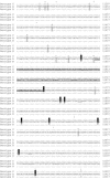
Comparative sequence analysis of the nsp6-nsp7 junction. Multiple alignment of the nucleotide sequences of CoV HKU1 genotypes A, B, and C. In CoV HKU1 genotype B and CoV HKU1 genotype C, only the nucleotides differing from those in CoV HKU1 genotype A are depicted. The nucleotides in CoV HKU1 genotype C that are the same as those in CoV HKU1 genotype A but different from those in CoV HKU1 genotype B are highlighted in black, and those in CoV HKU1 genotype B that are the same as those in CoV HKU1 genotype A but different from those in CoV HKU1 genotype C are highlighted in gray. The putative template switching region is underlined and bold. The first (TCA) and last (CAG) codons of nsp7 are also underlined. The arrows denote positions with nucleotide polymorphism (at position 11414, N16, N17, N20, N21, and N22 [genotype C] were T instead of C; at 11422, N16 and N20 [genotype C] were C instead of T; at 11449, N25 [genotype B] was C instead of T; at 11528, N16, N17, N20, and N22 [genotype C] were C instead of T; at 11740, N6, N7, N9, N10, N11, N13, N14, N18, N23, and N24 [genotype A] were T instead of C; at 12095, N6 and N7 [genotype A] were T instead of C; at 12140, N23 and N24 [genotype A] were C instead of T; at 12367, N15 and N25 [genotype C] were A instead of G; and at 12400, N6, N7, N9, N10, N11, N13, N14, N18, N23, and N24 [genotype A] were C instead of T).
FIG.4.
FIG.4.
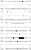
Comparative sequence analysis of the nsp16-HE gene junction. Multiple alignments of the nucleotide sequences of CoV HKU1 genotypes A, B, and C. In CoV HKU1 genotype A and CoV HKU1 genotype B, only the nucleotides differing from those in CoV HKU1 genotype C are depicted. The nucleotides in CoV HKU1 genotype C that are the same as those in CoV HKU1 genotype A but different from those in CoV HKU1 genotype B are highlighted in gray, and those in CoV HKU1 genotype C that are the same as those in CoV HKU1 genotype B but different from those in CoV HKU1 genotype A are highlighted in black. The putative template switching region is underlined and bold. The stop codon of ORF1ab (TAG) and the start codon of the HE gene (ATG) are also underlined. The arrows denote positions with nucleotide polymorphisms (at 21297, N6, N7, N9, N10, N11, N13, N14, N23, and N24 [genotype A] were T instead of G; at 21429, N6 [genotype A] was C instead of T; at 21576, N15 [genotype B] was C instead of T; at 21908, N15 and N25 [genotype B] were G instead of A; and at 21949, N14 was T instead of C).

Similar articles

Cited by

References

    1. Allander, T., M. T. Tammi, M. Eriksson, A. Bjerkner, A. Tiveljung-Lindell, and B. Andersson. 2005. Cloning of a human parvovirus by molecular screening of respiratory tract samples. Proc. Natl. Acad. Sci. USA 102:12891-12896. - PMC - PubMed
    1. Banner, L. R., and M. M. Lai. 1991. Random nature of coronavirus RNA recombination in the absence of selection pressure. Virology 185:441-445. - PMC - PubMed
    1. Bierne, H., M. Seigneur, S. D. Ehrlich, and B. Michel. 1997. uvrD mutations enhance tandem repeat deletion in the Escherichia coli chromosome via SOS induction of the RecF recombination pathway. Mol. Microbiol. 26:557-567. - PubMed
    1. Copper, P. D., A. Steiner-Pryor, P. D. Scotti, and D. Delong. 1974. On the nature of poliovirus genetic recombinants. J. Gen. Virol. 23:41-49. - PubMed
    1. East, M. L., K. Moestl, V. Benetka, C. Pitra, O. P. Honer, B. Wachter, and H. Hofer. 2004. Coronavirus infection of spotted hyenas in the Serengeti ecosystem. Vet. Microbiol. 102:1-9. - PMC - PubMed

Publication types

MeSH terms

LinkOut - more resources