. 2006 Jul;80(14):7199-207.
doi: 10.1128/JVI.00321-06.

Hepatitis C virus triggers mitochondrial permeability transition with production of reactive oxygen species, leading to DNA damage and STAT3 activation

Affiliations

Hepatitis C virus triggers mitochondrial permeability transition with production of reactive oxygen species, leading to DNA damage and STAT3 activation

Keigo Machida et al. J Virol. 2006 Jul.

Abstract

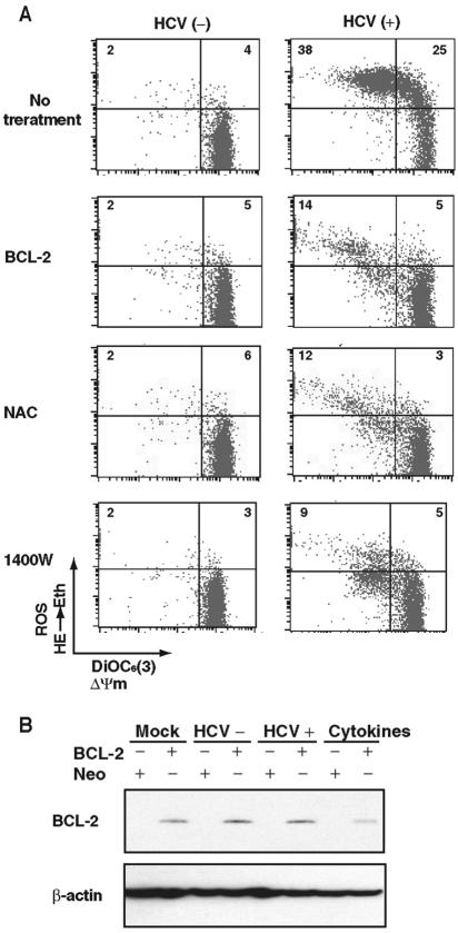
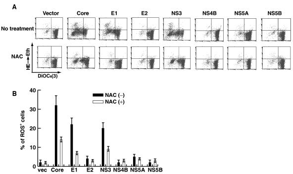
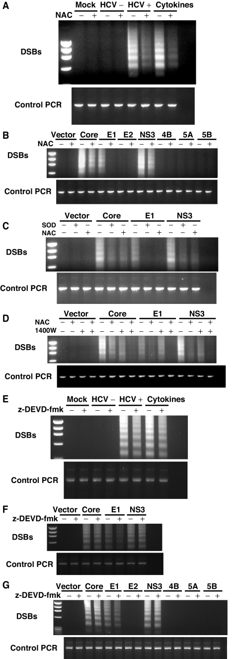
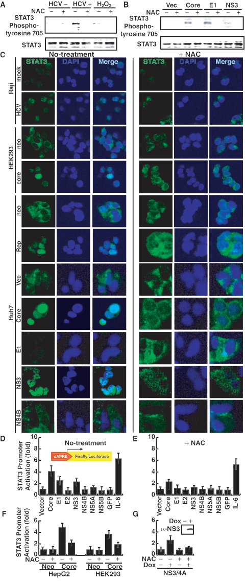
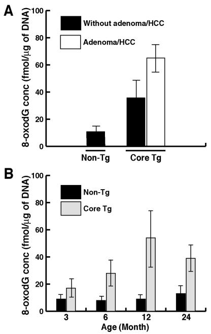
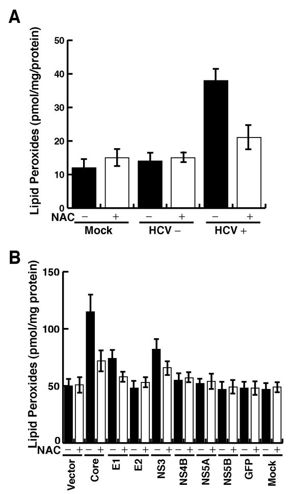
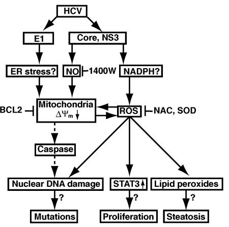
Hepatitis C virus (HCV) infection is frequently associated with the development of hepatocellular carcinomas and non-Hodgkin's B-cell lymphomas. Previously, we reported that HCV infection causes cellular DNA damage and mutations, which are mediated by nitric oxide (NO). NO often damages mitochondria, leading to induction of double-stranded DNA breaks (DSBs) and accumulation of oxidative DNA damage. Here we report that HCV infection causes production of reactive oxygen species (ROS) and lowering of mitochondrial transmembrane potential (DeltaPsi(m)) in in vitro HCV-infected cell cultures. The changes in membrane potential could be inhibited by BCL-2. Furthermore, an inhibitor of ROS production, antioxidant N-acetyl-L-cysteine (NAC), or an inhibitor of NO, 1,400W, prevented the alterations of DeltaPsi(m). The HCV-induced DSB was also abolished by a combination of NO and ROS inhibitors. These results indicated that the mitochondrial damage and DSBs in HCV-infected cells were mediated by both NO and ROS. Among the HCV proteins, core, E1, and NS3 are potent ROS inducers: their expression led to DNA damage and activation of STAT3. Correspondingly, core-protein-transgenic mice showed elevated levels of lipid peroxidation and oxidatively damaged DNA. These HCV studies thus identified ROS, along with the previously identified NO, as the primary inducers of DSBs and mitochondrial damage in HCV-infected cells.


Figures

FIG. 1.
FIG. 1.
(A) HCV-induced changes in mitochondrial membrane potential ΔΨm and ROS production in Raji cells. To measure mitochondrial membrane potential and ROS production, cells were incubated with DiOC6(3) and HE, respectively, at 37°C for 15 min. An experiment representative of four experiments is shown. In some experiments, the cells were treated with different inhibitors during virus infection as indicated. For BCL-2 expression, the cells were stably transfected with the BCL-2 expression plasmids before HCV infection. The numbers in each quadrant represent percentages of total cell population. (B) BCL-2 expression was confirmed by immunoblotting. β-Actin served as a loading control.
FIG. 2.
FIG. 2.
(A) HCV core, E1, and NS3 induce ROS. Raji cells were transfected with plasmids expressing the individual viral proteins in the presence or absence of NAC and analyzed as explained in the legend to Fig. 1. (B) Quantitative measurement of viral-protein-induced ROS in the presence or absence of NAC from panel A. vec, vector.
FIG. 3.
FIG. 3.
(A) HCV- or (B) HCV protein-induced DSBs. DSBs were detected by linker-ligation PCR as previously reported (31). HCV-infected cells were analyzed 12 days after infection. The viral-protein-expressing cells were analyzed 5 days after transfection. Some experiments were performed in the presence of ROS and NO inhibitors, respectively. The positive and negative controls used are indicated. Cytokines (TNF-α, gamma interferon, and IL-1β) were used as positive controls for double-stranded DNA breaks. HCV+, HCV; HCV−, UV-inactivated HCV. (B) ROS inhibitor (NAC) prevented viral-protein-induced ROS. (C) NAC or SOD treatment completely inhibited E1-induced DSBs, but partially inhibited core or NS3-induced DSBs. (D) NAC and iNOS inhibitor (1400W) together completely inhibited viral-protein-induced DSBs. (E to G) Caspase inhibitor (Z-DEVD-FMK) did not inhibit viral-protein-induced DSBs.
FIG. 4.
FIG. 4.
(A and B) HCV infection constitutively activates STAT3 tyrosine phosphorylation. Immunoblot analysis of STAT3 protein in cell extracts infected with HCV or transfected with individual HCV proteins. Cellular lysates were immunoprecipitated with anti-STAT3 polyclonal serum and immunoblotted with antiphosphotyrosine monoclonal antibody. Vec, vector. (C) Immunostaining of STAT3 under different conditions. Cells were stained with anti-STAT3 antibody (green) and DAPI (4′,6′-diamidino-2-phenylindole) (blue). neo, neomycin resistance gene transfected; mock, mock infected. (D and E) Transcriptional activation of STAT3 by individual HCV proteins in the absence of NAC (D) or in the presence of NAC (E) by using a luciferase reporter assay under the STAT3-regulated promoter. Huh7 cells were transfected with a STAT3 reporter gene construct (pLucTKS3), an internal control plasmid (pRL-null), and various viral-protein constructs. To serve as a control, a control plasmid (pLucTK) (vector) was used instead of the reporter gene construct. Forty-eight hours after transfection, cells were lysed and analyzed for luciferase activity. Vertical bars represent standard deviations (n = 4). GFP, green fluorescent protein. (F) Core stable transformants (HepG2 and HEK293 cells) versus neomycin resistance gene-transfected (Neo) cells in the luciferase assay. (G) Inducible (by doxycycline [Dox]) expression of NS3 in a stable transformant NS3 expression was confirmed by Western blotting using an anti-NS3 antibody (inset). Doxycycline was added in the culture supernatant of the cell line.
FIG. 5.
FIG. 5.
(A) HCV infection induces lipid peroxidation. Total cellular lipid peroxidation products 4-hydroxyalkenals and malondialdehyde were determined by colorimetric lipid peroxidation assay from cellular extracts of HCV-infected and uninfected Raji cells. HCV+, HCV; HCV−, UV-inactivated HCV; mock, mock infected. (B) Huh7 cells transfected with individual-viral-protein expression constructs were analyzed for lipid peroxides at 48 h posttransfection.
FIG. 6.
FIG. 6.
(A) Oxidative DNA damage in HCV core transgenic mice. The 8-oxodG in the livers of the core-transgenic (Core Tg) mice and age-matched nontransgenic littermates at 12 months of age with or without evidence of adenoma or HCC was measured. (B) Oxidative DNA damage as a function of age (n = 15). DNA was isolated from livers of transgenic mice or age-matched nontransgenic controls from 3 to 24 months of age.
FIG. 7.
FIG. 7.
Hypothetical pathways mediated by HCV-induced ROS.

Similar articles

Cited by

References

    1. Benali-Furet, N. L., M. Chami, L. Houel, F. De Giorgi, F. Vernejoul, D. Lagorce, L. Buscail, R. Bartenschlager, F. Ichas, R. Rizzuto, and P. Paterlini-Brechot. 2005. Hepatitis C virus core triggers apoptosis in liver cells by inducing ER stress and ER calcium depletion. Oncogene 24:4921-4933. - PubMed
    1. Bromberg, J. F., M. H. Wrzeszczynska, G. Devgan, Y. Zhao, R. G. Pestell, C. Albanese, and J. E. Darnell, Jr. 1999. Stat3 as an oncogene. Cell 98:295-303. - PubMed
    1. Brown, G. C. 2001. Regulation of mitochondrial respiration by nitric oxide inhibition of cytochrome c oxidase. Biochim. Biophys. Acta 1504:46-57. - PubMed
    1. Bureau, C., J. Bernad, N. Chaouche, C. Orfila, M. Beraud, C. Gonindard, L. Alric, J. P. Vinel, and B. Pipy. 2001. Nonstructural 3 protein of hepatitis C virus triggers an oxidative burst in human monocytes via activation of NADPH oxidase. J. Biol. Chem. 276:23077-23083. - PubMed
    1. Butel, J. S. 2000. Viral carcinogenesis: revelation of molecular mechanisms and etiology of human disease. Carcinogenesis 21:405-426. - PubMed

Publication types

MeSH terms