. 2006 Sep 1;575(Pt 2):603-15.
doi: 10.1113/jphysiol.2006.112995. Epub 2006 Jun 29.

Recurrent inhibition of the bladder C fibre reflex in the cat and its response to naloxone

Affiliations

Recurrent inhibition of the bladder C fibre reflex in the cat and its response to naloxone

L Mazières et al. J Physiol. .

Abstract

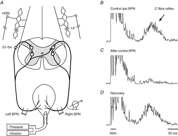
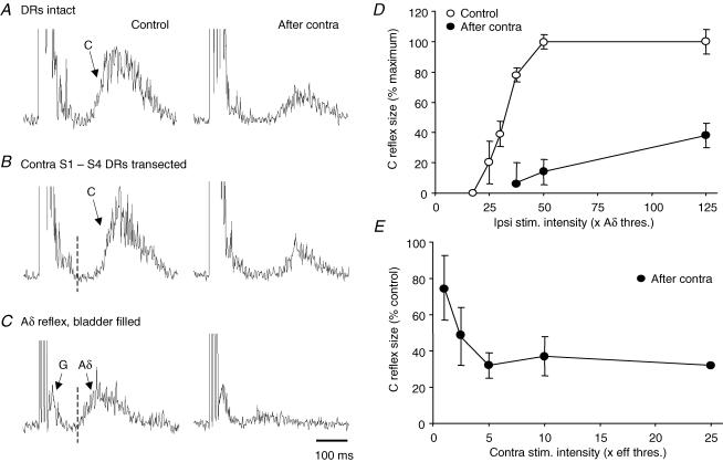
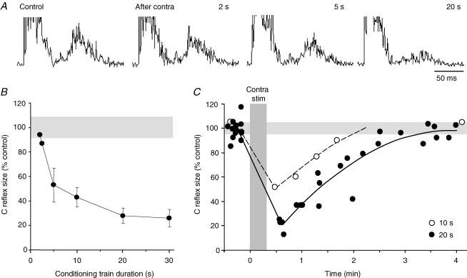
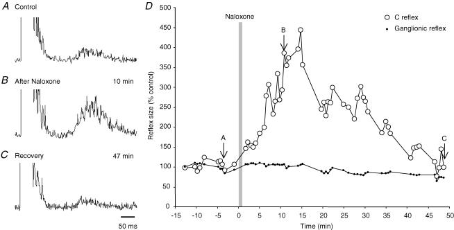
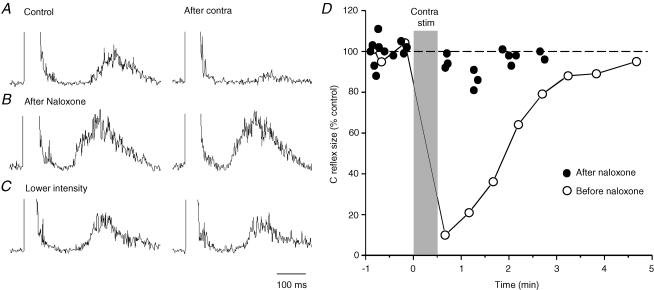
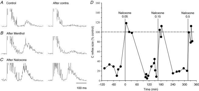
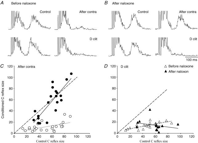
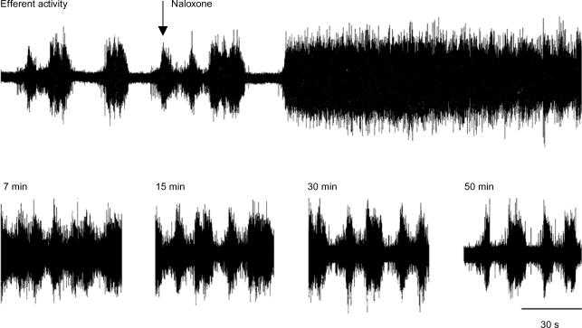
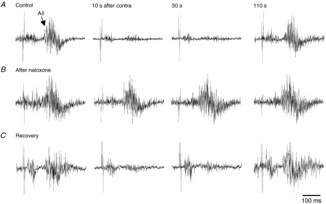
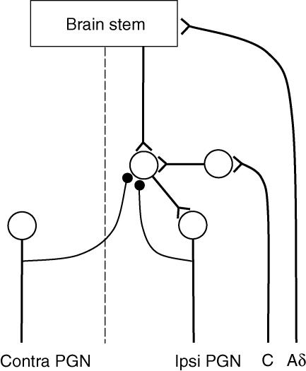
Recurrent inhibition of the bladder C fibre reflex was studied in adult female cats anaesthetized with alpha-chloralose. Test reflexes were evoked by electrical stimulation of bladder Adelta and C afferents in the right pelvic nerve and were recorded from the proximal end of a small ipsilateral pelvic nerve branch, transected close to the bladder. Such test reflexes were consistently depressed by repetitive electrical stimulation of the contralateral bladder pelvic nerve (20 Hz, 20 s) at intensities sufficient to recruit axons of bladder preganglionic neurones. The inhibition could be evoked after transection of the left dorsal roots S1-S4 and the sympathetic supply to the bladder but was abolished by transection of the pelvic nerve central to the site of stimulation. Hence, it most likely involved central recurrent collaterals of antidromically activated bladder preganglionic neurones. The reflex suppression was quite considerable - maximal C fibre reflexes were reduced to a group mean of 25% (+/- 9% confidence interval) of their control size. The effect had a slow onset, requiring a few seconds of conditioning stimulation to be revealed, and was very long lasting (minutes). Naloxone (0.01-0.5 mg kg(-1) i.v.) abolished the recurrent inhibition of both the C fibre and Adelta bladder reflexes, while inhibition from afferents in the dorsal clitoris nerve remained unchanged. It is concluded that the segmental bladder C fibre reflex and the spino-ponto-spinal Adelta micturition reflex are both targets of recurrent inhibition from bladder parasympathetic preganglionic neurones and that the effect involves an enkephalinergic mechanism.


Figures

Figure 1
Figure 1. Inhibitory effect of contralateral pelvic nerve stimulation on the bladder C fibre reflex
A, schematic diagram of the experimental arrangement. BD, C fibre reflex discharges evoked in a small ipsilateral bladder branch by stimulation of the remaining right bladder pelvic nerve (BPN) at an intensity maximal for C fibres (3 × 0.5 ms at 10 ms intervals, 50 × Aδ threshold, repetition rate 1 Hz). Each trace is an average of 20 rectified responses. B, the reflex response in the control situation; C, depression of the reflex immediately after conditioning stimulation of the contralateral left BPN at optimal parameters (5 × efferent threshold, 20 Hz, 20 s); D, recovery of the reflex 5 min after the end of conditioning stimulation. The left S1–S4 dorsal roots and the sympathetic supply to the bladder were transected, as indicated in A. Time calibration in D refers to all traces.
Figure 2
Figure 2. Recurrent inhibition of bladder Aδ and C fibre reflexes after transection of contralateral S1–S4 dorsal roots
A and B, inhibition of bladder C fibre reflex by stimulation of contralateral BPN before (A) and after (B) transection of the contralateral dorsal roots (DRs) S1–S4 (left, control responses; right, conditioned responses; other details as in Fig. 1). The strength of inhibition was unchanged by this transection. C, similar recurrent inhibition of a bladder Aδ reflex, same experiment after DR transection. The test reflex was evoked at maximal intensity for Aδ afferents (5 × Aδ threshold) with the bladder partly filled. Note shorter latency of the Aδ reflex compared to that of the C fibre reflex (vertical dotted lines in B and C). Arrow G indicates a small ganglionic component, similar responses of much larger size merge with the stimulus artefacts in all records with C fibre reflexes, as in A and B. Time calibration in C refers to all records. D, bladder C fibre reflex size plotted against test stimulation intensity; control (○) and corresponding response with maximal recurrent inhibition (•). E, increase in strength of recurrent inhibition with conditioning stimulation intensity; the inhibitory effect appeared at threshold intensity for bladder preganglionic fibres and was maximal at 5 × eff. threshold. Reflex size in D and E was measured as the area under the curve of the averaged reflex response (time window 150–450 ms after onset of test stimulation). Points show the mean (± 95%c.i.) of data pooled from several experiments (4 in D, n = 141 reflex responses; 2 in E, n = 25).
Figure 3
Figure 3. Recurrent inhibition by conditioning trains of different duration
Sample records in A show C fibre test reflexes before and after conditioning stimulation of the contralateral BPN by trains of 2, 5 and 20 s duration (20 Hz, 5 × efferent threshold). The pooled results of similar trials in 3 experiments are plotted in B with the size of the conditioned C fibre reflex expressed as a percentage of the control reflex (± c.i.; other details as in Fig. 2); the recurrent inhibition became apparent with 2 s long trains and was optimal with trains of 20 s. C, change in time course of recurrent inhibition induced by conditioning trains, 10 s (○) and 20 s (•) long. Each point is the average of 20 consecutive reflex responses (1 Hz) and is plotted at half the collecting period; data points for 20 s trains are from 4 conditioning trials. Gray bands in B and C mark 95%c.i. range of control reflexes. The time of conditioning stimulations (20 s long) of the contralateral BPN is indicated by the vertical bar.
Figure 4
Figure 4. Effect of naloxone on the size of the bladder C fibre reflex
AC, bladder C fibre reflexes obtained before (A) and at indicated times after systemic naloxone (0.025 mg kg−1i.v.; B and C). Time scale in C is for all records. D, time course of naloxone induced enhancement of the bladder C fibre reflex (○) and lack of effect on peripheral ganglionic discharge (dots). Reflex sizes are expressed as a percentage of the mean value before naloxone. Vertical bar marks the time of naloxone injection, arrows the sample records AC.
Figure 5
Figure 5. Suppression of recurrent inhibition after systemic naloxone
AC, recurrent inhibition of bladder C fibre reflexes as observed before (A) and after naloxone (0.05 mg kg−1i.v.; B and C); left control, right conditioned response. Test stimulation intensity was the same for records in A and B (50 × threshold for the Aδ reflex) but decreased in C (20 × threshold) to match the control response in A; other stimulation parameters remained the same throughout. Note that the recurrent inhibition was abolished by naloxone, even after the decrease in test stimulation intensity. D, graphic illustration of the same effect showing conditioned reflex sizes (as a percentage of control) before (○) and after naloxone (•). The latter points were obtained during 4 conditioning trials in the 30 min period following naloxone administration. The time of conditioning stimulations of the contralateral BPN is indicated by the grey bar.
Figure 6
Figure 6. Time course of naloxone suppression of recurrent inhibition
A–C, test bladder C fibre reflexes conditioned by contralateral BPN stimulation. In B, test reflexes were enhanced to match the response expected after naloxone (C), using a preceding slow urethral perfusion with menthol (0.06 mm in saline) to sensitize urethral cold receptors. This enhanced bladder C fibre reflex was virtually abolished by the recurrent inhibition while the similar sized response after naloxone was unaffected by the conditioning stimulation. D, plot showing consistent suppression by naloxone of the recurrent inhibition of bladder C fibre reflexes (three consecutive doses 0.05, 0.15 and 0.5 mg kg−1i.v.). The size of conditioned reflexes, as a percentage of their controls, is plotted against time from the first naloxone injection. The conditioned reflexes attained the size of their controls after each naloxone injection, implying abolished recurrent inhibition. The illustrated points represent a subset of reflexes evoked at identical test stimulation intensity, taken from a larger series of trials illustrated in Fig. 7.
Figure 7
Figure 7. Selective naloxone effect on recurrent inhibition
A, sample records of bladder C fibre reflexes inhibited by antidromic activation of contralateral preganglionic neurones (After contra; upper pair) or orthodromic activation of ipsilateral dorsal clitoris afferents (D clit, lower pair). The latter induced a shortlasting inhibition, so each test reflex was conditioned by a brief stimulus train to the dorsal clitoris nerve (50 Hz, 200 ms, starting 50 ms before test stimulation). The recurrent inhibition was assed as usual in the period after contralateral BPN stimulation (20 Hz, 20 s). The two conditioning stimuli were adjusted to give roughly similar inhibition of the bladder C fibre reflex. B, similar responses after naloxone (0.5 mg kg−1i.v.). The recurrent inhibition was abolished by the drug while the afferent inhibition remained unchanged. C and D, effects of the two conditioning stimuli on bladder C fibre reflexes of different sizes evoked by varying the test stimulation intensity. Each point represents the size of the conditioned reflex plotted against the size of its control reflex, before (○) and after naloxone (•). Hatched lines indicate conditioned reflexes equal in size to their controls, thin dotted and continuous lines are best fits for data points from before and after naloxone. The diagrams show pooled data from three naloxone sessions in the same experiment (same as in Fig. 6). For the naloxone effect, only reflexes sampled during the first 20 min after injections are included. Naloxone clearly suppressed the recurrent inhibition at all test reflex sizes while leaving the inhibition from dorsal clitoris afferents unchanged.
Figure 8
Figure 8. Effect of naloxone on phasic BPN efferent discharges
Upper trace is a continuous recording of typical phasic efferent activity in a small BPN branch, obtained with the bladder partly filled. Systemic naloxone (0.1 mg kg−1i.v.; arrow) rapidly (< 2 min) converted this phasic activity into a sustained tonic discharge, an activity that gradually returned to the control pattern during the following 50 min (lower traces).
Figure 9
Figure 9. Effect of naloxone on recurrent inhibition of bladder Aδ reflexes
Bladder Aδ reflexes recorded with the bladder filled (same experiment as Fig. 8). Each horizontal row of single traces illustrates a conditioning trial with reflex responses taken before and at indicated times after conditioning stimulation of the contralateral BPN (20 s, 20 Hz). Conditioning trials were performed before (A), 17 min (B) and 65 min (C) after naloxone (0.1 mg kg−1i.v.) The test stimulation intensity was reduced in B (from 4 to 2 × threshold for the Aδ reflex) to obtain an Aδ reflex of about the same size as in A, yet the recurrent inhibition was virtually abolished.
Figure 10
Figure 10. Schematic diagram of proposed neuronal pathways
Bladder Aδ and C fibre reflex pathways are shown to converge onto spinal prepreganglionic interneurones, which are also the target of enkephalinergic recurrent inhibitory connections from bladder preganglionic neurones (•).

Similar articles

Cited by

References

    1. Blok BF, Holstege G. Ultrastructural evidence for a direct pathway from the pontine micturition center to the parasympathetic preganglionic motoneurons of the bladder of the cat. Neuroscience Lett. 1997;222:195–198. - PubMed
    1. Cruz F. Mechanisms involved in new therapies for overactive bladder. Urology. 2004;63:65–73. - PubMed
    1. De Groat WC. Mechanisms underlying recurrent inhibition in the sacral parasympathetic outflow to the urinary bladder. J Physiol. 1976;257:503–513. - PMC - PubMed
    1. De Groat WC, Booth AM, Milne RJ, Roppolo JR. Parasympathetic preganglionic neurons in the sacral spinal cord. J Autonomic Nervous System. 1982;5:23–43. - PubMed
    1. De Groat WC, Kawatani M. Enkephalinergic inhibition in parasympathetic ganglia of the urinary bladder of the cat. J Physiol. 1989;256:13–29. - PMC - PubMed

Publication types

LinkOut - more resources