

doi: 10.1126/science.1128004.
Lhx2 maintains stem cell character in hair follicles
Affiliations
- PMID: 16809539
- PMCID: PMC2405918
- DOI: 10.1126/science.1128004
Item in Clipboard
Lhx2 maintains stem cell character in hair follicles
Science.
.
Abstract
During embryogenesis, stem cells are set aside to fuel the postnatal hair cycle and repair the epidermis after injury. To define how hair follicle stem cells are specified and maintained in an undifferentiated state, we developed a strategy to isolate and transcriptionally profile embryonic hair progenitors in mice. We identified Lhx2 as a transcription factor positioned downstream of signals necessary to specify hair follicle stem cells, but upstream from signals required to drive activated stem cells to terminally differentiate. Using gain- and loss-of-function studies, we uncovered a role for Lhx2 in maintaining the growth and undifferentiated properties of hair follicle progenitors.
Figures

Isolation of embryonic hair follicle progenitors. (A) P-cadherin is up-regulated at sites of hair follicle morphogenesis. This differential expression was used to isolate PCAD+ hair progenitors and PCAD− interfollicular basal cells by FACS (figs. S2 and S3). Scale bar, 40 μm. (B) Summary of cytospin analyses for epidermal markers K1, K5, and β4-integrin. (C) The differential activity of the Wnt reporter gene TOPGAL was assessed by chemiluminescence. β-gal, β-galactosidase; WT, wild type. (D) Semi-quantitative RT-PCR shows differential expression of known placode mRNAs. K5 and K14 are known to be down-regulated, but still expressed, in hair germs (15). Abbreviations are as follows: epi, epidermis; der, dermis; Pc, hair placode; HG, hair germ; HF, hair follicle; β4, β4-integrin.
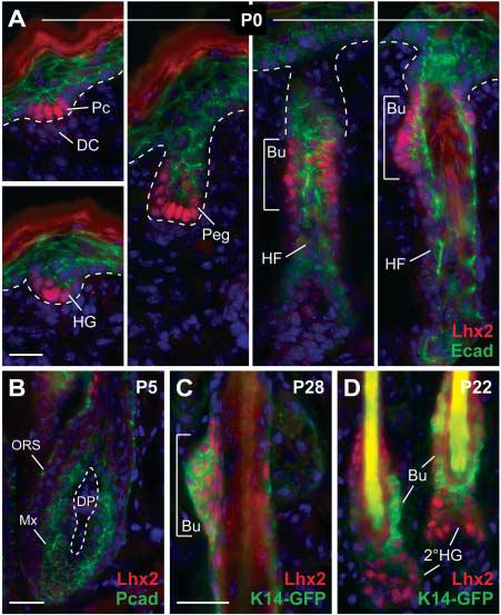
Lhx2 is expressed in early hair progenitors and postnatal stem cells. (A to D) Back skin sections from mice at indicated ages were stained with antibodies as color-coded and counterstained with DAPI (blue). Lhx2 is expressed in cells at the leading front of invaginating follicles and in the postnatal bulge compartment, but is diminished in mature proliferative hair progenitors (matrix). Abbreviations are as follows: Peg, hair peg; Bu, presumptive bulge; Mx, matrix cells; DC, dermal condensate; DP, dermal papilla; 2°HG, secondary hair germ emerging at the start of the postnatal hair cycle. Scale bars, 20 μm.
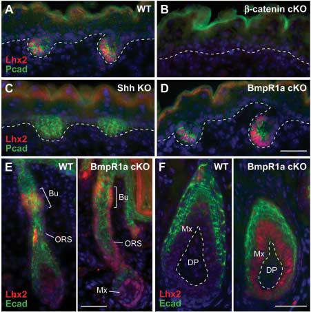
Lhx2 functions downstream of follicle stem cell specification but upstream of their differentiation. (A) Lhx2 is expressed in P-cadherin–positive hair germs. Shown is a representative wild-type littermate at E17.5. (B) Lhx2 is not expressed in the absence of hair follicle induction, as reflected in the β-catenin null skin epithelium (5). K14-Cre was used to conditionally target β-catenin. (C) Lhx2 is reduced in Shh null hair germs. Follicles are specified in the absence of Shh but fail to progress further (8, 10). (D) Lhx2 is expressed in BmpR1a conditional null hair germs. (E and F) In addition to expression in the presumptive bulge, Lhx2 persists in the lower ORS and matrix of neonatal and adult BmpR1a null follicles. Unable to undergo terminal differentiation, these BmpR1a null, Lhx2-expressing cells appear to be undifferentiated follicle stem cells (27, 28). Scale bars, 40 μm.
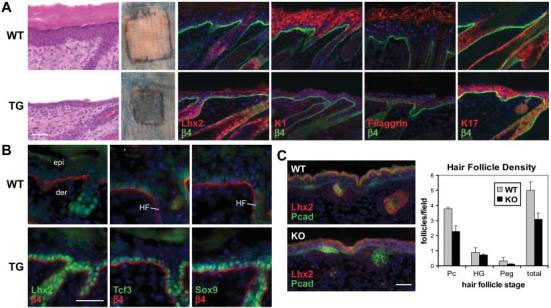
Gain- and loss-of-function studies reveal a role for Lhx2 in promoting follicle stem cell maintenance. (A) Transgenic expression of Lhx2 in the interfollicular epidermis suppresses terminal differentiation. Morphological and biochemical features of spinous, granular, and stratum corneum stages of epidermal differentiation are diminished in d8 (day 8 after graft) skin from K14-Lhx2 transgenic mice (TG) relative to wild-type littermates. Patches outlined in black denote skins grafted onto Nude mice and subjected to a β-galactosidase substrate exclusion assay to test for an intact epidermal barrier. The background absorption of dye in the surrounding Nude skin arises from defective hair follicle orifices. (B) Immunofluorescence reveals an induction of follicle stem cell markers in d4 K14-Lhx2 epidermis. (C) Loss of Lhx2 reduces hair morphogenesis. Representative skin sections from E16 littermates show comparable epidermal differentiation but fewer follicles at all stages in Lhx2 null embryos. The graph provides quantification from multiple sections of three embryos. Scale bars, 40 μm.
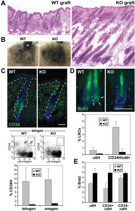
Lhx2 maintains follicle stem cells in a quiescent, inactive state. (A) Histology of wild-type and Lhx2 KO skins at 11 weeks after graft. Wild-type follicles were still in telogen, but KO follicles prematurely entered anagen. (B) Upon shaving at 8 weeks, wild-type hairs did not grow back, confirming their resting state. In contrast, KO hairs grew back within 3 weeks. A small portion (*) of the wild-type graft grew hairs because of a wound. (C) CD34 expression is dramatically reduced in KO follicle bulges, irrespective of whether they are in telogen or anagen. Shown are representative immunofluorescence images and FACS profiles of telogen follicles at 8 weeks, and CD34 quantification by flow cytometry in telogen and anagen follicles during the first post-natal cycle. (D) Loss of BrdU label retention in KO follicles. After a 3-day BrdU pulse on days 26 to 28 at the onset of anagen in both wild-type and KO skin grafts, and a 4-week chase when follicles had entered telogen (fig. S9), LRCs concentrated in the infrequently dividing bulge stem cells of wild-type follicles, but LRCs were diminished in Lhx2 KO skin. Shown are skin sections stained with antibody to BrdU and results of quantification by flow cytometry. (E) Increased BrdU incorporation by KO follicle stem cells. After a 4-hour BrdU pulse at day 40, when wild-type and KO follicles were in mid-anagen of their first postnatal hair cycle (fig. S9), cells were isolated and α6-integrin–expressing S-phase cells were quantified by flow cytometry. Scale bars, 40 μm.
References
-
- Hardy MH. Trends Genet. 1992;8:55. - PubMed
-
- Millar SE. J. Invest. Dermatol. 2002;118:216. - PubMed
-
- Schmidt-Ullrich R, Paus R. Bioessays. 2005;27:247. - PubMed
-
- DasGupta R, Fuchs E. Development. 1999;126:4557. - PubMed
-
- Huelsken J, Vogel R, Erdmann B, Cotsarelis G, Birchmeier W. Cell. 2001;105:533. - PubMed
Publication types
MeSH terms
Substances
Grants and funding
LinkOut - more resources
Full Text Sources
Other Literature Sources
Medical
Molecular Biology Databases
