. 2006 Oct 15;108(8):2695-702.
doi: 10.1182/blood-2006-05-021790. Epub 2006 Jun 29.

BCL11B participates in the activation of IL2 gene expression in CD4+ T lymphocytes

Affiliations

BCL11B participates in the activation of IL2 gene expression in CD4+ T lymphocytes

Valeriu B Cismasiu et al. Blood. .

Abstract

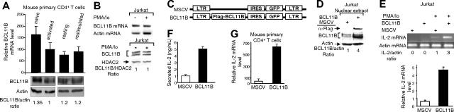
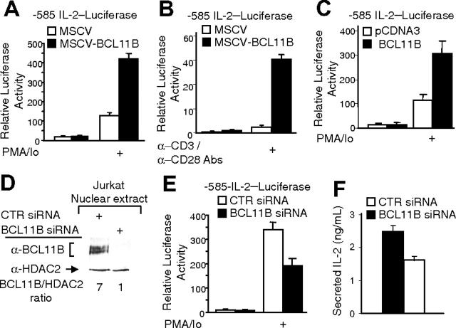
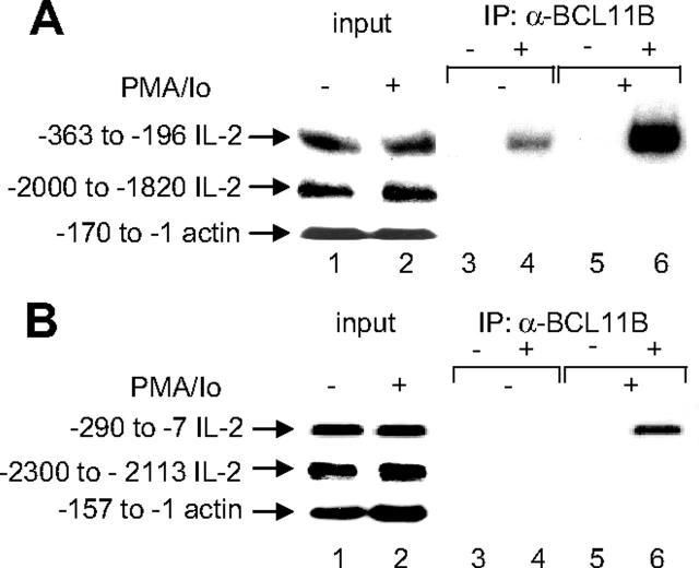
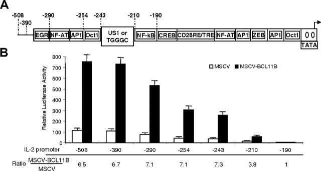
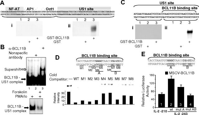
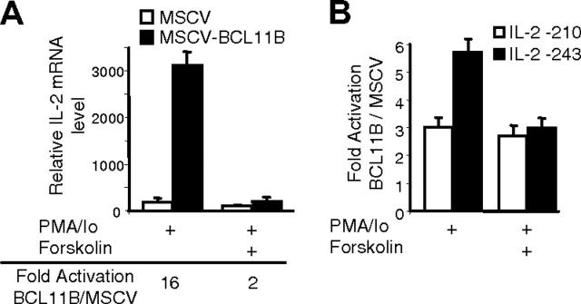
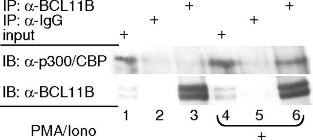
BCL11A and BCL11B are transcriptional regulators important for lymphopoiesis and previously associated with hematopoietic malignancies. Ablation of the mouse Bcl11b locus results in failure to generate double-positive thymocytes, implicating a critical role of Bcl11b in T-cell development. However, BCL11B is also expressed in CD4+ T lymphocytes, both in resting and activated states. Here we show both in transformed and primary CD4+ T cells that BCL11B participates in the control of the interleukin-2 (IL2) gene expression following activation through T-cell receptor (TCR). BCL11B augments expression from the IL2 promoter through direct binding to the US1 site. In addition, BCL11B associates with the p300 coactivator in CD4+ T cells activated through TCR, which may account for its transcriptional activation function. These results provide the first evidence that BCL11B, originally described as a transcriptional repressor, activates transcription of a target gene in the context of T-cell activation.


Figures

Figure 1.
Figure 1.
BCL11B up-regulates the levels of IL-2 mRNA in transformed and primary CD4+ T lymphocytes. (A, top) Expression of BCL11B evaluated by qRT-PCR in primary CD4+ T cells in the following conditions: freshly isolated (naive), stimulated for 24 hours with anti-CD3/anti-CD28 (activated), rested for 72 hours following the anti-CD3/anti-CD28 treatment (resting), or rested and restimulated with 50 ng/mL PMA and 1 μM ionomycin for 4 hours (restimulated). The relative abundance of BCL11B message was normalized to actin and calculated as described in “Reverse transcription and real-time PCR—quantitative RT-PCR (qRT-PCR).” (Bottom) Western blot analysis of BCL11B in primary CD4+ T cells treated in the conditions shown in the top panel; BCL11B levels were quantified by densitometric analysis and normalized to actin. (B) BCL11B expression evaluated by RT-PCR (top) and Western blot analysis (bottom) in Jurkat cells grown in the presence or absence of PMA (50 ng/mL) and ionomycin (1 μM) for 4 hours. BCL11B levels were quantified by densitometric analysis and normalized to HDAC2. (C) Schematic representation of the MSCV and MSCV-BCL11B retroviral constructs. LTR represents long terminal repeat; IRES, internal ribosomal entry site. (D) Western blot analysis demonstrating expression of Flag-BCL11B in retrovirally transduced Jurkat cells. (E) Levels of IL-2 mRNA evaluated by RT-PCR (top) and qRT-PCR (bottom) before (bottom) and after (both subpanels) treatment for 4 hours with PMA/ionomycin in MSCV and MSCV-BCL11B Jurkat cells. (F) Secretion of IL-2 determined by ELISA in media from MSCV and MSCV-BCL11B Jurkat cells after treatment with PMA/ionomycin for 16 hours. (G) Levels of IL-2 mRNA in MSCV and MSCV-BCL11B primary mouse CD4+ T cells after retroviral transduction and sorting of populations of GFP-positive cells. Cells were stimulated with PMA/ionomycin for 5 hours and the levels of IL-2 message were evaluated by qRT-PCR. The relative abundance of IL-2 message was normalized to actin and calculated as described in “Reverse transcription and real-time PCR—quantitative RT-PCR (qRT-PCR).” Three to 5 experiments were conducted, and the quantifications represent means ± SD.
Figure 2.
Figure 2.
BCL11B regulates IL-2 promoter activity. (A-B) MSCV-BCL11B and MSCV Jurkat cells were electroporated with the -585 IL-2 promoter-Firefly luciferase and CMV-Renilla luciferase constructs. Twenty-four hours after transfection, the cells were treated with PMA/ionomycin for 8 hours (A) or grown for 8 hours in the presence of plate-bound anti-CD3 and soluble anti-CD28 antibodies (B). The luciferase activity was measured using the Promega luciferase dual system. (C) Jurkat cells were electroporated with pCDNA3::BCL11B or pCDNA3 vectors and -585 IL-2 promoter-Firefly luciferase and CMV-Renilla luciferase constructs. Luciferase activity was measured as in panels A and B. (D) Western blot analysis of BCL11B after transfection of Jurkat cells with BCL11B specific (lane 2) or nontargeting (lane 1) siRNAs. HDAC2 was used as loading control. (E-F) Jurkat cells were transfected with the -585 IL-2 promoter-Firefly luciferase, CMV-Renilla luciferase constructs, and BCL11B specific or nontargeting siRNAs. (E) Luciferase activity was measured after PMA/ionomycin treatment for 8 hours; control cells lacked PMA/ionomycin treatment. (F) Secretion of IL-2 was determined by ELISA in media after treatment with PMA/ionomycin for 16 hours. The quantifications represent means of 3 independent experiments ± SD.
Figure 3.
Figure 3.
Endogenous BCL11B associates with the IL-2 promoter in Jurkat and primary CD4+ T cells. Immunoprecipitation (IP) of chromatin from Jurkat cells (A) or primary CD4+ T cells (B) stimulated with PMA/ionomycin (lanes 5 and 6) or no stimulation (lanes 3 and 4) using anti-BCL11B (lanes 4 and 6) or nonspecific (lanes 3 and 5) antibodies. Eluted DNA was analyzed by PCR using human (A) or mouse (B) IL-2 promoter-specific primers or actin promoter primers covering the indicated regions. In each case, lanes 1 and 2 represent input from untreated and treated cells, respectively.
Figure 4.
Figure 4.
IL-2 promoter activation by BCL11B requires the region between -243 and -190. (A) Schematic representation of the IL-2 promoter deletion mutants. (B) Luciferase reporter assays from MSCV-BCL11B (▪) and MSCV (□) Jurkat cells transfected with the IL-2 promoter deletion mutants. Ratios of luciferase activity between MSCV-BCL11B and MSCV Jurkat cells for each deletion mutant are shown. Reporter assays were conducted after treatment of the cells with PMA/ionomycin for 8 hours. The quantifications represent means of 3 independent experiments ± SD.
Figure 5.
Figure 5.
BCL11B binds and activates the IL-2 promoter through the US1 site. (A) EMSA using purified recombinant GST-BCL11B (lane 3) or GST (lane 2) and US1 (ii) or NFAT-AP1-Oct DNA probes (i). Lane 1 is free probe. (B) EMSA using nuclear extracts from Jurkat cells and the US1 probe. In the top panel the US1 probe was incubated with 5 μg nuclear extracts from Jurkat cells treated with PMA and ionomycin (lanes 1-3) and anti-BCL11B (lane 3) or nonspecific antibodies (lane 2). BCL11B-US1 complexes are indicated by the black arrow, and the antibody-supershifted BCL11B-US1 complex is indicated by the white arrow. In the bottom panel the US1 probe was incubated with extracts from cells treated with PMA/ionomycin or forskolin plus PMA/ionomycin. Treatments were conducted as described in Figure 6. (C) EMSA using purified GST-BCL11B (lanes 3) or GST (lanes 2) and proximal (ii) or distal (i) US1 DNA fragments. Lane 1 is free probe. (D) Mutational analysis of the BCL11B binding site. Top panel shows the sequence of the BCL11B binding site. Underlined are the bases mutated in each mutant (M1-M8). Middle panel shows EMSAs with purified recombinant GST-BCL11B, 32P-labeled BCL11B binding site probe plus excess of unlabeled WT or mutant DNA competitors (M1-M8). The bottom panel represents the quantification of the relative intensity of the corresponding bands. Maximal binding of the probe was determined in the sample lacking competitor DNA. For simplicity EMSAs in panels A-D present only protein-DNA complexes. (E) Reporter assays from MSCV-BCL11B Jurkat cells transfected with the following IL-2 promoter-luciferase constructs: wild-type -210 (lane 1), wild-type -243 (lane 2), mutant A -243 (lane 3), and mutant AB -243 (lane 4) (see top panel). In each case cells were treated with PMA/ionomycin for 8 hours before harvesting. Three experiments were conducted, and the quantifications represent means ± SD.
Figure 6.
Figure 6.
Forskolin blocks augmentation of IL-2 promoter activity by BCL11B. (A) IL-2 mRNA levels in MSCV (□) and MSCV-BCL11B (▪) Jurkat cells stimulated for 4 hours with PMA/ionomycin and pretreated or not with 10 μM forskolin for 30 minutes before stimulation. IL-2 mRNA levels were evaluated by qRT-PCR and normalized by actin in each sample. (B) Ratios of luciferase reporter assays between MSCV-BCL11B and MSCV Jurkat cells transfected with -210 IL-2 promoter (□) or -243 IL-2 promoter (▪). The cells were stimulated with PMA/ionomycin for 8 hours and pretreated or not with 10 μM forskolin for 30 minutes.
Figure 7.
Figure 7.
BCL11B associates with the p300 coactivator in activated Jurkat cells. Immunoprecipitation (IP) with anti-BCL11B (lanes 3 and 6) or nonspecific (lanes 2 and 5) antibodies and Western blot (IB) analysis with anti-p300 and anti-BCL11B antibodies. Nuclear extracts were prepared from Jurkat cells treated (lanes 4-6) or not (lanes 1-3) with PMA/ionomycin. Lanes 1 and 4 represent input.

Similar articles

Cited by

References

    1. Avram D, Fields A, Pretty On Top K, Nevrivy DJ, Ishmael JE, Leid M. Isolation of a novel family of C(2)H(2) zinc finger proteins implicated in transcriptional repression mediated by chicken ovalbumin upstream promoter transcription factor (COUP-TF) orphan nuclear receptors. J Biol Chem. 2000;275: 10315-10322. - PMC - PubMed
    1. Avram D, Fields A, Senawong T, Topark-Ngarm A, Leid M. COUP-TF (chicken ovalbumin upstream promoter transcription factor)-interacting protein 1 (CTIP1) is a sequence-specific DNA binding protein. Biochem J. 2002;368: 555-563. - PMC - PubMed
    1. Arlotta P, Molyneaux BJ, Chen J, Inoue J, Kominami R, Macklis JD. Neuronal subtype-specific genes that control corticospinal motor neuron development in vivo. Neuron. 2005;45: 207-221. - PubMed
    1. Wakabayashi Y, Watanabe H, Inoue J, et al. Bcl11b is required for differentiation and survival of αβ T lymphocytes. Nat Immunol. 2003;4: 533-539. - PubMed
    1. Miyazawa T, Sato H, Hatakeyama K, Kitagawa T, Kominami R. Allelic losses in mouse skin tumors induced by gamma-irradiation of p53 heterozygotes. Jpn J Cancer Res. 2002;93: 994-999. - PMC - PubMed

Publication types

MeSH terms