. 2006 Oct 15;108(8):2712-9.
doi: 10.1182/blood-2006-03-014001. Epub 2006 Jun 29.

B-chronic lymphocytic leukemia cells and other B cells can produce granzyme B and gain cytotoxic potential after interleukin-21-based activation

Affiliations

B-chronic lymphocytic leukemia cells and other B cells can produce granzyme B and gain cytotoxic potential after interleukin-21-based activation

Bernd Jahrsdörfer et al. Blood. .

Abstract

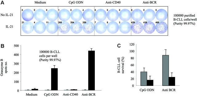
B cells currently are not viewed as being capable of producing granzyme B or being cytotoxic. We found that B-chronic lymphocytic leukemia (B-CLL) cells treated with interleukin-21 (IL-21) produce low levels of granzyme B. The addition of either CpG oligodeoxynucleotide (ODN) or anti-B-cell-receptor antibody (anti-BCR) to IL-21 results in enhanced production of functional granzyme B by B-CLL cells. B-CLL cells treated with IL-21 and CpG ODN undergo apoptosis and are able to induce apoptosis of untreated bystander B-CLL cells. This effect can be inhibited by anti-granzyme B antibody. Benign human B cells, Epstein-Barr virus (EBV)-transformed lymphoblasts, and many standard lymphoma cell lines produce high levels of granzyme B in response to IL-21 and anti-BCR. Our results suggest that the ability to induce production of functional granzyme B by B cells could open new approaches to the therapy of B-CLL and other B-cell malignancies. Our findings also have significant implications for our understanding of the role of B cells for immune regulation and for a variety of immune phenomena, including cancer immunity, autoimmunity, and infectious immunity.


Figures

Figure 1.
Figure 1.
IL-21 plus CpG ODN induces apoptosis of B-CLL cells. PBMCs from 9 patients with B-CLL were cultured for 4 days in the presence of CpG ODN (2.5 μg/mL) and IL-21 (100 ng/mL) or IL-2 (100 U/mL). Cell survival of B-CLL cells was determined using annexin V and PI staining and counterstaining with antibodies to CD19. (A) Shown are annexin V/PI dot plots from 1 representative experiment. Gated are CD19+ B-CLL cells. Numbers in the gates represent percentages of displayed events. (B) The mean B-CLL cell-survival rates from 9 independent experiments are plotted. Error bars indicate SEM.
Figure 2.
Figure 2.
The proapoptotic effect of IL-21 and CpG ODN on B-CLL cells is synergistic. PBMCs from 3 patients with B-CLL were cultured with CpG ODN and IL-21 at different concentration ratios for 4 days, and the percentage of apoptotic cells was determined. (A) Three individual experiments demonstrating a synergistic interaction between CpG ODN and IL-21 are shown. A horizontal line indicates an additive interaction, while the observed convex curve demonstrates synergy. (B) The effect of varying concentrations of CpG ODN and IL-21 demonstrated that both agents induced greater apoptosis than either agent alone, even at concentrations that give peak effects for each individual agent. Plotted are the mean cell-survival rates (n = 3). Error bars indicate SEM.
Figure 3.
Figure 3.
Proapoptotic effect of IL-21 and CpG ODN on B-CLL cells is direct. PBMCs from 3 patients with B-CLL were isolated and divided into 2 fractions. One fraction was purified to a percentage of more than 99% CD5+, CD19+ B-CLL cells. Both fractions were incubated for 3 days with IL-21, CpG ODN, or both agents, and apoptosis was determined flow cytometrically. Data are from 1 representative experiment of 3. Dot plots demonstrate the purity of B-cell populations based on CD19 expression. Numbers in the gates represent percentages of displayed events. Bar graphs illustrate the mean B-CLL cell-survival rates in response to treatment. Error bars indicate SEM from experimental replicates.
Figure 4.
Figure 4.
IL-21 and CpG ODN induce B-CLL cell expression of lysosome-associated molecular protein 1 (LAMP-1, CD107a) and granzyme B. PBMCs from 7 patients with B-CLL were cultured in the presence of CpG ODN, IL-21, or both. Expression of CD107a (LAMP-1) on CD19+ B-CLL cells was determined using FACS analysis. (A-B) Dot plots from 1 representative experiment of 7 show percentages of CD19+ B-CLL cells with increased side scatter and CD107a expression. The bar graph illustrates relative median fluorescence intensities (MFI) for CD107a expression compared with unstimulated cells. Error bars indicate SEM. (C) PBMCs from 5 patients with B-CLL were isolated and cultured in the presence of IL-21, CpG ODN, or both. For the last 4 hours, 1 μg/mL brefeldin A was added to the cells. Cells were then fixed, permeabilized, and stained with antibodies to anti-CD19 and granzyme B. The boldfaced number in Figure 4C represents the percentage of granzyme B-positive cells when treated with IL-21 and CpG ODN. One representative experiment of 5 is shown. (D) PBMCs from 5 patients with B-CLL were stained with a granzyme B colorimetric substrate, then cultured as outlined for panel C. The bar graph shows the percentage of CD19+ cells with substrate that was activated by granzyme B. Error bars indicate SEM.
Figure 5.
Figure 5.
IL-21 induces granzyme B secretion by B-CLL cells, which is synergistically enhanced by CpG ODN and anti-BCR. B-CLL cells from 3 patients were isolated and purified to a percentage of at least 99.9% based on CD19 expression. The cells were cultured at 37°C on 96-well ELISPOT plates for granzyme B detection at 100 000 cells/well and in the presence of different B-cell activators alone or with IL-21. After 16 hours, plates were developed and dots counted. Every condition was run in triplicate. (A) Individual ELISPOT plate from 1 representative experiment of 3 is shown. (B) Average spot numbers from 1 representative experiment of 3 are depicted. Error bars indicate SD. (C) B-CLL cell survival after 4 days of incubation with anti-BCR or CpG ODN alone (formula image
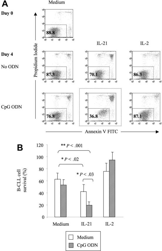
) or with IL-21 (▪) was flow cytometrically detected using annexin V, anti-CD19, and PI staining. Averages from 3 independent experiments are shown. Error bars indicate SEM.
Figure 6.
Figure 6.
B-CLL cells treated with IL-21 plus CpG ODN can induce apoptosis of untreated, bystander B-CLL cells. Anti-granzyme B antibodies inhibit bystander B-CLL cell killing. (A) Purified B-CLL cells were split into 2 fractions. One fraction was stained with PKH-26, then incubated for 24 hours in IL-21 with or without CpG ODN. Unstained cells were maintained in culture without stimulus. The stained, treated cells were washed, added to the untreated cells, and cocultured for 2 days. Survival of the untreated cells (as indicated by lack of membrane dye) was analyzed by flow cytometry. Plotted are the B-CLL cell-survival rates for the untreated (ie, bystander) cells cultured at different ratios with treated cells. One representative experiment of 3 with similar results is shown. (B) Average B-CLL cell-survival rates for the untreated (ie, bystander) B-CLL cells cultured at a ratio of 1:2 with treated cells. Results are from 3 independent experiments. Error bars indicate SEM. (C) B-CLL cells from 3 patients were cultured for 4 days in the presence of IL-21 (100 ng/mL), CpG ODN (2.5 μg/mL), and anti-human granzyme B antibody (formula image
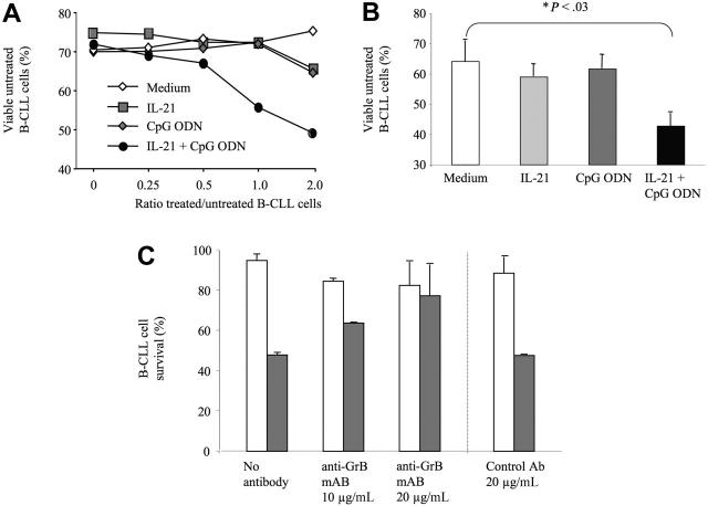
)ora control antibody (□). B-CLL cell survival was determined by FACS analysis using annexin V/PI staining and counterstaining with antibodies to CD19. Plotted are the mean B-CLL cell-survival rates in percent from 1 representative experiment of 3 with similar results. Error bars indicate SD.
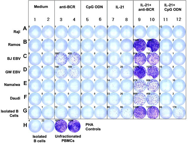
Figure 7.
Figure 7.
IL-21 induces de novo granzyme B synthesis and secretion by benign peripheral B cells and different B-cell lines. Standard cell lines, EBV transformed lymphoblasts, and B cells from healthy subjects (> 99.5% CD19+) were cultured in medium alone, anti-BCR, CpG ODN, IL-21, or combinations of these agents for 16 hours at a concentration of 100 000 cells/well. ELISPOT analysis for granzyme B was then performed. Controls included isolated B cells and unfractionated PBMCs treated with PHA.

Similar articles

Cited by

References

    1. Bossi G, Trambas C, Booth S, Clark R, Stinchcombe J, Griffiths GM. The secretory synapse: the secrets of a serial killer. Immunol Rev. 2002; 189: 152-160. - PubMed
    1. Wowk ME, Trapani JA. Cytotoxic activity of the lymphocyte toxin granzyme B. Microbes Infect. 2004;6: 752-758. - PubMed
    1. Trapani JA, Sutton VR. Granzyme B: proapoptotic, antiviral and antitumor functions. Curr Opin Immunol. 2003;15: 533-543. - PubMed
    1. Lord SJ, Rajotte RV, Korbutt GS, Bleackley RC. Granzyme B: a natural born killer. Immunol Rev. 2003;193: 31-38. - PubMed
    1. Froelich CJ, Orth K, Turbov J, et al. New paradigm for lymphocyte granule-mediated cytotoxicity: target cells bind and internalize granzyme B, but an endosomolytic agent is necessary for cytosolic delivery and subsequent apoptosis. J Biol Chem. 1996;271: 29073-29079. - PubMed

Publication types

MeSH terms