. 2006 Oct 15;108(8):2814-20.
doi: 10.1182/blood-2006-01-010363. Epub 2006 Jun 29.

Rac1 links leading edge and uropod events through Rho and myosin activation during chemotaxis

Affiliations

Rac1 links leading edge and uropod events through Rho and myosin activation during chemotaxis

Kersi N Pestonjamasp et al. Blood. .

Abstract

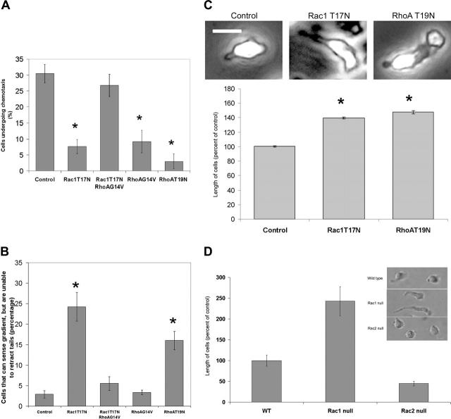
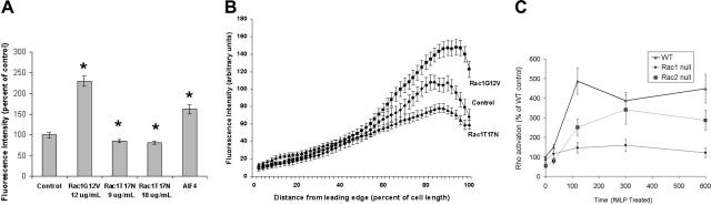
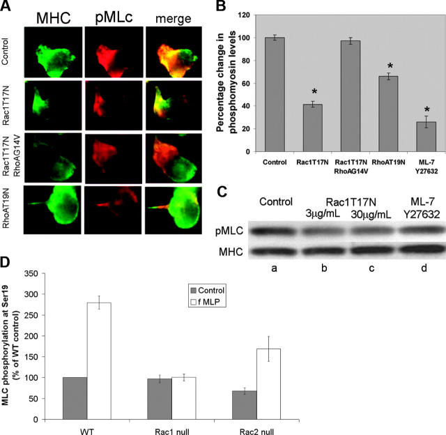
Chemotactic responsiveness is crucial to neutrophil recruitment to sites of infection. During chemotaxis, highly divergent cytoskeletal programs are executed at the leading and trailing edge of motile neutrophils. The Rho family of small GTPases plays a critical role in cell migration, and recent work has focused on elucidating the specific roles played by Rac1, Rac2, Cdc42, and Rho during cellular chemotaxis. Rac GTPases regulate actin polymerization and extension of the leading edge, whereas Rho GTPases control myosin-based contraction of the trailing edge. Rac and Rho signaling are thought to crosstalk with one another, and previous research has focused on mutual inhibition of Rac and Rho signaling during chemotaxis. Indeed, polarization of neutrophils has been proposed to involve the activity of a negative feedback system where Rac activation at the front of the cell inhibits local Rho activation, and vice versa. Using primary human neutrophils and neutrophils derived from a Rac1/Rac2-null transgenic mouse model, we demonstrate here that Rac1 (and not Rac2) is essential for Rho and myosin activation at the trailing edge to regulate uropod function. We conclude that Rac plays both positive and negative roles in the organization of the Rhomyosin "backness" program, thereby promoting stable polarity in chemotaxing neutrophils.


Figures

Figure 1.
Figure 1.
Rac1 and RhoA regulate neutrophil chemotaxis. (A-B) Chemotaxis in human neutrophils expressing dominant-negative Rac1. Human neutrophils were treated with GTPase mutants at 9 μg/mL (dominant negative) or 12 μg/mL (constitutively active) in the presence of Bioporter reagent as described in “Materials and methods.” The cells were then stimulated with a point source of chemoattractant, and the movement of the cells was recorded at 30-second intervals for 15 minutes. At these relatively low levels of expression of the dominant-negative mutants, inhibition of either Rac1 or RhoA activity results in marked reduction in the ability of human neutrophils to chemotax toward a point source of fMLP (A). Positive values represent the percentage of cells that moved freely toward the point source over a 15-minute interval. This was associated with a marked defect in tail retraction (quantified in B). Cells that sensed the gradient but exhibited elongated tails (inset in panel C; bar = 10 μM) and the inability to move toward the chemoattractant point source over the course of the 15-minute period were counted. Cells expressing Rac1T17N that were treated with RhoAG14V were now able to chemotax effectively (A), associated with a restoration of the cell's ability to retract the uropod, quantified in panel B. Data are derived from 4 to 8 experiments, and for each treatment in each experiment the movement of 40 to 100 cells was tracked. (C) Cell morphology during chemotaxis following Rac inhibition in human neutrophils. Cell length measurements were made on live human neutrophils undergoing chemotaxis as described in “Materials and methods.” The values are derived from 4 to 7 experiments and are an average ± standard error from 150 to 350 cells per condition. Measurements were made at random times during the 15-minute duration of the experiment, and thus represent an “average” over this time frame. The differences in cell length were highly significant (P < .001). Inset images were visualized using a 60×/1.45 NA objective lens. (D) Cell morphology during chemotaxis in Rac-deficient mouse neutrophils. Quantification of perturbed tail retraction in Rac1-null mouse neutrophils, as shown in the representative photomicrograph of neutrophils in an fMLP gradient (inset). Rac1-null neutrophils display poor tail retraction compared with wild-type and Rac2-null neutrophils. The average Rac1-null neutrophil length in fMLP-stimulated cells (head to tail) is more than twice as long as in stimulated wild-type cells (mean of 50 cells per genotype). Bar = 10 μm.
Figure 2.
Figure 2.
Rac1 regulates Rho activation. (A) Rac1 increases RhoA activity in human neutrophils. Constitutively active or dominant-negative Rac1 mutants were expressed in human neutrophils, stimulated with 1 μM fMLP, fixed, and stained with phalloidin (green) and the Rhotekin RBD (red), as described in “Materials and methods.” Rhotekin RBD mutated at R37,39A,D40A was used as an inactive control and showed only background staining (not shown). Fluorescence intensities of more than 300 cells from at least 3 samples were measured for each treatment. Error bars show standard error, and significant differences (at least P < .05) are indicated by asterisks. Cells were treated with 20μM aluminum fluoride (AIF4) for 15 minutes as a positive control for Rho activation. (B) Distribution of active Rho from front to back in human neutrophils. Rhotekin RBD staining was mostly confined to the uropod region in control cells following fMLP stimulation. The intensity of Rho activation was greater in the uropod of cells treated with Rac1G12V, and substantially less intense in Rac1T17N-treated cells. Linescans of control (vector treated), Rac1G12V-, and Rac1T17N-expressing cells reveal significant differences in levels of active Rho along the length of these cells, particularly at the rear, compared with controls. Fluorescence intensities were measured from the leading edge to the tail, with cell lengths normalized to 100% to allow comparison. Results shown are the mean ± standard error of at least 30 cells per condition. (C) Rac1-null mouse neutrophils fail to activate Rho GTPase following fMLP stimulation. FMLP-mediated Rho activation in murine neutrophils requires Rac1 but not Rac2. Using the Rhotekin assay (“Materials and methods”), we demonstrate that in cells stimulated globally with 1 μM fMLP, RhoA activation increases in a similar pattern in wild-type and Rac2-null neutrophils over 600 seconds, but is severely perturbed in Rac1-null neutrophils. Rac1 is significantly different from wild-type and Rac2 for all time points greater than 100 seconds (P < .01).
Figure 3.
Figure 3.
Chemoattractant-mediated phosphorylation of myosin light chain requires Rac1. (A) Effect of GTPase mutants on phosphorylation of myosin light chain in human neutrophils. Human neutrophils were transduced with the indicated GTPases using BioPorter reagent, globally stimulated with 1 μM fMLP, fixed, and simultaneously stained with antibodies to myosin heavy chain (green) and phosphorylated MLC (red). There is a significant reduction in myosin phosphorylation upon treatment with Rac1T17N compared with control. In contrast, phosphorylation is significantly restored by RhoAG14V. Inhibition of RhoA by RhoAT19N also suppresses MLC phosphorylation. Cells shown are representative of more than 20 cells screened per condition. (B) Suppression of myosin light chain phosphorylation by Rac1T17N. Human neutrophils transduced (using BioPorter reagent) with the dominant-negative Rac1T17N mutant (3 μg/mL), with dominant-negative RhoA (9 μg/mL), with Rac1T17N (3 μg/mL) plus constitutively active RhoAG14V (12 μg/mL), or with a combination of the drugs ML-7 and Y27632 (at 20 μM each) to simultaneously inactivate MLCK and Rho kinase, respectively, were stimulated with fMLP and stained with antibodies to MHC and pMLC. After normalization for total myosin, the relative intensity of pMLC, as determined from the immunofluorescent images, was compared with untreated control in the bar graph shown. Values represent an average of at least 100 cells. (C) Biochemical analysis of MLC Ser19 phosphorylation. In addition to single-cell measurements, overall changes in myosin phosphorylation were also measured by immunostaining of blots made from cell lysates. Lane a represents controls; b and c, cells treated with Rac1T17N at 3 and 30 μg/mL, respectively; and d, cells treated with a combination of ML-7 and Y27632 (20-μM each). (D) Myosin II phosphorylation requires Rac1 activity in mouse neutrophils. Quantitation of immunoblots of phospho-myosin II regulatory light chain (MLC) in bone marrow neutrophils exposed to fMLP (1 μM). FMLP-mediated phospho-MLC level increases in a similar pattern in wild-type and Rac2-null neutrophils over 120 seconds, but is severely perturbed in Rac1-null neutrophils.

Similar articles

Cited by

References

    1. Fenteany G, Glogauer M. Cytoskeletal remodeling in leukocyte function. Curr Opin Hematol. 2004;11: 15-24. - PubMed
    1. Niggli V. Signaling to migration in neutrophils: importance of localized pathways. Int J Biochem Cell Biol. 2003;35: 1619-1638. - PubMed
    1. Worthylake RA, Lemoine S, Watson JM, Burridge K. RhoA is required for monocyte tail retraction during transendothelial migration. J Cell Biol. 2001;154: 147-160. - PMC - PubMed
    1. Niggli V. Rho-kinase in human neutrophils: a role in signalling for myosin light chain phosphorylation and cell migration. FEBS Lett. 1999;445: 69-72. - PubMed
    1. Bokoch GM. Regulation of innate immunity by Rho GTPases. Trends Cell Biol. 2005;15: 163-171. - PubMed

Publication types

MeSH terms