. 2006 Jul;26(14):5270-83.
doi: 10.1128/MCB.02137-05.

Differential binding of replication proteins across the human c-myc replicator

Affiliations

Differential binding of replication proteins across the human c-myc replicator

Maloy Ghosh et al. Mol Cell Biol. 2006 Jul.

Abstract

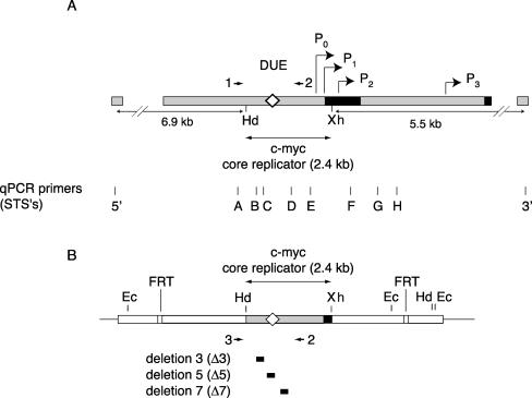
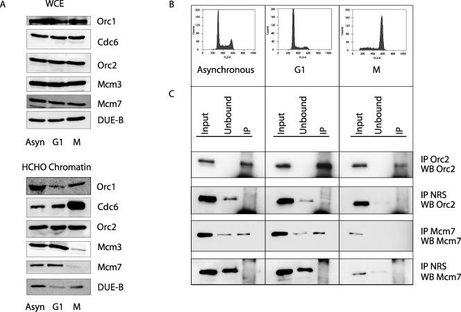
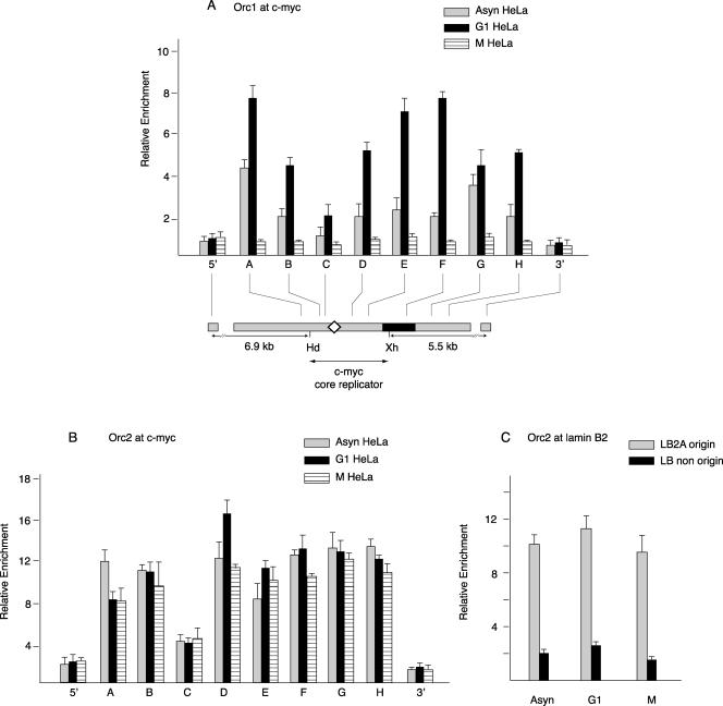
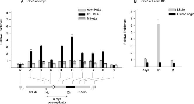
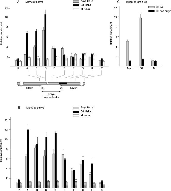
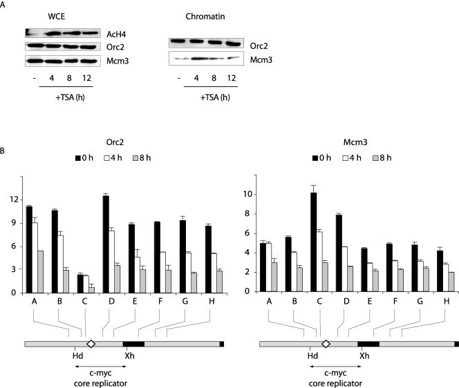
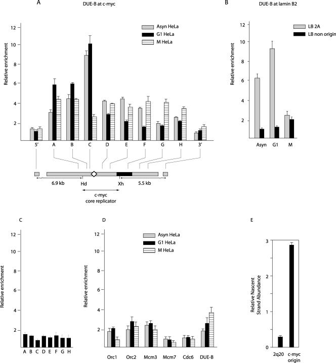
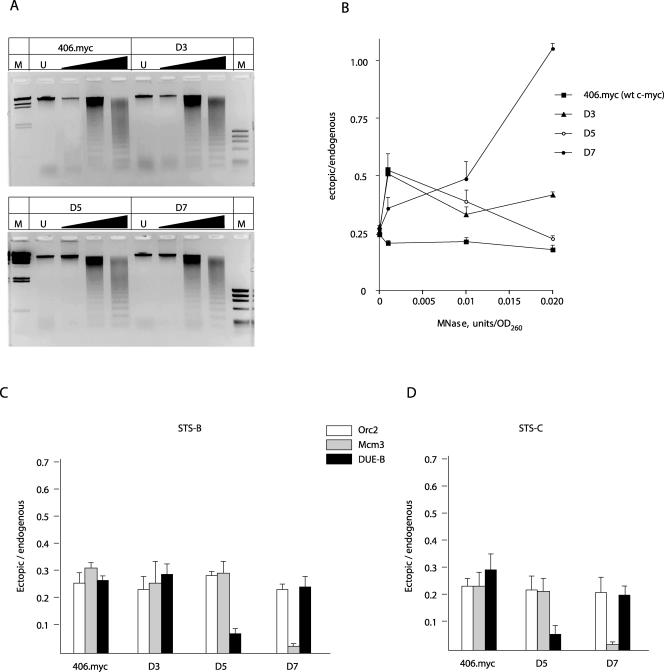
The binding of the prereplication complex proteins Orc1, Orc2, Mcm3, Mcm7, and Cdc6 and the novel DNA unwinding element (DUE) binding protein DUE-B to the endogenous human c-myc replicator was studied by chromatin immunoprecipitation. In G(1)-arrested HeLa cells, Mcm3, Mcm7, and DUE-B were prominent near the DUE, while Orc1 and Orc2 were least abundant near the DUE and more abundant at flanking sites. Cdc6 binding mirrored that of Orc2 in G(1)-arrested cells but decreased in asynchronous or M-phase cells. Similarly, the signals from Orc1, Mcm3, and Mcm7 were at background levels in cells arrested in M phase, whereas Orc2 retained the distribution seen in G(1)-phase cells. Previously shown to cause histone hyperacetylation and delocalization of replication initiation, trichostatin A treatment of cells led to a parallel qualitative change in the distribution of Mcm3, but not Orc2, across the c-myc replicator. Orc2, Mcm3, and DUE-B were also bound at an ectopic c-myc replicator, where deletion of sequences essential for origin activity was associated with the loss of DUE-B binding or the alteration of chromatin structure and loss of Mcm3 binding. These results show that proteins implicated in replication initiation are selectively and differentially bound across the c-myc replicator, dependent on discrete structural elements in DNA or chromatin.


Figures

FIG. 1.
FIG. 1.
Maps of DNA sequences used in this study. (A) Endogenous human c-myc locus. The first c-myc exon and the 5′ part of the second exon are shown as black boxes. The HindIII/XhoI fragment constitutes the wild-type c-myc core replicator (Hd, HindIII; Xh, XhoI). The diamond denotes the DUE region. The c-myc promoters (bent arrows) are indicated as P0, P1, P2, and P3. Arrowheads indicate primers 1 and 2 used in nested PCR (see text). 5′, 3′, and A through H denote qPCR STS primer sets. (B) Structure of the ectopic c-myc replicator locus in cell lines containing the integrated wild-type (map) or deleted (black boxes) core replicator constructs (45). Arrowheads indicate primers 2 and 3 used in nested PCR (see text). The two qPCR primer sites STS-B and STS-C used in nested PCR are indicated. Ec, EcoRI; FRT, FLP recombination target.
FIG. 2.
FIG. 2.
Cell synchrony. (A) Whole-cell extract (WCE) or formaldehyde-cross-linked (HCHO) chromatin was prepared from asynchronous (Asyn) cells or cells arrested with mimosine (G1) or nocodazole (M) and immunoblotted for the indicated proteins. Protein from equal numbers of asynchronous or G1- or M-phase cells was loaded. (B) Asynchronous HeLa cells, HeLa cells synchronized in G1 with mimosine, or HeLa cells synchronized in M with nocodazole were stained with propidium iodide and analyzed by flow cytometry. (C) ChIP chromatin from asynchronous or G1- or M-phase HeLa cultures analyzed by Western blotting (WB), using the indicated antibodies. Input, cross-linked chromatin from asynchronous, G1, and M cells before ChIP (5% aliquot); unbound, supernatant fraction after ChIP (5% aliquot); IP, immunoprecipitate (50% aliquot). NRS, normal rabbit serum.
FIG. 3.
FIG. 3.
Orc1 and Orc2 binding. Shown are results from ChIP using chromatin from asynchronous (Asyn), mimosine-arrested G1-phase, or nocodazole-arrested M-phase HeLa cells with antibodies against (A) Orc1 or (B) Orc2 proteins. The ChIP DNA was used for qPCR of STSs in the c-myc replicator. The relative enrichment (ordinate) at each STS is the copy number of the DNA precipitated with the specific antibody divided by the copy number of the DNA precipitated by normal rabbit serum at the same STS. Map details are described in the legend for Fig. 1. (C) Orc2 protein binding at the lamin B2 replicator. ChIP DNA was used to measure the abundance of STS LB2A sequences (110 bp from the transition point at the lamin B2 origin) and lamin B2 nonorigin sequences (5 kb downstream of the lamin B2 origin) by qPCR.
FIG. 4.
FIG. 4.
Cdc6 binding. Cross-linked chromatin from asynchronous (Asyn), mimosine-arrested G1-phase, or nocodazole-arrested M-phase HeLa cells was precipitated with affinity-purified antibodies against Cdc6 protein, and the ChIP DNA was used for qPCR of STSs in the (A) c-myc or (B) lamin B2 replicator locus. Map details are described in the legend for Fig. 1.
FIG. 5.
FIG. 5.
Mcm3 and Mcm7 binding. ChIP on chromatin from asynchronous (Asyn), mimosine-arrested G1-phase, or nocodazole-arrested M-phase HeLa cells with antibodies against (A) Mcm3 or (B) Mcm7 proteins, quantitated at the c-myc replicator by qPCR. Map details are described in the legend for Fig. 1. (C) Mcm3 protein binding at the lamin B2 replicator.
FIG. 6.
FIG. 6.
Redistribution of Mcm3 following TSA treatment. (A) Whole-cell extract (WCE) or chromatin (51) was prepared from cells treated with TSA for 0 h, 4 h, 8 h, or 12 h and immunoblotted for the indicated proteins. (B) ChIP on chromatin from HeLa cells treated with TSA for 0 h, 4 h, or 8 h, using antibodies against (left) Orc2 or (right) Mcm3. The ChIP DNA was used for qPCR of STSs in the c-myc replicator. Map details are described in the legend for Fig. 1. AcH4, acetylated histone H4.
FIG. 7.
FIG. 7.
DUE-B binding and origin activity. Shown are results of ChIP on chromatin from asynchronous (Asyn), G1-phase, or M-phase HeLa cells with polyclonal antibodies against DUE-B protein. The ChIP DNA was used for qPCR at STSs in the (A) c-myc or (B) lamin B2 replicator. Map details are described in the legend for Fig. 1. (C) Cross-linked chromatin was isolated from G1-arrested Daudi cells, which do not express DUE-B, and immunoprecipitated with polyclonal antibodies against DUE-B, and the precipitated DNA was used for qPCR of STSs in the c-myc replicator. (D) Decreased pre-RC protein binding at a site selected for low DUE-B binding. The chromosome 2p20 DNA sequence underrepresented in the anti-DUE-B antibody ChIP was isolated from the input HeLa chromatin pool (see Materials and Methods). qPCR analysis of ChIP DNA with Orc1, Orc2, Mcm3, Mcm7, Cdc6, or DUE-B antibody was carried out at the 2p20 site. (E) Replication origin activity (1- to 2-kb nascent-strand abundance) at the DUE-B depleted 2q20 site compared to that at the endogenous c-myc origin.
FIG. 8.
FIG. 8.
Effect of sequence deletion on protein binding to the c-myc replicator. The 406.myc cell line containing the ectopic wild-type (wt) c-myc replicator and the three cell lines containing c-myc replicator deletion mutant constructs (Δ3, Δ5, and Δ7 [Fig. 1]) (45) were compared. (A) MNase digestion dose response. Gels were stained with ethidium bromide. M, markers of HindIII-digested λ DNA (left) and HaeIII-digested ΦX174 DNA (right). (B) Nested-PCR quantitation at STS-B of MNase resistance at the ectopic and endogenous replicators. OD260, optical density at 260 nm. (C and D) Orc2, Mcm3, and DUE-B binding at the ectopic and endogenous c-myc replicators assayed by ChIP at (C) STS-B and (D) STS-C (Fig. 1).

Similar articles

Cited by

References

    1. Abdurashidova, G., M. B. Danailov, A. Ochem, G. Triolo, V. Djeliova, S. Radulescu, A. Vindigni, S. Riva, and A. Falaschi. 2003. Localization of proteins bound to a replication origin of human DNA along the cell cycle. EMBO J. 22:4294-4303. - PMC - PubMed
    1. Aggarwal, B. D., and B. R. Calvi. 2004. Chromatin regulates origin activity in Drosophila follicle cells. Nature 430:372-376. - PubMed
    1. Aladjem, M. I., L. W. Rodewald, J. L. Kolman, and G. M. Wahl. 1998. Genetic dissection of a mammalian replicator in the human beta-globin locus. Science 281:1005-1009. - PubMed
    1. Alexandrow, M. G., and J. L. Hamlin. 2004. Cdc6 chromatin affinity is unaffected by serine-54 phosphorylation, S-phase progression, and overexpression of cyclin A. Mol. Cell. Biol. 24:1614-1627. - PMC - PubMed
    1. Alexandrow, M. G., M. Ritzi, A. Pemov, and J. L. Hamlin. 2002. A potential role for mini-chromosome maintenance (MCM) proteins in initiation at the dihydrofolate reductase replication origin. J. Biol. Chem. 277:2702-2708. - PubMed

Publication types

MeSH terms

LinkOut - more resources