. 2006 Jul;26(14):5300-9.
doi: 10.1128/MCB.00273-06.

Mechanistic studies of the mitotic activation of Mos

Affiliations

Mechanistic studies of the mitotic activation of Mos

Jianbo Yue et al. Mol Cell Biol. 2006 Jul.

Abstract

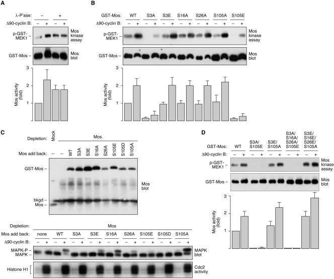
The protein kinase Mos is responsible for the activation of MEK1 and p42 mitogen-activated protein kinase during Xenopus oocyte maturation and during mitosis in Xenopus egg extracts. Here we show that the activation of Mos depends upon the phosphorylation of Ser 3, a residue previously implicated in the regulation of Mos stability; the dephosphorylation of Ser 105, a previously unidentified phosphorylation site conserved in Mos proteins; and the regulated dissociation of Mos from CK2beta. Mutation of Ser 3 to alanine and/or mutation of Ser 105 to glutamate produces a Mos protein that is defective for M-phase activation, as assessed by in vitro kinase assays, and defective for induction of oocyte maturation and maintenance of the spindle assembly checkpoint in extracts. Interestingly, Ser 105 is situated at the beginning of helix alphaC in the N-terminal lobe of the Mos kinase domain. Changes in the orientation of this helix have been previously implicated in the activation of Cdk2 and Src family tyrosine kinases. Our work suggests that Ser 105 dephosphorylation represents a novel mechanism for reorienting helix alphaC.


Figures

FIG. 1.
FIG. 1.
Mitotic activation of Mos. (A) Activation of recombinant GST-Flag-Mos by Δ90-cyclin B-treated Xenopus egg extracts. The top blot shows Mos activity as indicated by an in vitro kinase assay using recombinant GST-MEK1 as a substrate and phospho-MEK1 immunoblotting to detect the amount of MEK1 phosphorylation. The bottom blot shows that equal levels of GST-Flag-Mos protein were present in the two samples. The bar graph shows pooled data from four independent experiments, with data expressed as means ± standard deviations. (B) Electrophoretic mobility shift of recombinant GST-Flag-Mos incubated with Δ90-cyclin B-treated Xenopus egg extracts. Samples were immunoprecipitated with Flag antibody and immunoblotted with Mos antibody.
FIG. 2.
FIG. 2.
Mos phosphorylation sites. (A) GST-Flag-Mos was treated with Δ90-cyclin B-treated Xenopus egg extracts or interphase extracts and subjected to immunoprecipitation with Flag antibody, followed by Coomassie blue staining (top) or immunoblotting with Mos antibody (bottom). “ns” designates a nonspecific band that ran just above Mos. (B) Schematic depiction of the Mos domain structure and the mapped phosphorylation sites. We have highlighted the kinase domain, helix αC, the region of Mos important for interaction with CK2β (21), and the T loop, or activation loop.
FIG. 3.
FIG. 3.
Electrophoretic mobility shifts in Mos phosphorylation site mutants. Samples of wild-type GST-Flag-Mos (Mos-WT) and various Mos mutants were incubated with Δ90-cyclin B-treated Xenopus egg extracts or interphase extracts and were then immunoprecipitated with Flag antibody and immunoblotted with Mos antibody.
FIG. 4.
FIG. 4.
Effects of phosphorylation on Mos activity. (A) Effect of λ phosphatase treatment on Mos activity. GST-Flag-Mos was treated with Δ90-cyclin B-treated Xenopus egg extracts or interphase extracts and subjected to immunoprecipitation with Flag antibody. Samples were then treated with λ phosphatase (λ P'ase) or were mock phosphatase treated and were subjected to an in vitro kinase assay using recombinant GST-MEK1 as a substrate and phospho-MEK1 immunoblotting to detect the amount of MEK1 phosphorylation. The phospho-MEK1 and Mos blots are taken from one experiment. The bar graph shows data from three independent experiments expressed as means ± standard deviations. (B) Activities of various phosphorylation site mutants. Kinase assays and blot analyses were carried out as described for panel A. The bar graph shows data from three independent experiments expressed as means ± standard deviations. WT, wild type. (C) Reconstitution of the mitotic phosphorylation of p42 MAPK by adding Mos mutants back to Mos-depleted extracts. Extracts were depleted of Mos by two rounds of sequential immunodepletion. A nominal concentration of 30 nM wild-type GST-Flag-Mos and various Mos mutants was added back to the Mos-depleted extracts. The top panel shows a Mos blot of the extracts. The bottom panels show p42 MAPK blots and histone H1 kinase assay results for the Mos-depleted extracts and Mos-reconstituted extracts after incubation with or without Δ90-cyclin B. Other studies showed that concentrations of Mos-S3A and Mos-S105E as high as 120 nM still failed to restore mitotic phosphorylation of p42 MAPK, as judged by the electrophoretic mobility shift (data not shown). bkgd, background bard. (D) Activities of various multisite mutants. Kinase assays and blot analyses were carried out as described for panel A. The bar graph shows data from three independent experiments expressed as means ± standard deviations.
FIG. 5.
FIG. 5.
Regulated dissociation of CK2β from Mos. (A) Cyclin-dependent phosphorylation of p42 MAPK in CK2β-depleted extracts. Extracts were subjected to two rounds of immunodepletion with nonspecific immunoglobulin G or anti-CK2β. To one aliquot of the CK2β-depleted extract, we added back recombinant CK2β to restore normal levels of the protein. The resulting levels of CK2β were determined by immunoblotting (left). The mock-depleted, CK2β-depleted, and CK2β-depleted/restored extracts were then treated with or without Δ90-cyclin B and subjected to phospho-MAPK immunoblotting and histone H1 kinase assays (right). (B) Cyclin-dependent phosphorylation of p42 MAPK in CK2β-supplemented extracts. Extracts were treated with buffer or CK2β (1 μM) for 30 min and then were treated with Δ90-cyclin B. Samples were taken at various times and subjected to phospho-MAPK immunoblotting and histone H1 kinase assays. (C) Dissociation of CK2β from Mos in M phase. Recombinant GST-Flag-CK2β (20 nM) and either wild-type GST-Flag-Mos (Mos-WT) or one of two quadruple-phosphorylation-site Mos mutants (20 nM) were added to Xenopus egg extracts with or without Δ90-cyclin B. Lysates were immunoblotted with Flag antibody (left) or were subjected to immunoprecipitation with Mos antibody, followed by immunoblotting with Flag antibody (right). Note that different Mos mutants were pulled down with differing efficiencies (e.g., compare lanes 9 and 10 with lanes 11 and 12), but cyclin treatment had no effect on the efficiency of the Mos pulldown (lane 7 versus lane 8; lane 9 versus lane 10; lane 11 versus lane 12).
FIG. 6.
FIG. 6.
Biological function of Mos phosphorylation site mutants. (A) Oocyte maturation. Oocytes were incubated with progesterone or buffer or were microinjected with one of the seven GST-Flag-Mos proteins as indicated. Maturation was scored as a function of time, using the appearance of a white dot at the animal pole of the oocyte as an indicator of maturation. (B) Spindle assembly checkpoint. CSF-arrested Xenopus egg extracts were subjected to two rounds of Mos immunodepletion or mock depletion. Sperm chromatin was added at a concentration of 15,000 per μl, and calcium (1 mM) was added in the absence and presence of nocodazole (10 ng/μl). Histone H1 kinase assays were monitored as functions of time. WT, wild type.
FIG. 7.
FIG. 7.
Phosphorylation of Mos at Ser 3 and Ser 105 during oocyte maturation and after egg activation. (A) Specificities of the pS3-Mos and pS105-Mos antibodies. Wild-type (WT) or mutant GST-Flag-Mos was treated with Δ90-cyclin B-treated Xenopus egg extracts or interphase extracts and subjected to immunoprecipitation with Flag antibody, followed by immunoblotting with pS3-Mos and pS105-Mos antibodies. (B) Mos accumulation and phosphorylation during oocyte maturation. Oocytes were treated with progesterone for various lengths of time. Lysates were subjected to immunoblotting with Mos, pS3-Mos, and pS105-Mos antibodies. The asterisks denote nonspecific protein bands in the Mos blots. p42 MAPK blots and H1 kinase (H1K) assay results are shown for comparison. (C) Mos degradation and phosphorylation after egg activation. Dejellied eggs were treated with calcium ionophore and blotted for Mos, pS3-Mos, and pS105-Mos. p42 MAPK blots and H1 kinase assay results are shown for comparison. (D) Schematic depiction of the changes in Mos phosphorylation and CK2β-association between interphase and M phase.

Similar articles

Cited by

References

    1. Abrieu, A., D. Fisher, M. N. Simon, M. Doree, and A. Picard. 1997. MAPK inactivation is required for the G2 to M-phase transition of the first mitotic cell cycle. EMBO J. 16:6407-6413. - PMC - PubMed
    1. Andrésson, T., and J. V. Ruderman. 1998. The kinase Eg2 is a component of the Xenopus oocyte progesterone-activated signaling pathway. EMBO J. 17:5627-5637. - PMC - PubMed
    1. Bitangcol, J. C., A. S. Chau, E. Stadnick, M. J. Lohka, B. Dicken, and E. K. Shibuya. 1998. Activation of the p42 mitogen-activated protein kinase pathway inhibits Cdc2 activation and entry into M-phase in cycling Xenopus egg extracts. Mol. Biol. Cell 9:451-467. - PMC - PubMed
    1. Castro, A., M. Peter, L. Magnaghi-Jaulin, S. Vigneron, S. Galas, T. Lorca, and J. C. Labbe. 2001. Cyclin B/cdc2 induces c-Mos stability by direct phosphorylation in Xenopus oocytes. Mol. Biol. Cell 12:2660-2671. - PMC - PubMed
    1. Chen, M., and J. A. Cooper. 1995. Ser-3 is important for regulating Mos interaction with and stimulation of mitogen-activated protein kinase kinase. Mol. Cell. Biol. 15:4727-4734. - PMC - PubMed

Publication types

MeSH terms

LinkOut - more resources