. 2006 Jul;26(14):5310-24.
doi: 10.1128/MCB.00084-06.

Prostate-specific membrane antigen regulates angiogenesis by modulating integrin signal transduction

Affiliations

Prostate-specific membrane antigen regulates angiogenesis by modulating integrin signal transduction

Rebecca E Conway et al. Mol Cell Biol. 2006 Jul.

Abstract

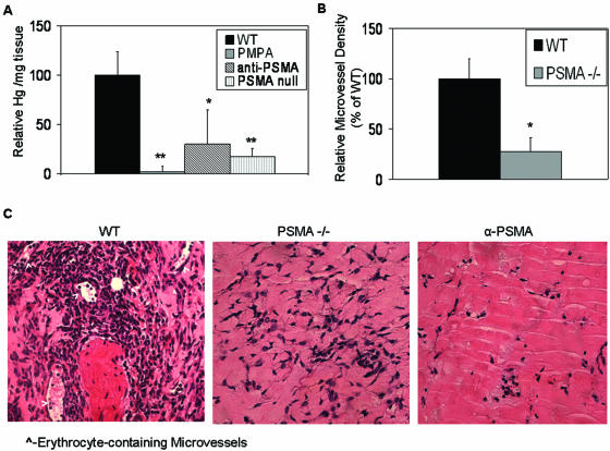
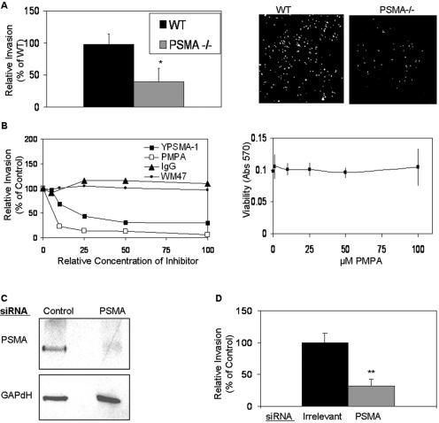
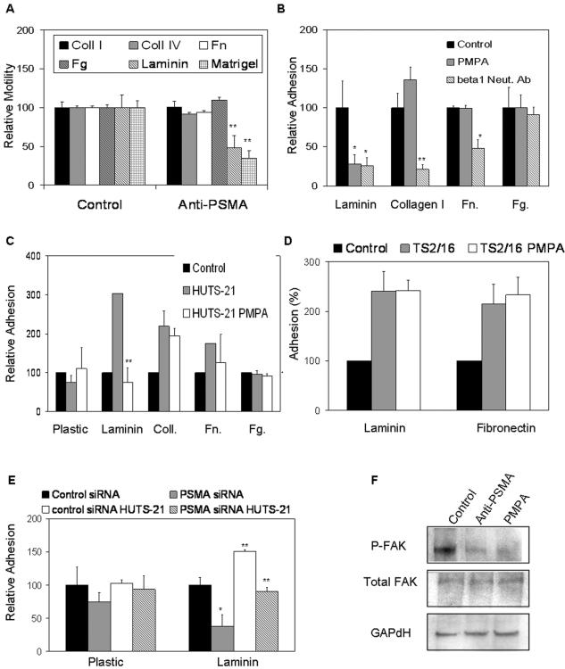
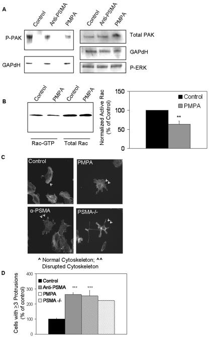
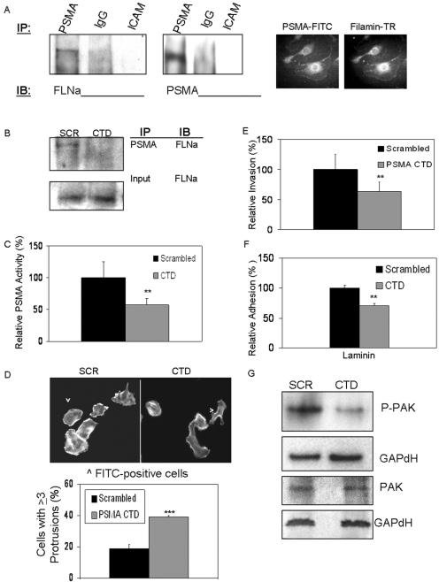
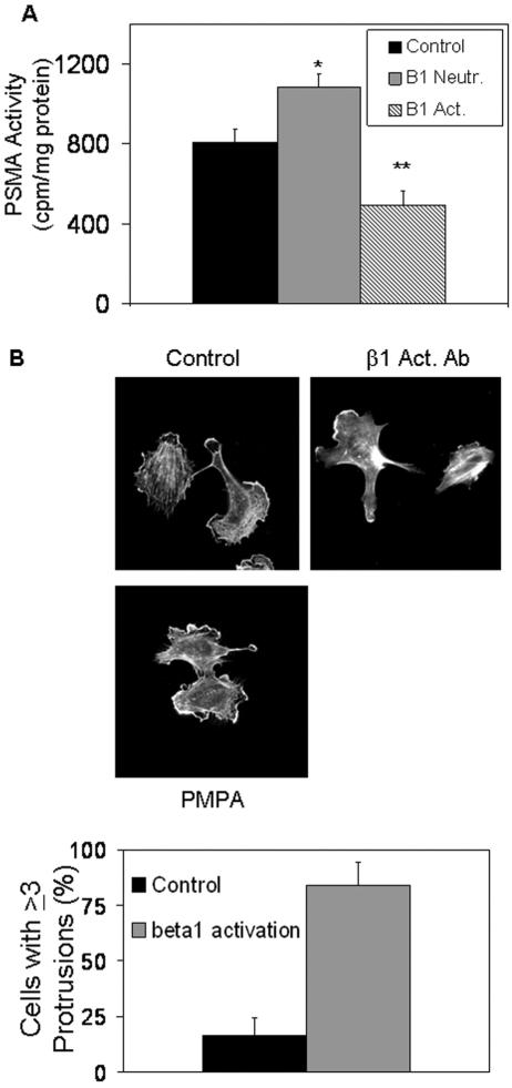
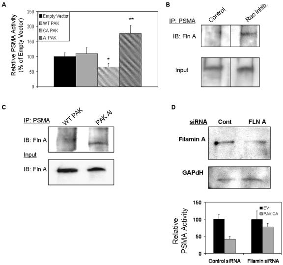
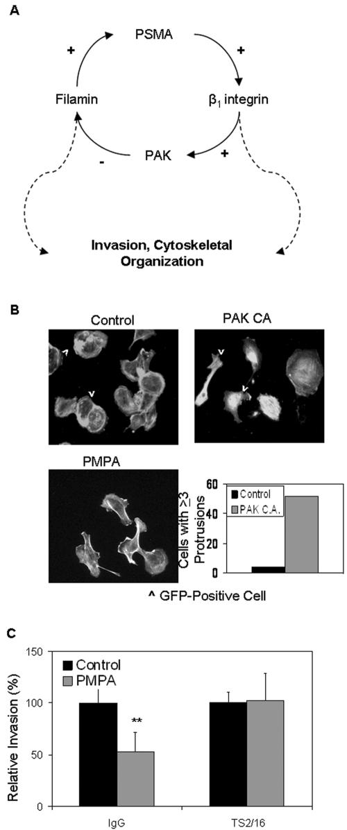
The transmembrane peptidase prostate-specific membrane antigen (PSMA) is universally upregulated in the vasculature of solid tumors, but its functional role in tumor angiogenesis has not been investigated. Here we show that angiogenesis is severely impaired in PSMA-null animals and that this angiogenic defect occurs at the level of endothelial cell invasion through the extracellular matrix barrier. Because proteolytic degradation of the extracellular matrix is a critical component of endothelial invasion in angiogenesis, it is logical to assume that PSMA participates in matrix degradation. However, we demonstrate a novel and more complex role for PSMA in angiogenesis, where it is a principal component of a regulatory loop that is tightly modulating laminin-specific integrin signaling and GTPase-dependent, p21-activated kinase 1 (PAK-1) activity. We show that PSMA inhibition, knockdown, or deficiency decreases endothelial cell invasion in vitro via integrin and PAK, thus abrogating angiogenesis. Interestingly, the neutralization of beta(1) or the inactivation of PAK increases PSMA activity, suggesting that they negatively regulate PSMA. This negative regulation is mediated by the cytoskeleton as the disruption of interactions between the PSMA cytoplasmic tail and the anchor protein filamin A decreases PSMA activity, integrin function, and PAK activation. Finally, the inhibition of PAK activation enhances the PSMA/filamin A interaction and, thus, boosts PSMA activity. These data imply that PSMA participates in an autoregulatory loop, wherein active PSMA facilitates integrin signaling and PAK activation, leading to both productive invasion and downregulation of integrin beta(1) signaling via reduced PSMA activity. Therefore, we have identified a novel role for PSMA as a true molecular interface, integrating both extracellular and intracellular signals during angiogenesis.


Figures

FIG. 1.
FIG. 1.
Loss of PSMA in vivo abrogates angiogenesis. Results of in vivo Matrigel plug assay show that (A) hemoglobin (Hg) content is decreased in PSMA-inhibited (n = 6; P = 0.002) or PSMA-null plugs (n = 6; P = 0.0001), (B) microvessel density is decreased in PSMA-null plugs (n = 6; P = 0.014), and (C) hematoxylin and eosin staining of Matrigel plug sections reveals a significant decrease in erythrocyte-containing microvessels in PSMA-null or -inhibited plugs (indicated by arrowheads). WT, wild type; α-PSMA, PSMA-neutralizing antibody; PSMA −/−, PSMA null. *, 0.05 < P > 0.01; **, 0.01 < P > 0.001. Error bars indicate standard deviations.
FIG. 2.
FIG. 2.
PSMA is required for endothelial cell invasion. (A) Lung endothelial cells isolated from PSMA-null (PSMA −/−) animals have decreased abilities to invade in vitro (n = 3; P = 0.02). Right panels show images of invading cells stained with calcein-AM. WT, wild type. Error bars indicate standard deviations. *, 0.05 < P > 0.01. (B) Dose dependency of inhibition of HUVEC invasion in response to YPSMA-1 monoclonal antibody (doses range from 0 to 0.1 mg/ml) or PMPA (doses range from 0 to 100 μM). The right panel shows HUVEC cell viability assay in the presence of 100 μM PMPA. IgG, immunoglobulin G; Abs, antibodies. (C) KS1767 cells transfected with PSMA siRNA have decreased PSMA protein levels. (D) Transfection of KS1767 cells with PSMA siRNA decreases invasion, (n = 3, P = 0.009). **, 0.01 < P > 0.001. Error bars indicate standard deviations.
FIG. 3.
FIG. 3.
PSMA increases β1 integrin signaling in endothelial cells. (A) PSMA-dependent in vitro motility of HUVECs is specifically mediated through laminin (n = 3; P = 0.005) but not other matrix proteins. (B) PSMA antagonists inhibit basal adhesion of HUVECs to laminin equally as well as neutralizing β1 antibodies (Neut. Ab) do, but have no significant effect on binding to fibrinogen, fibronectin, or collagen. (C) Inhibition of PSMA interferes with activated β1 integrin (activated with HUTS-21 antibody) binding to laminin but not other substrates (n = 3; P = 0.002). (D) PSMA inhibition in the presence of the consitutively activating β1 antibody TS2/16 does not alter adhesion to laminin or fibronectin. (E) HUVECs transfected with siRNA targeted against PSMA show reduced basal (n = 3; P = 0.028) and β1-mediated (n = 3; P = 0.001) adhesion compared to that of cells transfected with control siRNA; (F) HUVECs treated with PMPA or anti-PSMA antibody have decreased levels of phosphorylated FAK. *, 0.05 < P > 0.01; **, 0.01 < P > 0.001. Error bars indicate standard deviations.
FIG. 4.
FIG. 4.
PSMA regulates PAK phosphorylation and cytoskeletal morphology. (A) HUVECs treated with PSMA inhibitor or neutralizing antibody have decreased levels of phosphorylated PAK, while total PAK and phosphorylated extracellular signal-regulated kinase levels are unaffected. P-ERK, phospho-extracellular signal-regulated kinase (ERK). (B) HUVECs treated with PMPA show reduced levels of activated Rac, while total Rac levels remain unchanged. Quantification of the average of three Rac activation experiments is shown by the graph (n = 3; P = 0.002). **, 0.01 < P > 0.001. (C) HUVECs treated with either the PMPA inhibitor, the PSMA neutralizing antibody (α-PSMA), or siRNA-treated endothelial cells isolated from PSMA-null (PSMA−/−) animals display actin-containing protrusions extending in multiple directions. (D) quantitation of cells with protrusions extending in at least three directions reveals an approximate twofold increase in PSMA-inhibited or PSMA-null (PSMA −/−) cells (P < 0.001). *** = 0.001< P > 0.0001. Error bars indicate standard deviations.
FIG. 5.
FIG. 5.
PSMA interaction with filamin A in endothelial cells regulates PSMA function. (A) Immunoprecipitation (IP) of PSMA but not immunoglobulin G (IgG) or ICAM-1 pulls down filamin A in HUVECs and filamin, and PSMA colocalizes in migrating HUVECs (PSMA-FITC and filamin-Texas Red [TR] antibodies). Control photobleaching data are provided in Fig. S1 of the supplemental material. (B) PSMA CTD peptide transfection in HUVECs disrupts PSMA/filamin A interaction. Disrupting PSMA/filamin interaction by transfecting the PSMA CTD peptide (C) decreases PSMA enzyme activity (P = 0.003), (D) disrupts endothelial cell morphology, resulting in an increase in membrane protrusions (P < 0.001), (E) decreases endothelial cell invasion (P = 0.002), (F) decreases endothelial cell adhesion to laminin (P = 0.001), and (G) decreases PAK activation as measured by phosphorylated-PAK levels. SCR, scrambled peptides; IB, immunoblot. **, 0.01 < P > 0.001; ***, 0.001 < P > 0.0001. Error bars indicate standard deviations.
FIG. 6.
FIG. 6.
Integrin β1 regulates PSMA activity. (A) β1 integrin inhibition (Neutr.) increases PSMA activity (n = 3; P = 0.01), while β1 activation (Act.) decreases PSMA activity (n = 3; P = 0.001). *, 0.05 < P > 0.01; **, 0.01 < P > 0.001. (B) HUVECs incubated on laminin-coated cell culture dishes with a β1-activating antibody (β1-Act. Ab) display protrusions in multiple directions, similar to those of PSMA inhibited cells (PMPA treatment). Error bars indicate standard deviations.
FIG. 7.
FIG. 7.
PAK regulates PSMA activity. (A) PSMA activity is decreased in cells transfected with a construct encoding a constitutively active (CA) PAK mutant (P = 0.045) and increased in cells transfected with a plasmid encoding the PAK autoinhibitory (AI) domain (P = 0.001). Results are the average of three independent experiments performed in triplicate. WT, wild type. *, 0.05 < P > 0.01; **, 0.01 < P > 0.001. Error bars indicate standard deviations. (B) Filamin/PSMA interaction is increased in cells treated with the Rac inhibitor; a nonspecific band is shown as a loading control. Fln A, filamin A; IP, immunoprecipitate; IB, immunoblot. (C) HUVECs transfected with autoinhibitory (AI) PAK also show increased PSMA/filamin interaction. Experiments controlling for the expression of the PAK constructs transfected in HUVECs are provided in Fig. S2 of the supplemental material. Fln A, filamin A; IP, immunoprecipitate; IB, immunoblot; WT, wild type. (D) HUVECs transfected with filamin A (FLN A) siRNA show reduced levels of filamin A protein compared to cells transfected with control siRNA (Cont). Additionally, reducing filamin A levels in HUVECs rescues loss of PSMA activity caused by transfecting constitutively active (CA) PAK constructs into HUVECs (for control siRNA, P < 0.001; for filamin A siRNA, P = 0.149). EV, empty vector. Error bars indicate standard deviations.
FIG. 8.
FIG. 8.
PSMA-mediated PAK and integrin activation affect endothelial cell function. (A) Hypothetical mechanism for PSMA regulation of endothelial cell invasion. PSMA positively regulates β1 integrin signaling and PAK phosphorylation in endothelial cells. Because PAK activation/deactivation must be precisely regulated for productive motility, we predict that PAK activation negatively regulates PSMA activity. Therefore, these components constitute a regulatory loop of PSMA regulation in endothelial cells, where β1 integrin signal trans duction and subsequent PAK activation leads to both productive invasion and downregulation of integrin β1 signaling via reduction in PSMA activity. (B) Cells transfected with plasmids encoding constitutively active (CA) PAK display increased protrusions in multiple directions. GFP, green fluorescent protein. (C) β1 activation by the constitutively activating antibody TS2/16 rescues PSMA-dependent invasion in the presence of the PMPA inhibitor. IgG, immunoglobulin G. **, 0.01< P > 0.001. Error bars indicate standard deviations.

Similar articles

Cited by

References

    1. Anilkumar, G., S. A. Rajasekaran, S. Wang, O. Hankinson, N. H. Bander, and A. K. Rajasekaran. 2003. Prostate-specific membrane antigen association with filamin A modulates its internalization and NAALADase activity. Cancer Res. 63:2645-2648. - PubMed
    1. Arroyo, A. G., A. Garcia-Pardo, and F. Sanchez-Madrid. 1993. A high affinity conformational state on VLA integrin heterodimers induced by an anti-β1 chain monoclonal antibody. J. Biol. Chem. 268:9863-9868. - PubMed
    1. Bacich, D. J., E. Ramadan, D. S. O'Keefe, N. Bukhari, I. Wegorzewska, O. Ojeifo, R. Olszewski, C. C. Wrenn, T. Bzdega, B. Wroblewska, W. D. Heston, and J. H. Neale. 2002. Deletion of the glutamate carboxypeptidase II gene in mice reveals a second enzyme activity that hydrolyzes N-acetylaspartylglutamate. J. Neurochem. 83:20-29. - PubMed
    1. Barker, J. N. 1991. The pathophysiology of psoriasis. Lancet 338:227-230. - PubMed
    1. Bauvois, B. 2004. Transmembrane proteases in cell growth and invasion: new contributors to angiogenesis? Oncogene 23:317-329. - PubMed

Publication types

MeSH terms