. 2006 Jul;26(14):5348-59.
doi: 10.1128/MCB.02351-05.

BAL1 and BBAP are regulated by a gamma interferon-responsive bidirectional promoter and are overexpressed in diffuse large B-cell lymphomas with a prominent inflammatory infiltrate

Affiliations

BAL1 and BBAP are regulated by a gamma interferon-responsive bidirectional promoter and are overexpressed in diffuse large B-cell lymphomas with a prominent inflammatory infiltrate

Przemyslaw Juszczynski et al. Mol Cell Biol. 2006 Jul.

Abstract

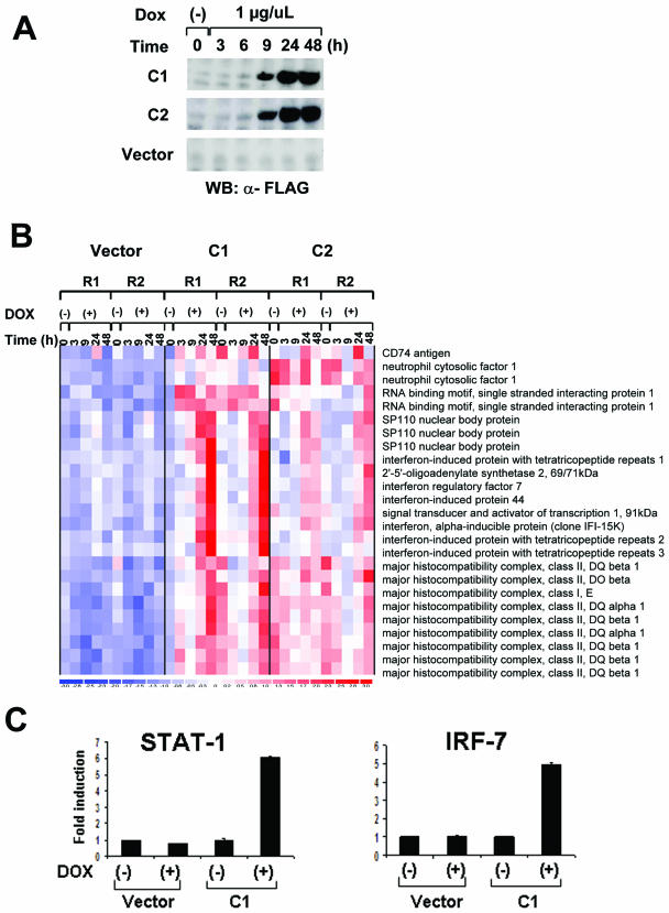
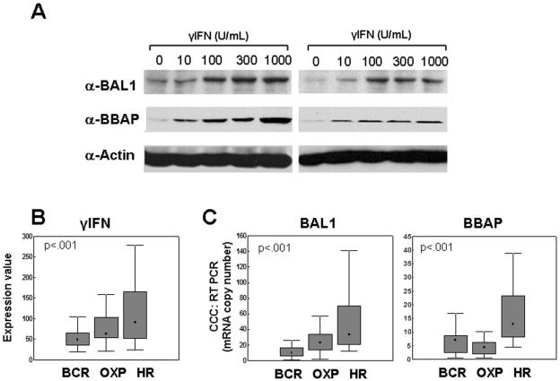
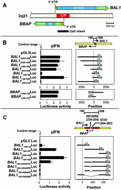
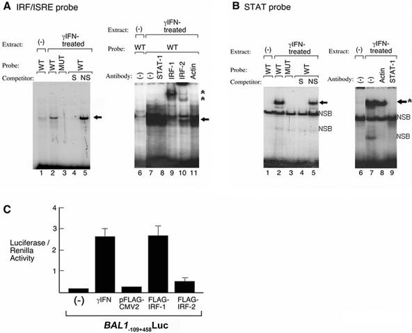
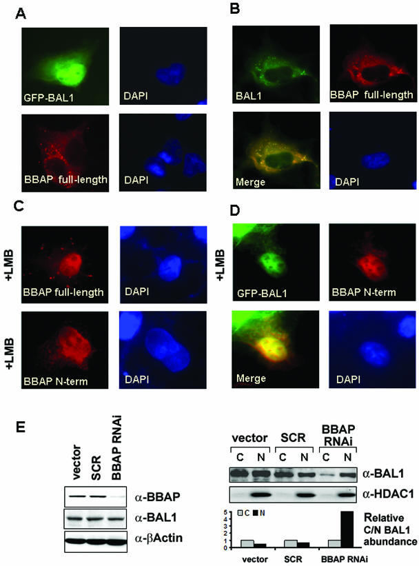
BAL1 is a transcription modulator that is overexpressed in chemoresistant, diffuse large B-cell lymphomas (DLBCLs). BAL1 complexes with a recently described DELTEX family member termed BBAP. Herein, we characterized BAL1 and BBAP expression in primary DLBCL subtypes defined by their comprehensive transcriptional profiles. BAL1 and BBAP were most abundant in lymphomas with a brisk host inflammatory response, designated host response (HR) tumors. Although these DLBCLs include significant numbers of tumor-infiltrating lymphocytes and interdigitating dendritic cells, BAL1 and BBAP were expressed primarily by malignant B cells, prompting speculation that the genes might be induced by host-derived inflammatory mediators such as gamma interferon (IFN-gamma). In fact, IFN-gamma induced BAL1 and BBAP expression in DLBCL cell lines; doxycycline-induced BAL1 also increased the expression of multiple IFN-stimulated genes, directly implicating BAL1 in an IFN signaling pathway. We show that BAL1 and BBAP are located on chromosome 3q21 in a head-to-head orientation and are regulated by a IFN-gamma-responsive bidirectional promoter. BBAP regulates the subcellular localization of BAL1 by a dynamic shuttling mechanism, highlighting the functional requirement for coordinated BBAP and BAL1 expression. IFN-gamma-induced BAL1/BBAP expression contributes to the molecular signature of HR DLBCLs and highlights the interplay between the inflammatory infiltrate and malignant B cells in these tumors.


Figures

FIG. 1.
FIG. 1.
BAL1 induction of ISG. (A) BAL1 expression in a doxycycline-inducible lymphoma cell line. BAL1-inducible and empty vector control lines were treated with doxycycline (Dox) for the indicated periods of time. Thereafter, whole-cell lysates were size fractionated, immunoblotted, and analyzed with anti-FLAG antibody. WB, Western blotting; α-FLAG, anti-FLAG. (B) ISG induced by BAL1. Seventeen genes (represented by 25 probe sets) were bona fide type 1 and 2 ISG or genes indirectly modulated by IFN-γ. (C) Quantitative RT-PCR analysis of STAT-1 and IRF-7 induction by BAL. RNAs from a BAL1-inducible clone (C1) and control vector-only cells that were untreated (−) or treated with doxycycline (+) for 36 h were analyzed for STAT-1 and IRF-7 abundance by quantitative RT-PCR.
FIG. 2.
FIG. 2.
BAL1 and BBAP induction by IFN-γ in lymphoma cell lines and overexpression in primary DLBCLs. (A) Induction of BAL1 and BBAP by IFN-γ in two lymphoma cell lines (DHL4 [right panel] and DHL10 [left panel]). Cells were treated with the indicated doses of IFN-γ (0 to 1,000 U/ml) for 24 h. Thereafter, whole-cell lysates were size fractionated, immunoblotted, and analyzed with anti-BAL1 or anti-BBAP antibodies (α-BAL1 or α-BBAP, respectively). (B and C) Overexpression of IFN-γ, BAL1, and BBAP transcript abundance in DLBCL comprehensive clusters. Primary DLBCLs were identified as BCR, OXP, and HR tumors, and levels of IFN-γ, BAL1, and BBAP transcript abundance in the clusters were compared using a Kruskal-Wallis test. (B) IFN-γ transcript abundances were compared using microarray data (U133A probe set 210354_at [arbitrary units]). (C) BAL1 and BBAP transcript abundance was evaluated using real-time quantitative RT-PCR. Data are developed using graphs which denote the medians (▪), 25 to 75% values (shaded bars), and nonoutlier ranges (I bars).
FIG. 3.
FIG. 3.
Identification and characterization of the IFN-γ-inducible, bidirectional BAL1/BBAP promoter. (A) Schematic representation of the bidirectional genomic organization of BAL1 and BBAP genes and their shared, CpG-related promoter (common promoter [CP], indicated by a red bar). In, intron; Ex, exon; 5′UTR, 5′ untranslated region. (B) Identification of a minimal, bidirectionally active BAL1/BBAP promoter. (Upper panel) BAL1 promoter fragments including or lacking the predicted minimal promoter sequence (+80 to +307, represented by a red bar) were cloned into a luciferase reporter vector, transiently transfected into HEK293 cells, and assayed for responsiveness to IFN-γ. (Upper right panel) Respective BAL1 promoter constructs (black lines) were cloned upstream of the firefly luciferase gene (gray bar with arrow). (Upper left panel) Representative luciferase activities from three independent experiments are normalized to Renilla luciferase activity and are presented as black bars ± standard deviations. (Lower panel) The reversed minimal promoter construct (BBAP+458+109Luc) or constructs with an additional 1.2 kb of 5′ upstream sequence (BBAP+1648109Luc) were transiently transfected into HEK293 cells and assessed as for the upper panel. (C) The bidirectional BAL1/BBAP promoter requires intact IRF-1 and STAT binding sites for IFN-γ-induced activity. A series of deletion constructs lacking predicted individual IRF or STAT binding sites or both sites (dIRF, dSTAT, and dIRE, respectively) were cloned into the pGL3 vector, transiently transfected into HEK293 cells, and assessed as described for panel B. ISRE, IFN-stimulated response element.
FIG. 4.
FIG. 4.
BAL1/BBAP IRF and STAT promoter elements directly bind IFN-γ-induced proteins. (A) IRF shift assay. (Left panel) Nuclear extracts from IFN-γ-treated (lanes 2 to 5) or untreated (lane 1) cells were incubated with a wild-type (WT) (lanes 1, 2, 4, and 5) or mutant (MUT) (lane 3) 32P-labeled, double-stranded DNA probe corresponding to the IRF binding site in the BAL1/BBAP promoter. Specific (S) or nonspecific (NS) competitor was included in certain assays (lane 4 or 5, respectively). The gel shift band corresponding to the probe-protein complex is indicated with an arrow. (Right panel) Nuclear extracts from IFN-γ-treated (lanes 7 to 11) or untreated (lane 6) cells were incubated with the 32P-labeled IRF probe. Antibodies against STAT-1, IRF-1, IRF-2, or β-actin were included in lane 8, 9, 10, or 11, respectively. The gel shift band corresponding to probe-protein complex is indicated with an arrow, and supershift bands corresponding to probe-protein-antibody complex are noted with asterisks. (B) STAT-1 shift assay. (Left panel) Nuclear extracts from IFN-γ-treated (lanes 2 to 5) or untreated (lane 1) cells were incubated with a wild-type (lanes 1, 2, 4, and 5) 32P-labeled, double-stranded DNA probe corresponding to the STAT binding site in the BAL1/BBAP promoter. Specific (S) or nonspecific (NS) competitor was included in lane 4 or 5, respectively. The gel shift band corresponding to the formed probe-protein complex is indicated with an arrow; the lower shift band is nonspecific (NSB). (Right panel) Nuclear extracts from IFN-γ-treated (lanes 7 to 9) or untreated (lane 6) cells were incubated with the wild-type, 32P-labeled STAT probe. Antibodies against STAT-1 or β-actin were included in lane 9 or 8, respectively. The gel shift band corresponding to formed probe-protein complex is indicated by an arrow, and disruption of the complex by the STAT-1 antibody is noted by asterisks. (C) Roles of IRF-1 and -2 in BAL1 promoter activation. The BAL1 minimal promoter construct (BAL1109+458Luc) was transiently cotransfected with the empty pFLAG-CMV2 vector or with the IRF-1 or IRF-2 pFLAG-CMV2 expression vector. Control cells were transfected with BAL1109+458Luc (alone) and treated with IFN-γ or left untreated as indicated. Thereafter, cells were lysed and assessed for luciferase activity as described in the legend to Fig. 3.
FIG. 5.
FIG. 5.
Subcellular localization of BAL1 and BBAP in primary HR DLBCLs. (A) Cytoplasmic and nuclear (inset, right panel) staining of BAL1 in the tumor cells of two primary HR DLBCLs (left and right panels). (B) Cytoplasmic and nuclear (inset, right panel) staining of BBAP in tumor cells of the same two primary HR DLBCLs. Magnification, ×400; inset, ×1,000.
FIG. 6.
FIG. 6.
Subcellular localization of transfected BAL1 and BBAP in COS-7 cells. COS-7 cell monolayers were grown on coverslips and transfected with the following expression vectors: either eGFP-BAL1 or full-length FLAG-BBAP (A), eGFP-BAL1 and FLAG-BBAP (B), FLAG-tagged full-length or N-terminal BBAP (C), and eGFP-BAL1 and FLAG-tagged N-terminal BBAP (D). Cells in panels C and D were treated with LMB (10 ng/ml) as indicated. After 24 h, cells were fixed and subjected to indirect immunofluorescence. Nuclei were visualized by counterstaining with DAPI. (E) shRNA-mediated knockdown of BBAP. HeLa cells were transduced with a retrovirus expressing BBAP-specific shRNA (BBAP-RNAi) or virus encoding no shRNA (vector) or expressing a negative-control scrambled sequence (SCR). Seventy-two hours after retroviral transduction, whole-cell lysates or nuclear and cytosolic fractions were obtained, size fractionated, and analyzed by immunoblotting (left panel) or immunoblotting and scanning densitometry (right panel). (Left panel) Immunoblots of whole-cell lysates analyzed with anti-BBAP (a-BBAP), anti-BAL1, and anti-β-actin (loading control). (Upper right panel) Immunoblots of nuclear and cytosolic fractions analyzed with anti-BAL1 and anti-HDAC1 (nuclear-protein control). (Lower right panel) Densitometric analysis of relative nuclear and cytosolic BAL1 protein abundances in vector-, SCR-, or BBAP RNAi-treated cells.

Similar articles

Cited by

References

    1. Abramson, J. S., and M. A. Shipp. 2005. Advances in the biology and therapy of diffuse large B-cell lymphoma: moving toward a molecularly targeted approach. Blood 106:1164-1174. - PubMed
    1. Abu-Shaar, M., H. D. Ryoo, and R. S. Mann. 1999. Control of the nuclear localization of Extradenticle by competing nuclear import and export signals. Genes Dev. 13:935-945. - PMC - PubMed
    1. Aguiar, R. C., K. Takeyama, C. He, K. Kreinbrink, and M. A. Shipp. 2005. B-aggressive lymphoma family proteins have unique domains that modulate transcription and exhibit poly(ADP-ribose) polymerase activity. J. Biol. Chem. 280:33756-33765. - PubMed
    1. Aguiar, R. C., Y. Yakushijin, S. Kharbanda, R. Salgia, J. A. Fletcher, and M. A. Shipp. 2000. BAL is a novel risk-related gene in diffuse large B-cell lymphomas that enhances cellular migration. Blood 96:4328-4334. - PubMed
    1. Albig, W., P. Kioschis, A. Poustka, K. Meergans, and D. Doenecke. 1997. Human histone gene organization: nonregular arrangement within a large cluster. Genomics 40:314-322. - PubMed

MeSH terms