. 2006 Jul;26(14):5382-93.
doi: 10.1128/MCB.00369-06.

Role of p38 in replication of Trypanosoma brucei kinetoplast DNA

Affiliations

Role of p38 in replication of Trypanosoma brucei kinetoplast DNA

Beiyu Liu et al. Mol Cell Biol. 2006 Jul.

Abstract

Trypanosomes have an unusual mitochondrial genome, called kinetoplast DNA, that is a giant network containing thousands of interlocked minicircles. During kinetoplast DNA synthesis, minicircles are released from the network for replication as theta-structures, and then the free minicircle progeny reattach to the network. We report that a mitochondrial protein, which we term p38, functions in kinetoplast DNA replication. RNA interference (RNAi) of p38 resulted in loss of kinetoplast DNA and accumulation of a novel free minicircle species named fraction S. Fraction S minicircles are so underwound that on isolation they become highly negatively supertwisted and develop a region of Z-DNA. p38 binds to minicircle sequences within the replication origin. We conclude that cells with RNAi-induced loss of p38 cannot initiate minicircle replication, although they can extensively unwind free minicircles.


Figures
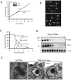
FIG. 1.
FIG. 1.
Effects of p38 RNAi. (A) Effect of RNAi (induced on day 0) on cell growth. The number of parasites/milliliter on the y axis is the measured value times the dilution factor. (Inset) Northern blot showing the effect of RNAi (40 h) on p38 mRNA level. Ethidium bromide-stained rRNA is a loading control. The slight increase in cell density at day 11 is due to recovery from RNAi. (B) Effect of RNAi on kinetoplast size. DAPI staining shows kinetoplasts from cells without RNAi or after 6 days of RNAi. n, nucleus; k, kinetoplast. Bar, 5 μm. (C) Kinetics of kDNA loss as determined by visual analysis of images like those in panel B (>300 randomly selected cells were analyzed each day). The increase in normal-size kDNA network at days 7 and 8 is due to recovery from RNAi. (D) Kinetics of kDNA loss determined by Southern blotting of total minicircles and maxicircles. After digestion with HindIII/XbaI, total cellular DNA (5 × 10⁵ cell equivalents/lane) was fractionated on a 1.5% agarose gel containing ethidium bromide. Southern blots were probed for a maxicircle fragment (Maxi), a minicircle fragment (Mini), and a hexose transporter fragment as a loading control (Load). The 1-kb minicircle species was the dominant band in the digestion product of the heterogeneous minicircles; smaller fragments disappeared at the same rate as the 1-kb fragment. The positions of 1.4- and 1.0-kb molecular size markers are shown to the left of the gels. (E) EM of cells without RNAi or after 4 days of RNAi. k, kinetoplast, b.b., basal body. Bar, 200 nm.
FIG. 2.
FIG. 2.
Localization of p38-GFP. Live cells stably expressing p38-GFP fusion protein were stained with DAPI and analyzed by fluorescence microscopy. (Inset) Enlargement of merged picture showing localization of p38-GFP. Bar, 2 μm.
FIG. 3.
FIG. 3.
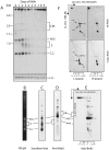
Effect of RNAi on free minicircle replication intermediates. (A) Total DNA (106 cell equivalents/lane) was fractionated on a 1.5% agarose gel in TBE buffer (in this experiment, the gel but not the running buffer contained 1 μg/ml ethidium bromide). After Southern blotting, free minicircles were detected by hybridization with a pJN6 probe and autoradiography. The reappearance of free minicircles at day 9 is due to recovery from RNAi. The positions of molecular size markers (in kilobases) are shown to the left of the gels. (B to E) Cells (after 2 days of RNAi) were labeled for 40 min with 50 μM BrdU. Free minicircles from a 500-ml culture were used for the single lane in the gels shown in panels B, D, and E. (B) Ethidium bromide (EB) staining of total free minicircles. (C) Southern blot (106 cell equivalents/lane) of total free minicircles. (D and E) BrdU label was detected by probing with anti-BrdU antibody. 1st Dim. (TBE-EB), first dimension (TBE-ethidium bromide); 2nd dim., second dimension; a, genomic DNA; b and c, catenated minicircles; d, θ-structures; e, gapped/nicked minicircles; f, knotted minicircles; g, branch migration products from θ structures. (F) Total DNA from 3 × 10⁷ uninduced (no RNAi) or induced cells (4 days of RNAi) were fractionated on a two-dimensional gel. Strand-specific hybridizations were conducted with synthetic oligonucleotide probes. The scales below the panels indicate the sizes of linear markers in the second dimension. θ, θ-structure; S, fraction S; ccD, covalently closed dimer; N/G, nicked/gapped minicircles; C.C., covalently closed minicircles; C.M., catenated minicircles; M.G., multiply gapped minicircles; L.C., loading control.
FIG. 4.
FIG. 4.
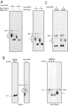
Analysis of fraction S. Samples (gel purified from 1 × 10⁷ to 2 × 10⁷ cells) were run on a 1.5% agarose gel with 1 μg/ml ethidium bromide (unless otherwise indicated), and minicircle species were detected on a Southern blot. (A) Effects of topoisomerases. Gel-purified fraction S (after 2 or 4 days of RNAi) was treated with (+) or without (−) human topoisomerase II, wheat germ topoisomerase I, or E. coli topoisomerase I. H.s. Topo II, Homo sapiens topoisomerase II; W.g. Topo I, wheat germ topoisomerase I; E.c. Topo I, E. coli topoisomerase I. (B) Comparison of electrophoretic mobility of gyrase-treated minicircles with fraction S (the left-hand panels show gels with ethidium bromide [EB gel], and the right-hand panel shows a gel without ethidium bromide [Non-EB gel]). (C) Immunoprecipitation of fraction S. Total minicircles (T) were treated with anti-Z-DNA antibody and then with protein G-Sepharose FF. Minicircles were from the supernatant (S) and pellet (P). The positions of nicked/gapped minicircles (N/G), fraction S (S), and covalently closed minicircles (C.C.) are shown at the sides of the gels.
FIG. 5.
FIG. 5.
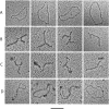
EM of minicircles. (A) Relaxed minicircles; (B) fraction S minicircles; (C) fraction S bound to anti-Z-DNA antibody; (D) fraction S bound to SSB. Bar, 50 nm.
FIG. 6.
FIG. 6.
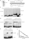
DNA binding assays (EMSA) using recombinant GST-p38. Samples were analyzed by nondenaturing 5% PAGE and autoradiography. (A) SDS-PAGE of purified GST-p38. The positions of molecular mass markers (in kilodaltons) are shown to the left of the gel. (B) Map of the conserved region of minicircles. UMS and HEX are the start sites for the leading and lagging strands (dashed lines). Solid lines with arrowheads represent single-stranded oligonucleotides used for EMSA. (C) The oligonucleotides and sequences are as follows: a, 5′-GTATTACACCAACCCCTTTCGAAGTACCTCGGACC-3′; a′, 5′-TTTGAGGTCCGAGGTACTTCGAAAGGGGTTGGTGTAAT-3′(UMS underlined); b, 5′-AAAAATGCCCCAAAATAGCACGGGATTTGTGTATG-3′; b′, 5′-CATACACAAATCCCGTGCTATTTTGGGGCATTTTT-3′; c, 5′-GGGATTTGTGTATGTGTATTTTTGCACGCCCTTAAGATTT-3′(hexamer underlined); c′, 5′-AAATCTTAAGGGCGTGCAAAAATACACATACACAAATCCC-3′; d, 5′-AGCATCTGGCTTACTGAAGCAGACCCTATCATCT-3′; e, dsDNA produced by annealing oligonucleotides a and a′; f, dsDNA produced by annealing oligonucleotides c and j (5′ TTAAATCTTAAGGGCGTGCAAAAATACACATACAC-3′); g, 5′-GGGGTTGGTGTA-3′; h, 5′-TTGCACGCCCTTAA-3′; and i, 5′-TGTGTGTGTGTGTGTGTGTGTGTGTGTGTG-3′. Oligonucleotides were 5′ labeled with polynucleotide kinase except for oligonucleotides e and f, which were labeled with [α-32P]dATP by filling in the ends using Klenow fragment. For EMSA, GST-p38 (0.8 pmol/assay) was used for oligonucleotides a, a′, b, b′, c, c′, d, and i (10 fmol/assay); GST-p38 (3.2 pmol, 6.4 pmol/assay) were used for oligonucleotides e and f (20 fmol/assay); GST-p38 (3.1 pmol/assay) were used for oligonucleotides g and h (20 fmol/assay). (D) Competition experiments. In these experiments, 0.8 pmol GST-p38 was used for each assay. Lane 1, oligonucleotide a′ or c only; lane 2, oligonucleotide a′ or c and GST (1.9 pmol); lane 3, oligonucleotide a′ or c and GST-p38; lanes 4 to 13, GST-p38 incubated with 32P-labeled oligonucleotide a′ or c (10 fmol/assay) in the presence of increasing concentrations of unlabeled competitors d, a′, or c (5-, 10-, 20-, 50-, and 100-fold). (E) Increasing amounts of GST-p38 (0, 0.2, 0.4, 0.8, 1.6, 3.2, and 6.4 pmol) were incubated with 32P-labeled oligonucleotide a′ (lanes 1 to 7) or c (lanes 8 to 14). (F) Scatchard plot (17) of binding of oligonucleotide a′ (containing UMS) and oligonucleotide c (containing hexamer). Binding reaction mixtures contained 3.1 pmol of GST-p38. The intercepts on the x axis were 700 pM for oligonucleotide a′ and 510 pM for oligonucleotide c. These values indicate that only about 0.4% of the GST-p38 protein was active in binding.
FIG. 7.
FIG. 7.
Model for p38's function in initiation of minicircle replication. (A) In normal replication (with p38 present), covalently closed free minicircle is partially unwound by helicase/topoisomerase, allowing synthesis of the leading and lagging strands. (B) When p38 is depleted by RNAi, replication cannot initiate, but unwinding of the parental strands still proceeds. Presumably the single strands are bound by SSB. When the DNA is deproteinized, the single strands wind up, introducing compensating negative supertwists and a region of left-handed Z-DNA. Topo I or II, topoisomerase I or II.

Similar articles

Cited by

References

    1. Abu-Elneel, K., D. R. Robinson, M. E. Drew, P. T. Englund, and J. Shlomai. 2001. Intramitochondrial localization of universal minicircle sequence-binding protein, a trypanosomatid protein that binds kinetoplast minicircle replication origins. J. Cell Biol. 153:725-734. - PMC - PubMed
    1. Brun, R., and M. Schonenberger. 1979. Cultivation and in vitro cloning of procyclic culture forms of Trypanosoma brucei in a semi-defined medium. Acta Trop. 36:289-292. - PubMed
    1. Downey, N., J. C. Hines, K. M. Sinha, and D. S. Ray. 2005. Mitochondrial DNA ligases of Trypanosoma brucei. Eukaryot. Cell 4:765-774. - PMC - PubMed
    1. Drew, M. E., and P. T. Englund. 2001. Intramitochondrial location and dynamics of Crithidia fasciculata kinetoplast minicircle replication intermediates. J. Cell Biol. 153:735-744. - PMC - PubMed
    1. Droge, P., J. M. Sogo, and H. Stahl. 1985. Inhibition of DNA synthesis by aphidicolin induces supercoiling in simian virus 40 replicative intermediates. EMBO J. 4:3241-3246. - PMC - PubMed

Publication types

MeSH terms

LinkOut - more resources