. 2006 Jul;26(14):5394-405.
doi: 10.1128/MCB.00519-06.

The X and Y chromosomes assemble into H2A.Z-containing [corrected] facultative heterochromatin [corrected] following meiosis

Affiliations

The X and Y chromosomes assemble into H2A.Z-containing [corrected] facultative heterochromatin [corrected] following meiosis

Ian K Greaves et al. Mol Cell Biol. 2006 Jul.

Erratum in

  • Mol Cell Biol. 2006 Oct;26(19):7343

Abstract

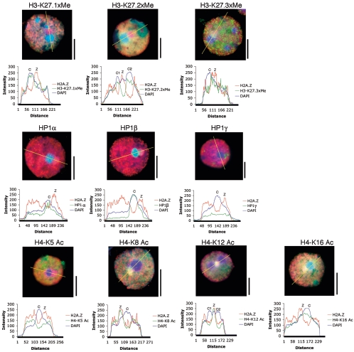
Spermatogenesis is a complex sequential process that converts mitotically dividing spermatogonia stem cells into differentiated haploid spermatozoa. Not surprisingly, this process involves dramatic nuclear and chromatin restructuring events, but the nature of these changes are poorly understood. Here, we linked the appearance and nuclear localization of the essential histone variant H2A.Z with key steps during mouse spermatogenesis. H2A.Z cannot be detected during the early stages of spermatogenesis, when the bulk of X-linked genes are transcribed, but its expression begins to increase at pachytene, when meiotic sex chromosome inactivation (MSCI) occurs, peaking at the round spermatid stage. Strikingly, when H2A.Z is present, there is a dynamic nuclear relocalization of heterochromatic marks (HP1beta and H3 di- and tri-methyl K9), which become concentrated at chromocenters and the inactive XY body, implying that H2A.Z may substitute for the function of these marks in euchromatin. We also show that the X and the Y chromosome are assembled into facultative heterochromatic structures postmeiotically that are enriched with H2A.Z, thereby replacing macroH2A. This indicates that XY silencing continues following MSCI. These results provide new insights into the large-scale changes in the composition and organization of chromatin associated with spermatogenesis and argue that H2A.Z has a unique role in maintaining sex chromosomes in a repressed state.


Figures

FIG. 1.
FIG. 1.
H2A.Z is first observed at the pachytene cell stage. Mouse seminiferous tubules were stained with propidium iodide (PI) and immunostained with H2A.Z (A and B) or H2A (C) affinity-purified antibodies. For the merged panel, the first and second panels are artificially colored red and green, respectively. Bar, 50 μm. (D) To determine at which stage of meiotic prophase H2A.Z becomes apparent, meiotic surface spread spermatocytes were stained with an H2A.Z antibody (red) and an antibody to the synaptonemal complex marker SCP3 (green). Also shown is an outline highlighting the basic steps during spermatogenesis. Colored arrows highlighting the different cell types are shown and used in the merged panel to illustrate the location of the major cell types in the seminiferous tubule. Bar, 10 μm.
FIG. 2.
FIG. 2.
H2A.Z protein expression peaks at the round spermatid stage. (A) The amount of indirect fluorescence generated by affinity-purified H2A.Z antibodies was determined and quantitated for germ cells at different stages of spermatogenesis, as described in Materials and Methods. To take into account changes in fluorescence intensity due to chromatin compaction, the mean ratio of H2A.Z fluorescence to DAPI staining was determined. (B) Transcript levels of H2A.Z, adenine phosphoribosyltransferase (APRT), and Y-linked zinc finger protein (ZFY) from 9-, 17-, and 30-day-old mice were determined by quantitative real-time PCR using glyceraldehyde phosphate dehydrogenase as an internal control.
FIG. 3.
FIG. 3.
Dynamic nuclear relocalization of di- and trimethylated H3 K9 when H2A.Z appears at pachytene/diplotene. Surface spread leptotene/zygotene and pachytene cells were stained with DAPI and immunostained with H2A.Z and H3 methylated K9 antibodies (A) or macroH2A antibodies (B) as shown. For the merged panel, the second and third panels are artificially colored red and green, respectively. White arrows indicate the location of the sex vesicle (SV). Shown are the paths used to determine fluorescence intensity in pachytene/diplotene nuclei. Bar, 10 μm. The scales are in arbitrary units.
FIG. 4.
FIG. 4.
HP1α and -β accumulate in constitutive heterochromatic domains at pachytene/diplotene. Surface spread leptotene/zygotene and pachytene cells were stained with DAPI and immunostained with H2A.Z, as well as HP1α, HP1β, or HP1γ antibodies, as shown. For the merged panel, the second and third panels are artificially colored red and green, respectively. White arrows show the location of the sex vesicle (SV). Shown are the paths used to determine fluorescence intensity in pachytene/diplotene nuclei. Bar, 10 μm.
FIG. 5.
FIG. 5.
Enrichment of histone H3 tri-methyl K27 at pachytene/diplotene. Surface spread leptotene/zygotene and pachytene cells were stained with DAPI and immunostained with H2A.Z and histone H3 methylated K27 antibodies as shown. For the merged panel, the second and third panels are artificially colored red and green, respectively. White arrows show the location of the sex vesicle (SV). Shown are the paths used to determine fluorescence intensity in pachytene/diplotene nuclei. Bar, 10 μm.
FIG. 6.
FIG. 6.
A unique chromosomal domain enriched H2A.Z. (A) Surface spread round and elongated spermatid cells were stained with DAPI and immunostained with H2A.Z (red) and histone H3 methylated K9 antibodies (green) as shown. (B) Round spermatids immunostained with antibodies against H2A.Z (red) and a phosphorylated form of RNA polymerase II (green). (C) Round spermatids were stained with DAPI and immunostained with H2A.Z (red) and macroH2A (green) antibodies. (D) Round spermatids were stained with propidium iodide (red) and immunostained with H2A (green) antibodies. White arrows show the location of the H2A.Z enriched domain. Shown are the paths used to determine fluorescence intensity in round spermatid nuclei. C, chromocenter; Z, the enriched H2A.Z domain next to the chromocenter. Bar, 10 μm.
FIG. 7.
FIG. 7.
Histone modification status of the distinct H2A.Z chromosomal domain. Round spermatids were immunostained with a battery of different antibodies as shown. The intensity of fluorescence for each antibody following a linear path across the nucleus are shown and plotted. C, chromocenter; Z, the enriched H2A.Z domain next to the chromocenter. Bar, 10 μm.
FIG. 8.
FIG. 8.
The unique chromosomal domain enriched with H2A.Z are the sex chromosomes. FISH analyses were performed on round spermatids using specific chromosome X and Y paints. White arrows indicate the location of the H2A.Z enriched domain. Shown are the paths used to determine fluorescence intensity. C, chromocenter; Z, the enriched H2A.Z domain next to the chromocenter. Bar, 10 μm.

Similar articles

Cited by

References

    1. Abbott, D. W., M. Laszczak, J. D. Lewis, H. Su, S. C. Moore, M. Hills, S. Dimitrov, and J. Ausio. 2004. Structural characterization of macroH2A containing chromatin. Biochemistry 43:1352-1359. - PubMed
    1. Armstrong, S. J., M. A. Hulten, A. M. Keohane, and B. M. Turner. 1997. Different strategies of X-inactivation in germinal and somatic cells: histone H4 underacetylation does not mark the inactive X chromosome in the mouse male germline. Exp. Cell Res. 230:399-402. - PubMed
    1. Bruce, K., F. A. Myers, E. Mantouvalou, P. Lefevre, I. Greaves, C. Bonifer, D. J. Tremethick, A. W. Thorne, and C. Crane-Robinson. 2005. The replacement histone H2A.Z in a hyperacetylated form is a feature of active genes in the chicken. Nucleic Acids Res. 33:5633-5639. - PMC - PubMed
    1. Bulynko, Y., L. C. Hsing, R. W. Mason, D. J. Tremethick, and S. A. Grigoryev. 2006. Cathepsin L stabilizes histone modification landscape on Y chromosome and pericentric heterochromatin. Mol. Cell. Biol. 26:4172-4184. - PMC - PubMed
    1. Byrne, J. A., S. Simonsson, P. S. Western, and J. B. Gurdon. 2003. Nuclei of adult mammalian somatic cells are directly reprogrammed to oct-4 stem cell gene expression by amphibian oocytes. Curr. Biol. 13:1206-1213. - PubMed

Publication types

MeSH terms

LinkOut - more resources