. 2006 Jul;26(14):5518-27.
doi: 10.1128/MCB.00625-06.

Constitutive ALK5-independent c-Jun N-terminal kinase activation contributes to endothelin-1 overexpression in pulmonary fibrosis: evidence of an autocrine endothelin loop operating through the endothelin A and B receptors

Affiliations

Constitutive ALK5-independent c-Jun N-terminal kinase activation contributes to endothelin-1 overexpression in pulmonary fibrosis: evidence of an autocrine endothelin loop operating through the endothelin A and B receptors

Xu Shi-Wen et al. Mol Cell Biol. 2006 Jul.

Abstract

The signal transduction mechanisms generating pathological fibrosis are almost wholly unknown. Endothelin-1 (ET-1), which is up-regulated during tissue repair and fibrosis, induces lung fibroblasts to produce and contract extracellular matrix. Lung fibroblasts isolated from scleroderma patients with chronic pulmonary fibrosis produce elevated levels of ET-1, which contribute to the persistent fibrotic phenotype of these cells. Transforming growth factor beta (TGF-beta) induces fibroblasts to produce and contract matrix. In this report, we show that TGF-beta induces ET-1 in normal and fibrotic lung fibroblasts in a Smad-independent ALK5/c-Jun N-terminal kinase (JNK)/Ap-1-dependent fashion. ET-1 induces JNK through TAK1. Fibrotic lung fibroblasts display constitutive JNK activation, which was reduced by the dual ETA/ETB receptor inhibitor, bosentan, providing evidence of an autocrine endothelin loop. Thus, ET-1 and TGF-beta are likely to cooperate in the pathogenesis of pulmonary fibrosis. As elevated JNK activation in fibrotic lung fibroblasts contributes to the persistence of the myofibroblast phenotype in pulmonary fibrosis by promoting an autocrine ET-1 loop, targeting the ETA and ETB receptors or constitutive JNK activation by fibrotic lung fibroblasts is likely to be of benefit in combating chronic pulmonary fibrosis.


Figures

FIG. 1.
FIG. 1.
TGF-β induces ET-1 production in primary human normal lung fibroblasts. (A) TGF-β induces ET-1 protein. Normal lung fibroblasts were cultured and treated with or without TGF-β1 (4 ng/ml) as described in Materials and Methods. Conditioned media were analyzed for ET-1 production using a specific ET-1 ELISA. TGF-β1 significantly (P < 0.05) induced ET-1 protein. Data for an average of six wells ± standard deviation are shown. (B) TGF-β induces the ET-1 promoter. Normal lung fibroblasts were transfected with an ET-1 promoter/luciferase reporter construct and treated with or without TGF-β1 (4 ng/ml) as described in Materials and Methods. Cell layers were analyzed for luciferase expression and adjusted for differences in transfection efficiency between samples revealed by β-galactosidase expression produced by a cotransfected construct containing a cytomegalovirus promoter-driven β-galactosidase gene. TGF-β1 significantly (P < 0.05) induced ET-1 promoter activity. Data for an average of six wells ± standard deviation in three independent experiments are shown.
FIG. 2.
FIG. 2.
TGF-β induces the ET-1 promoter in primary human normal lung fibroblasts in a Smad-independent JNK/Ap-1-dependent fashion. (A) Effect of mutating the Ap-1 or the Smad element of the ET-1 promoter on TGF-β induction of ET-1. Normal lung fibroblasts were transfected with a wild-type ET-1 promoter/luciferase reporter construct (WT) or an otherwise identical construct containing mutated Smad (Smad mut) or Ap-1 (Ap-1 mut) elements. These elements were previously shown to be functional in endothelial cells (43). Mutation of the Ap-1 element, but not the Smad element, significantly (P < 0.05) reduces the TGF-β1 induction of the ET-1 promoter. Data for an average of six wells ± standard deviation in three independent experiments are shown. (B) Effect of inhibition of Smad or c-jun/Ap-1 pathways on TGF-β1-induced ET promoter activity. A wild-type ET-1 promoter/luciferase reporter construct (WT) was transfected into normal lung fibroblasts with or without empty expression vector, expression vectors encoding Smad 7 or TAM-67 (dominant negative c-jun [dn c-jun]) or 10 μM JNK inhibitor SP-600125. Cells were treated with or without TGF-β1 (4 ng/ml). Inhibition of JNK/c-jun, but not Smad, signaling significantly (P < 0.05) reduced the TGF-β1 induction of the ET-1 promoter. As a control, a generic Smad3-responsive promoter (SBE-lux) was transfected into fibroblasts. Induction of this promoter by TGF-β1 was sensitive to overexpression of Smad7. Data for an average of six wells ± standard deviation in three independent experiments are shown. (C) Effect of loss of Smad3 expression on TGF-β1-induced ET-promoter activity. The wild-type ET-1 promoter containing a Smad3 recognition sequence functionally operative in endothelial cells (WT) (43) was transfected into Smad3+/+ or Smad3/ embryonic fibroblasts (Smad3 WT or Smad3 KO, respectively) and treated with or without TGF-β1 (4 ng/ml). By using these cells, Smad3 has previously been shown to be the Smad operating in driving Smad binding element (SBE)-dependent transcriptional activation in fibroblasts and induction of TGF-β/Smad-dependent protein expression (25, 41). Loss of Smad3 did not affect the TGF-β1 induction of the ET-1 promoter, but the induction of SBE-lux was impaired. Data for an average of six wells ± standard deviation in three independent experiments are shown. (D) Effect of bosentan treatment on TGF-β induction of α-SMA. Fibroblasts from three normal individuals (N1, N2, and N3) were treated with or without ET-1 (100 nM) or TGF-β1 (4 ng/ml) as indicated. Twenty-four hours later, cell extracts were prepared and subjected to Western blot analysis with the indicated antibodies.
FIG. 3.
FIG. 3.
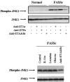
Fibrotic lung fibroblasts isolated from patients with lung involvement in scleroderma (FASSc) display elevated, constitutive JNK1 activation that is ALK5 independent. Normal and FASSc fibrotic lung fibroblasts were serum starved and treated with or without TGF-β1 (4 ng/ml) for the durations indicated. Cell layers were harvested and subjected to sodium dodecyl sulfate-polyacrylamide gel electrophoresis and Western blot analysis with anti-phospho-JNK1 and anti-JNK1 antibodies. Cells were also treated in the presence and absence of the ALK5 TGF-β type I receptor inhibitor SB 431542 (26). Whereas TGF-β1 induces JNK1 phosphorylation in an ALK5-dependent fashion, FASSc fibroblasts display elevated, ALK5-independent JNK phosphorylation.
FIG. 4.
FIG. 4.
Nuclear extracts from FASSc lung fibroblasts display elevated levels of Ap-1 binding activity. (A) FASSc nuclear extracts contain elevated Ap1 binding activity. A radiolabeled probe containing the consensus Ap-1 element of the ET-1 promoter (43) was incubated with nuclear extracts prepared from pulmonary fibroblasts isolated from two normal individuals (N1 and N2) or two individuals with pulmonary fibrosis associated with scleroderma (S1 and S2). Cells were treated in the presence or absence of TGF-β1 for 30 min prior to harvesting. TGF-β induces binding to the Ap-1 element of the ET-1 promoter; extracts from FASSc cells show elevated endogenous Ap-1 binding activity. To verify that protein binding was due to Ap-1 binding activity, protein binding to DNA was competed by addition of a 100-fold molar excess of cold oligomer bearing a consensus Ap-1 site, but not by oligomers containing consensus Ap-2 or Sp-1 sites. (B) Transcription factor c-jun binds the Ap1 element in the ET-1 promoter. Preincubation of extracts for 1 h with anti-c-jun, but not anti-HuR (control), reduced factor binding to probe, indicating that c-jun was required for protein/DNA complex formation. Individuals N2 and S2 from panel A were examined.
FIG. 5.
FIG. 5.
FASSc lung fibroblasts show elevated ET-1 expression in a Smad-independent c-jun/JNK-dependent fashion. (A) FASSc lung fibroblasts (LF) possess elevated levels of ET-1 mRNA. Normal and FASSc lung fibroblasts were cultured to confluence and treated for 18 h in the presence or absence of the dual ETA/ETB receptor antagonist bosentan. Cells were cultured from four normal individuals and four individuals with FASSc. Total RNA was harvested, reverse transcribed into cDNA, and subjected to real-time PCR analysis with primers recognizing human ET-1 and 28S RNA. Ratios of ET-1/28S RNA levels ± standard deviations are shown (n = 3). Note that in the presence of bosentan, the elevated level of ET-1 mRNA in FASSc fibroblasts is markedly reduced. (B) Elevated expression of ET-1 is JNK/c-jun dependent. Normal and FASSc lung fibroblasts were transfected with a wild-type ET-1 promoter/luciferase reporter construct (WT) or an otherwise identical construct containing mutated Smad (Smad mut) or Ap-1 (Ap-1 mut) elements. Mutation of the Ap-1 element, but not the Smad element, significantly (P < 0.05) reduced the TGF-β1-induction of the ET-1 promoter. Similarly, addition of the JNK inhibitor SP-600125 (SP; 10 μM) reduced ET-1 promoter activity in FASSc, but not normal, lung fibroblasts. Data for an average of six wells ± standard deviation in three independent experiments are shown.
FIG. 6.
FIG. 6.
ALK5 is not required for the overexpression of ET-1 in FASSc lung fibroblasts. Normal and FASSc lung fibroblasts were transfected with an ET-1 promoter/luciferase reporter construct and treated in the presence or absence of TGF-β1 (4 ng/ml) for 24 h. Cells were treated in the presence or absence of the ALK5 inhibitor SB-431542 or the JNK inhibitor SP600125. Whereas ALK5 inhibition blocked the TGF-β induction of the ET-1 promoter in FASSc lung fibroblasts (P < 0.05; compared by Student's t test to TGF-β-treated sample), ALK5 inhibition did not significantly decrease the elevated basal ET-1 promoter activity in FASSc lung fibroblasts (P < 0.05; compared by Student's t test to basal ET-1 promoter activity in FASSc fibroblasts).
FIG. 7.
FIG. 7.
ET-1 induces JNK1 phosphorylation and α-SMA production through TAK1. Wild-type fibroblasts (TAK WT) or fibroblasts in which TAK1 was deleted (TAK KO) (47) were treated with or without recombinant ET-1 (100 nM) for 30 min (for phospho-JNK1 blots) or 24 h (for α-SMA blots). Cell layers were harvested and subjected to Western blot analysis with the antibodies indicated. It is notable that ET-1 did not induce JNK1 phosphorylation or induce α-SMA expression in TAK1 knockout fibroblasts, and basal α-SMA expression was lower in TAK1 knockout fibroblasts.
FIG. 8.
FIG. 8.
An autocrine ET loop results in the elevated JNK1 activation observed in FASSc lung fibroblasts. Scleroderma lung fibroblasts were treated with the PDGF receptor inhibitor Gleevec, the angiotensin II receptor inhibitor losartan, a soluble IL-1 receptor antagonist (IL-1ra), the dual ETA/ETB receptor inhibitor bosentan (Anti-ETAr&Br), the ETA receptor inhibitor PD156707 (Anti-ETAr), or the ETB receptor inhibitor BQ788 (Anti-ETBr). Cell extracts were prepared and subjected to Western blot analyses with the indicated antibodies. Inhibition of autocrine ET signaling reduced the activation of JNK1 in FASSc fibroblasts to that of normal fibroblasts. It is notable that bosentan (Anti-ETAr&Br) treatment did not appreciably affect basal JNK1 activation in normal fibroblasts.
FIG. 9.
FIG. 9.
JNK inhibition reduces overexpression of ET-1 and α-SMA by FASSc lung fibroblasts. Scleroderma lung fibroblasts were treated with the JNK inhibitor SP600125 for 24 h. Cell extracts were harvested and subjected to Western blot analyses with the antibodies indicated. Lung fibroblasts from three normal individuals (N1, N2, and N3) and three individuals with FASSc (S1, S2, and S3) were examined. SP600125 inhibits α-SMA production by FASSc fibroblasts. Similarly, conditioned media from identical experiments were pooled and subjected to ELISA to detect ET-1 levels.
FIG. 10.
FIG. 10.
Schematic diagram of autocrine ET signaling, TGF-β, and JNK contribution to overexpression of ET-1 in fibrotic FASSc lung fibroblasts and hence to fibrosis. Constitutively activated ET-dependent JNK activates the ET-1 promoter in an ALK5-independent fashion. Exogenous TGF-β induces the ET-1 promoter through JNK, leading to additional production of ET-1. TGF-β also directly induces profibrotic proteins (31). These events result in enhanced production of profibrotic proteins and ECM production, remodeling, and contraction.

Similar articles

Cited by

References

    1. Abraham, D. J., R. Vancheeswaran, M. R. Dashwood, P. Pantelides, W. Shi-Wen, R. M. du Bois, and C. M. Black. 1997. Increased levels of endothelin-1 and differential endothelin type A and B receptor expression in scleroderma-associated fibrotic lung disease. Am. J. Pathol. 151:831-841. - PMC - PubMed
    1. Abraham, D. J., X. Shiwen, C. M. Black, S. Sa, Y. Xu, and A. Leask. 2000. Tumor necrosis factor alpha suppresses the induction of connective tissue growth factor by transforming growth factor-beta in normal and scleroderma fibroblasts. J. Biol. Chem. 275:15220-15225. - PubMed
    1. Anggard, E. E., R. M. Botting, and J. R. Vane. 1990. Endothelins. Blood Vessels 27:269-281. - PubMed
    1. Asano, Y., H. Ihn, K. Yamane, M. Kubo, and K. Tamaki. 2004. Impaired Smad7-Smurf-mediated negative regulation of TGF-beta signaling in scleroderma fibroblasts. J. Clin. Investig. 113:253-264. - PMC - PubMed
    1. Badylak, S. F. 2002. The extracellular matrix as a scaffold for tissue reconstruction. Semin. Cell Dev. Biol. 13:377-383. - PubMed

Publication types

MeSH terms

Substances