Comparative Study
. 2006 Jul;105(1):160-8.
doi: 10.1097/00000542-200607000-00026.

Painful peripheral nerve injury decreases calcium current in axotomized sensory neurons

Affiliations
Comparative Study

Painful peripheral nerve injury decreases calcium current in axotomized sensory neurons

J Bruce McCallum et al. Anesthesiology. 2006 Jul.

Abstract

Background: Reports of Ca(2+) current I(Ca) loss after injury to peripheral sensory neurons do not discriminate between axotomized and spared neurons. The spinal nerve ligation model separates axotomized from spared neurons innervating the same site. The authors hypothesized that I(Ca) loss is a result of neuronal injury, so they compared axotomized L5 dorsal root ganglion neurons to spared L4 neurons, as well as neurons from rats undergoing skin incision alone.

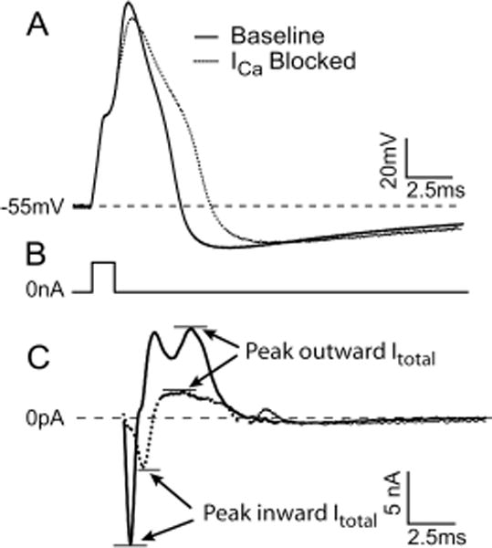
Methods: After behavioral testing, dissociated neurons from L4 and L5 dorsal root ganglia were studied in both current and voltage patch clamp modes. The biophysical consequence of I(Ca) loss on the action potential was confirmed using selective I(Ca) antagonists. Data were grouped into small, medium, and large cells for comparison.

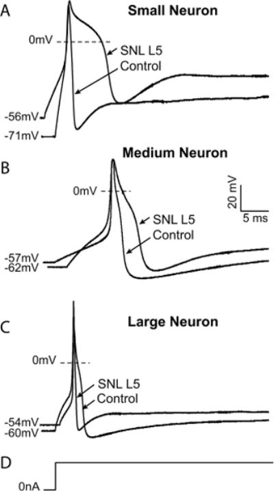
Results: Reduced I(Ca) was predominantly a consequence of axotomy (L5 after spinal nerve ligation) and was most evident in small and medium neurons. ICa losses were associated with action potential prolongation in small and medium cells, whereas the amplitude and duration of after hyperpolarization was reduced in medium and large neurons. Blockade with Ca(2+) channel antagonists showed that action potential prolongation and after hyperpolarization diminution were alike, attributable to the loss of I(Ca).

Conclusion: Axotomy is required for I(Ca) loss. I(Ca) loss correlated with changes in the biophysical properties of sensory neuron membranes during action potential generation, which were due to I(Ca) loss leading to decreased outward Ca(2+)-sensitive K currents. Taken together, these results suggest that neuropathic pain may be mediated, in part, by loss of I(Ca) and the cellular processes dependent on Ca(2+).


Figures

Fig. 1
Fig. 1
(A) Measures of action potential (AP) produced by 1-s current injection, shown below the voltage trace. An inflection on the descending limb of the AP is demonstrated. Threshold for initiation of the AP. RMP = resting membrane potential; AP50 = AP duration at 50% of peak. (B) Determination of afterhyperpolarization (AHP) dimensions after an AP initiated by a 2-ms current injection, shown below the voltage trace. AHP amplitude is measured from RMP to peak. AHP50 = AHP duration at 50% recovery; AHPτ = time constant for AHP recovery. (C) Measures of Ca2+ current (ICa) response to voltage command in the form of an AP waveform. Inward ICa is measured at peak and as the integral of total inward current (Ca2+ charge transfer) during AP waveform.
Fig. 2
Fig. 2
Exemplary traces showing effects of injury on action potential dimensions in control neurons and after ligation and axotomy of the fifth lumbar spinal nerve (SNL L5). (A) A typical small neuron displays prolonged action potential duration and a more depolarized resting membrane potential after injury. (B) A medium neuron exhibits prolonged action potential as well as decreased afterhyperpolarization amplitude and accelerated recovery of the afterhyperpolarization after spinal nerve ligation. (C) A large neuron develops decreased afterhyperpolarization amplitude and duration. (D) The stimulus in each case is a sustained current pulse of an amplitude just adequate for action potential generation. Scale bars apply to all traces.
Fig. 3
Fig. 3
Voltage clamp data comparing paired sensory neurons from the L4 and L5 levels after spinal nerve ligation (SNL) and neurons from control animals, including large and medium groups. No differences were discerned in large cells (A). Medium cells demonstrated significant difference in maximum Ca2+ conductance (Gmax) in response to 200-ms, 10-mV, stepwise, square wave pulses (B), and in peak amplitude (C) and total charge transfer (D) in response to a standardized action potential waveform. n = 7–13 neurons for each group. Peak current is normalized for cell capacitance; charge transfer is normalized for cell surface area. § P < 0.05 versus both L4 and control, by Kruskal-Wallis nonparametric analysis of variance with Dunn multiple comparison test for post hoc differences.
Fig. 4
Fig. 4
Correlations between neuronal Ca2+ conductance (Gmax) and action potential (AP) parameters. (A) Decreased Gmax after injury correlated with prolonged AP duration (AP50) in L5 dorsal root ganglia neurons of small cells (Pearson correlation r = −0.7, P = 0.0006), whereas Gmax and AP duration among neurons from control rats were not related (r = 0.3, P = 0.232). (B) AP duration positively correlated with Gmax in large cells from L4 and L5 dorsal root ganglia before injury (r = 0.78 ± 0.2, P = 0.0011), and this positive effect of increased Gmax on AP duration was not affected by injury in neurons from L5 ganglia (r = 0.8 ± 0.2, P = 0.0055). (C) Decreased Gmax after injury correlated to decreased afterhyperpolarization amplitude (AHPampl) in large neurons (r = −0.6, P = 0.0259), whereas changes in Gmax did not predict changes in AHPampl in neurons from control rats (r = −0.09, P = 0.7249). SNL = spinal nerve ligation.
Fig. 5
Fig. 5
Action potential parameters and whole cell currents (Itotal) before and after Ca2+ channel (ICa) blockade. (A) Action potential elicited in response to a 3-nA, 1-ms pulse. (B) The stimulus artifact is removed for clarity. (C) Corresponding whole cell currents in response to the same action potential used as a voltage command. Reduction of depolarization rate after Ca2+ channel blockade is due to reduced inward Itotal, whereas action potential prolongation is mediated by delayed repolarization due to reduced outward Itotal. See table 2 for summary of results. Blockade cocktail included 200 nm SNX-111, 200 nm ω-Aga IVA, 200 nm SNX-482, and 200 nm nisoldipine.
Fig. 6
Fig. 6
Schematic diagram of reduced Ca2+ current (ICa) contribution to neuropathic pain. Injured nerve tissue distal to the spinal nerve ligation (SNL) undergoes wallerian degeneration (dotted line), while neurons from the L5 dorsal root ganglion (DRG) are activated by movement, catecholamines, other algogenic agents and cross-excitation from adjacent intact neurons. Reduced ICa in Aα/Aß and Aδ fibers shortens afterhyperpolarizations (AHPs), which contributes to burst firing. Loss of ICa impairs natural signal filtering at the T-branch, where the stem axon splits into spinal nerve and dorsal root branches. Reduced ICa also prolongs action potential (AP) duration, which may increase excitatory synaptic transmission in the dorsal horn (DH). Spared nerves transmit activity evoked by stimulation in the receptive field, and these signals encounter DH neurons that are sensitized by L5 input. L4, L5 = neurons in the fourth and fifth lumbar ganglia.

References

    1. Hogan Q. Animal pain models. Reg Anesth Pain Med. 2002;27:385–401. - PubMed
    1. Elmslie KS. Calcium channel blockers in the treatment of disease. J Neurosci Res. 2004;75:733–41. - PubMed
    1. Bennett GJ, Xie YK. A peripheral mononeuropathy in rat that produces disorders of pain sensation like those seen in man. Pain. 1988;33:87–107. - PubMed
    1. Kim SH, Chung JM. An experimental model for peripheral neuropathy produced by segmental spinal nerve ligation in the rat. Pain. 1992;50:355–63. - PubMed
    1. Sommer C, Schafers M. Painful mononeuropathy in C57BL/Wld mice with delayed wallerian degeneration: differential effects of cytokine production and nerve regeneration on thermal and mechanical hypersensitivity. Brain Res. 1998;784:154–62. - PubMed

Publication types