. 2006 Jul 26;25(14):3367-76.
doi: 10.1038/sj.emboj.7601223. Epub 2006 Jun 29.

Large nucleotide-dependent movement of the N-terminal domain of the ClpX chaperone

Affiliations

Large nucleotide-dependent movement of the N-terminal domain of the ClpX chaperone

Guillaume Thibault et al. EMBO J. .

Abstract

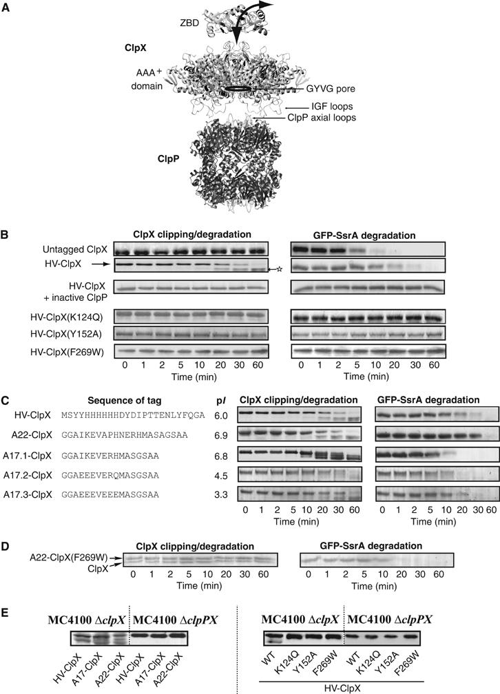
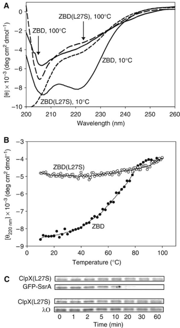
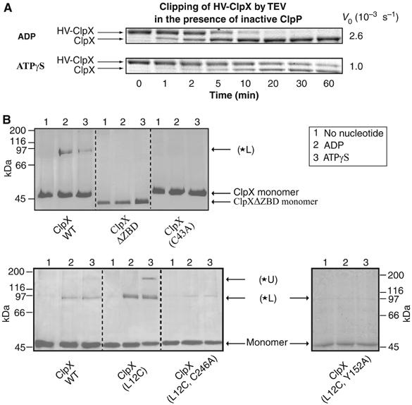
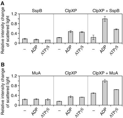
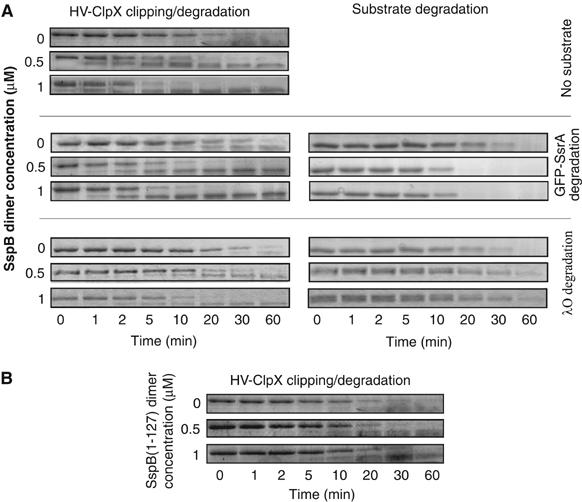
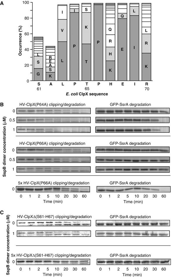
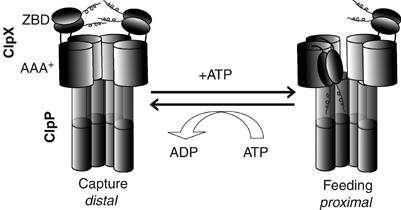
The ClpXP ATPase-protease complex is a major component of the protein quality control machinery in the cell. A ClpX subunit consists of an N-terminal zinc binding domain (ZBD) and a C-terminal AAA+ domain. ClpX oligomerizes into a hexamer with the AAA+ domains forming the base of the hexamer and the ZBDs extending out of the base. Here, we report that ClpX switches between a capture and a feeding conformation. ZBDs in ClpX undergo large nucleotide-dependent block movement towards ClpP and into the AAA+ ring. This motion is modulated by the ClpX cofactor, SspB. Evidence for this movement was initially obtained by the surprising observation that an N-terminal extension on ClpX is clipped by bound ClpP in functional ClpXP complexes. Protease-protection, crosslinking, and light scattering experiments further support these findings.


Figures

Figure 1
Figure 1
Clipping and degradation of N-terminally tagged ClpX by bound ClpP. (A) A model of the ClpXP complex. The structure shown is based on the solved structure of E. coli and Streptococcus pneumoniae ClpP (Wang et al, 1997; Gribun et al, 2005), a hexameric model of the AAA+ domain of E. coli ClpX based on the solved monomeric AAA+ domain of H. pylori ClpX (Kim and Kim, 2003) and the hexameric E. coli HslU structure (Bochtler et al, 2000), and on our proposed trimer-of-dimers model for the ZBD of E. coli ClpX (Donaldson et al, 2003). Note that ClpX oligomers can bind to both ends of the ClpP cylinder. (B) Degradation assays were carried out at 37°C by preincubating ClpP, GFP-SsrA, ATP, and ATP regenerating system, and then adding ClpX or ClpX mutant. Aliquots were removed at different time points and then visualized on SDS–PAGE gels. Mass spectrometry analysis was carried out on the band indicated by a star (refer to the text). (C) Degradation assays were carried out using ClpX with different N-terminal tags. The sequence and pI of the tags used are given. (D) Shown are degradation assays in which ClpXP complexes were preformed in the presence of ATP, then GFP-SsrA and A22-ClpX(F269W) were added to the mixture. A22-ClpX(F269W) does not get clipped, while GFP-SsrA is degraded. (E) MC4100 ΔclpX or ΔclpPX cells expressing different ClpX mutants were grown to midlog phase and then were subjected to Western blot analysis using anti-ClpX antibodies.
Figure 2
Figure 2
Disruption of ZBD structure blocks N-terminal clipping. (A) Circular dichroism scans of ZBD (30 μM) and ZBD(L27S) (111 μM) at 10 and 100°C. (B) Thermal melts of ZBD (30 μM) and ZBD(L27S) (111 μM) monitored by circular dichroism. (C) ClpP-dependent degradation of GFP-SsrA and of λO mediated by ClpX(L27S). Similar results were obtained using HV-ClpX(L27S) (data not shown).
Figure 3
Figure 3
Mapping the nucleotide-dependent conformational changes of ZBD in ClpX oligomer. (A) HV-ClpX was mixed with inactive ClpP(S111A) in the presence of ADP or ATPγS. At time zero, TEV protease was added and the cleavage of the HV-tag from HV-ClpX was monitored by SDS–PAGE analysis as a function of time. V0 refers to the initial rate of disappearance of HV-ClpX. (B) Different ClpX mutants were incubated with mPDM crosslinker in the absence or presence of different nucleotides; subsequently, samples were visualized on 10% SDS–PAGE gels. Similar profiles were obtained in the presence of ClpP.
Figure 4
Figure 4
Light scattering experiments demonstrate the presence of distinct nucleotide-dependent orientations of ZBD in ClpX oligomer. (A, B) Light scattering experiments are shown demonstrating nucleotide-dependent complex formation between ClpXP and SspB or MuA. An increase in the intensity of scattered light indicates the formation of larger particles. Similar results were obtained in the absence of ClpP.
Figure 5
Figure 5
SspB enhances the rate of clipping of HV-ClpX by bound ClpP. (A) Degradation assays were carried out using 0.17 μM HV-ClpX6 and 0.09 μM ClpP14 in the presence of different concentrations of SspB dimer. The concentration of GFP-SsrA or λO was 4 μM (for monomers). (B) Assays were carried out as in (A) in the absence of substrates, however, in the presence of different concentrations of SspB(1–127).
Figure 6
Figure 6
The presence of a conserved region between ZBD and AAA+ domains in ClpX that modulates the rate of clipping of tagged ClpX by bound ClpP. (A) ClpX sequences from 103 different bacteria were aligned using ClustalW. Residue numbering is according to SwissProt E. coli ClpX protein. Bars less than 100% are due to the absence of residues at the corresponding positions in some bacteria. (B, C) The ClpP-dependent degradation of GFP-SsrA mediated by HV-ClpX(P64A), HV-ClpX(P66A), or HV-ClpXΔ(S61-H67) was monitored by SDS–PAGE analysis.
Figure 7
Figure 7
Proposed model of the nucleotide-dependent movement of ZBD in the ClpX oligomer. The ZBDs are proposed to switch from a capture state in which the ZBDs are distal from ClpP and resting on top of the AAA+ ring to a feeding state in which at least one ZBD dimer moves into the AAA+ ring and becomes proximal to ClpP.

Similar articles

Cited by

References

    1. Bochtler M, Hartmann C, Song HK, Bourenkov GP, Bartunik HD, Huber R (2000) The structures of HsIU and the ATP-dependent protease HsIU-HsIV. Nature 403: 800–805 - PubMed
    1. Boeckmann B, Bairoch A, Apweiler R, Blatter MC, Estreicher A, Gasteiger E, Martin MJ, Michoud K, O'Donovan C, Phan I, Pilbout S, Schneider M (2003) The SWISS-PROT protein knowledgebase and its supplement TrEMBL in 2003. Nucleic Acids Res 31: 365–370 - PMC - PubMed
    1. Bolon DN, Grant RA, Baker TA, Sauer RT (2004a) Nucleotide-dependent substrate handoff from the SspB adaptor to the AAA+ ClpXP protease. Mol Cell 16: 343–350 - PubMed
    1. Bolon DN, Wah DA, Hersch GL, Baker TA, Sauer RT (2004b) Bivalent tethering of SspB to ClpXP is required for efficient substrate delivery: a protein-design study. Mol Cell 13: 443–449 - PubMed
    1. Burton RE, Siddiqui SM, Kim YI, Baker TA, Sauer RT (2001) Effects of protein stability and structure on substrate processing by the ClpXP unfolding and degradation machine. EMBO J 20: 3092–3100 - PMC - PubMed

Publication types

MeSH terms

LinkOut - more resources