. 2006 Jul 12;25(13):3144-55.
doi: 10.1038/sj.emboj.7601201. Epub 2006 Jun 29.

X-ray crystal structure of MENT: evidence for functional loop-sheet polymers in chromatin condensation

Affiliations

X-ray crystal structure of MENT: evidence for functional loop-sheet polymers in chromatin condensation

Sheena McGowan et al. EMBO J. .

Abstract

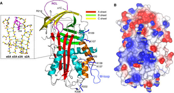
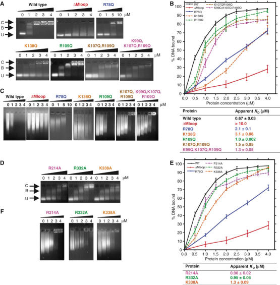
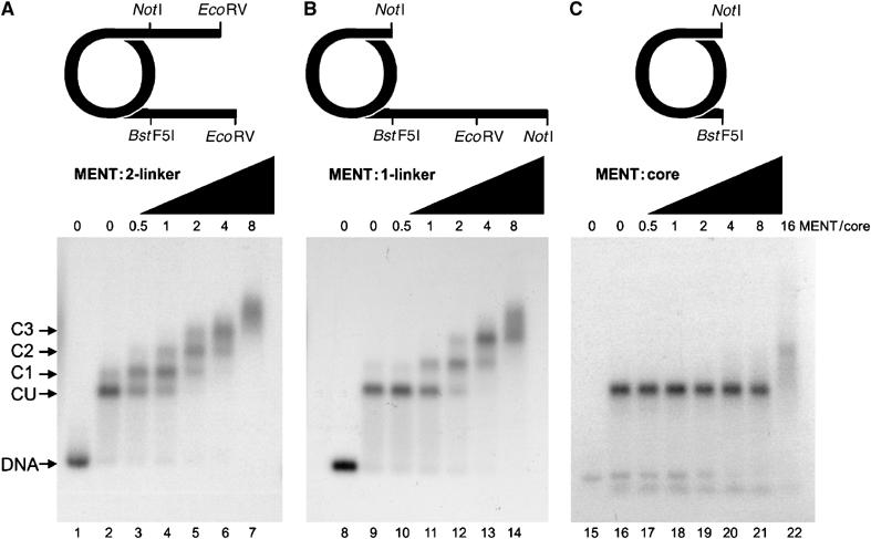
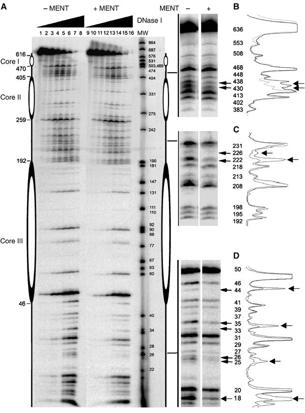
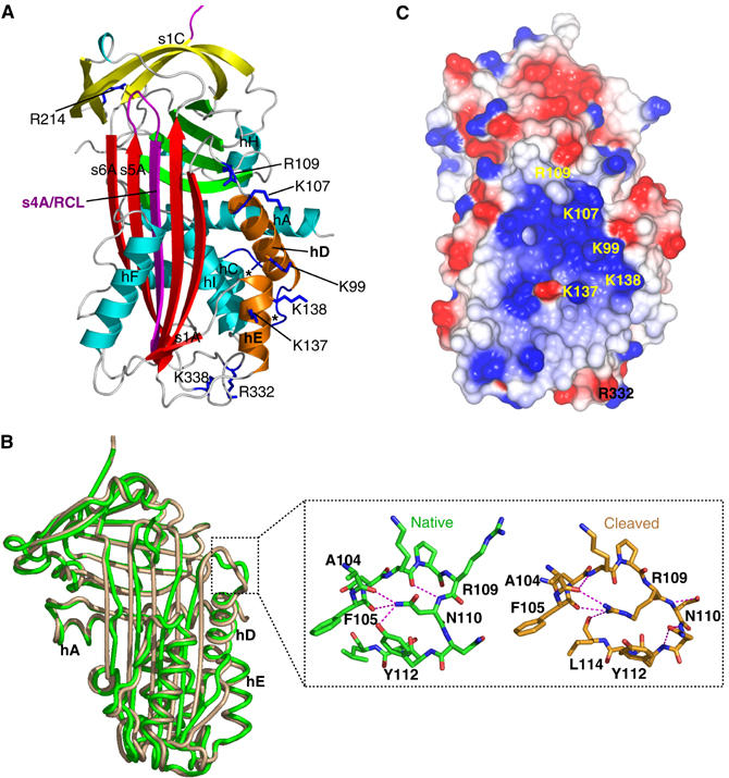
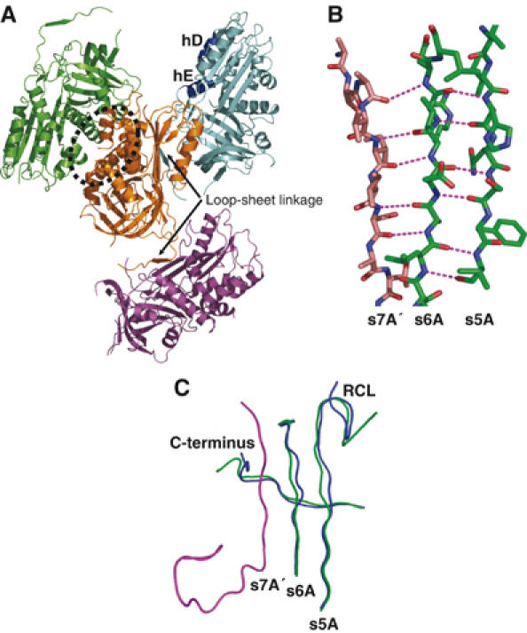
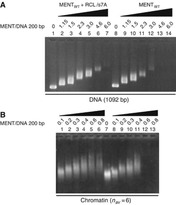
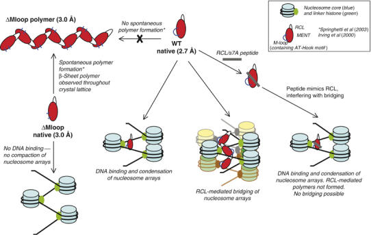
Most serpins are associated with protease inhibition, and their ability to form loop-sheet polymers is linked to conformational disease and the human serpinopathies. Here we describe the structural and functional dissection of how a unique serpin, the non-histone architectural protein, MENT (Myeloid and Erythroid Nuclear Termination stage-specific protein), participates in DNA and chromatin condensation. Our data suggest that MENT contains at least two distinct DNA-binding sites, consistent with its simultaneous binding to the two closely juxtaposed linker DNA segments on a nucleosome. Remarkably, our studies suggest that the reactive centre loop, a region of the MENT molecule essential for chromatin bridging in vivo and in vitro, is able to mediate formation of a loop-sheet oligomer. These data provide mechanistic insight into chromatin compaction by a non-histone architectural protein and suggest how the structural plasticity of serpins has adapted to mediate physiological, rather than pathogenic, loop-sheet linkages.


Figures

Figure 1
Figure 1
(A) Cartoon of native MENTWT showing the A β-sheet (red), B β-sheet (green), C β-sheet (yellow), RCL (purple) (the disordered region is dashed) and helices hA–hI (cyan). Residues K99, K107, R109, R332 and K338 are in dark blue bonds (labelled). The partial insertion of the RCL and the break in s6A (dotted rectangle) of the A-sheet are shown in the inset (hydrogen bonds as green broken lines). (B) CCP4MG (Potterton et al, 2002, 2004) electrostatic potential surface of native MENTWT. The positions of K99, K107, R109, K137, K138, R332 and K338 are shown.
Figure 2
Figure 2
Analysis of purified MENT proteins incubated with DNA and chromatin. Gel mobility shift analysis of D- and E-helix mutants (A) and alanine mutants (D) in comparison to MENTWT (WT) and two negative control proteins, MENTΔMloop (ΔMloop) and a single AT-hook mutant, MENTR78Q (R78Q) (control proteins only shown in panels A and C). Proteins were incubated with DNA and analysed by agarose gel electrophoresis. The final concentration (μM) of purified proteins, as indicated, is shown at the top of each gel panel. Arrows indicate the position of unbound DNA (U), bound DNA/complex (B) and large nucleoprotein species (C). Quantitative analysis of EMSAs of D- and E-helix (B) and alanine mutants (E) is shown graphically. Graphs represent the % of bound DNA at increasing MENT concentrations (as indicated at the bottom of graph). Graphed data of the control proteins MENTWT, MENTΔMloop and MENTR78Q are shown as solid lines and the mutant proteins as dashed lines. Affinities (KD) were calculated from gel electrophoresis experiments as described in Materials and methods and are defined as the concentration (μM) of protein required to bind 50% of DNA. (C, F) Chromatin association assays with increasing concentrations of D- and E-helix mutant (C) and alanine mutant proteins (F). The final concentration (μM) of purified protein, as indicated, added to soluble erythrocyte chromatin (OD260=1.6) is shown at the top of each gel panel.
Figure 3
Figure 3
Native agarose gel electrophoresis of MENT interactions with reconstituted mononucleosomes. Mononucleosomes with two linkers (A, lanes 2–7), one linker (containing the same quantity of free DNA as panel A) (B, lanes 9–14) and no linkers (C, lanes 16–22) were reconstituted from histones and clone 601 DNA (Lowary and Widom, 1998) labelled with [32P]ATP. Lanes 1, 8, and 15 contain free DNA used for the two-linker (lane 1), one-linker (lane 8) and no-linker (lane 15) nucleosomes, respectively. MENT/nucleosome core ratios are indicated at the top of the agarose gel panels. Arrows indicate the position of complexes corresponding to unbound nucleosomes (CU) and discrete MENT–nucleosome complexes (C1–C3).
Figure 4
Figure 4
DNase I footprinting of MENT reconstituted with trinuclesomes. (A) Reconstituted nucleosome trimers labelled with 32P-ATP without MENT (lanes 1–8) or reconstituted with two molecules of MENT per nucleosome (lanes 9–16) were incubated with DNase I for 0 (1,9), 1 (2, 10), 2 (3, 11), 5 (4, 12), 10 (5, 13), 20 (6, 14), 30 (7, 15) and 40 (8, 16) min and analysed on a 6% polyacrylamide/urea gel. The molecular size markers (lane MW) represent 32P-labelled DNA of pUC19 (GenBank accession no. L09137) and pEGFP-C3 (GenBank accession no. U57607) simultaneously digested with MspI restriction enzyme. (B–D) Magnified regions of the gel where MENT interferes with DNase I digestion. The molecular sizes of the DNA bands (bp) are indicated. On the left of panels B–D quantitation of DNase I footprints is shown. The extent of protection of each region in trinucleosome from DNase I without (heavy line) and with MENT (light line) was quantitated using ImageJ.
Figure 5
Figure 5
(A) The structure of cleaved MENTWT labelled as in Figure 1A. The termini of the M-loop (between hC and hD) are indicated by *. (B) Superposition of native (green) and cleaved (brown) MENTWT. The change in conformation at the top of the D-helix is indicated by a dotted square and shown in the inset. Hydrogen bonds are shown by dashed lines and R109, N110, Y112, F105 and A104 are labelled. (C) CCP4MG (Potterton et al, 2002, 2004) electrostatic potential surface of cleaved MENTWT, coloured as in Figure 1B.
Figure 6
Figure 6
(A) Structure of the native MENTΔMloop tetramer in the asymmetric unit. Each monomer is coloured differently; the orange and green monomers form a ‘back to back' dimer (indicated by a dotted oval); the orange molecule forms a loop–sheet linkage (arrow) to the cyan molecule and a loop–sheet linkage to the magenta molecule. hD and hE are labelled. (B) Loop–sheet hydrogen bonds formed by the RCL (brown) of one molecule with the s6A of an adjoining molecule (green). Hydrogen bonds are shown as magenta broken lines. (C) Comparison of native MENTWT (blue) and MENTΔMloop (green) reveals conformational change in the C-terminus (labelled) and s5A/s6A of the A β-sheet of MENTΔMloop in response to the interaction with the RCL of a neighbouring molecule (magenta). Note also the different trajectory of the RCL (labelled).
Figure 7
Figure 7
An RCL/s7A peptide interferes with MENT-induced self-association of naked DNA and chromatin. (A) Agarose gel electrophoresis of DNA (1092 bp long) reconstituted with control MENT (lanes 8–14) and MENT reconstituted with an RCL/s7A peptide (1–7). (B) Agarose gel electrophoresis of soluble erythrocyte chromatin reconstituted with control MENT (lanes 8–13) and MENT reconstituted with RCL/s7A peptide (lanes 1–6). The MENT/DNA ratios (molecule/200 bp) were as indicated at the top of the lane markers.
Figure 8
Figure 8
Model for MENT function. Studies on mutants of MENT (red ovals) reveal that deletion of the M-loop (and hence the AT-hook motif) (blue lines) results in the loss of DNA binding and therefore MENTΔMloop (ΔMloop) is unable to condense nucleosome arrays. Linker histone H5 is depicted with the nucleosome as MENT and linker histone meet together in chicken blood cells and act synergistically (Springhetti et al, 2003). Deletion of the M-loop in MENT leads to spontaneous polymerization of the molecule (Springhetti et al, 2003) demonstrating that the M-loop in the absence of DNA prevents MENT polymerization. In contrast, the MENTWT (WT) is unable to spontaneously form polymers; however, we have previously shown that oligomeric MENTWT structures form in the presence of DNA (Springhetti et al, 2003). It is suggested that MENTWT, utilizing dynamic s7A–s6A interactions to form oligomers, can bridge distinct nucleosome arrays. Consistent with this model, an RCL/s7A peptide (grey line) is able to disrupt the edge-strand interactions and partially block MENT function.

References

    1. Allain FH, Yen YM, Masse JE, Schultze P, Dieckmann T, Johnson RC, Feigon J (1999) Solution structure of the HMG protein NHP6A and its interaction with DNA reveals the structural determinants for non-sequence-specific binding. EMBO J 18: 2563–2579 - PMC - PubMed
    1. Aravind L, Landsman D (1998) AT-hook motifs identified in a wide variety of DNA-binding proteins. Nucleic Acids Res 26: 4413–4421 - PMC - PubMed
    1. Baglin TP, Carrell RW, Church FC, Esmon CT, Huntington JA (2002) Crystal structures of native and thrombin-complexed heparin cofactor II reveal a multistep allosteric mechanism. Proc Natl Acad Sci USA 99: 11079–11084 - PMC - PubMed
    1. Bulynko YA, Hsing LC, Mason RW, Tremethick D, Grigoryev SA (2006) Cathepsin L stabilizes histone modification landscape on Y chromosome and pericentromeric heterochromatin. Mol Cell Biol 26: 4172–4184 - PMC - PubMed
    1. Carrell RW, Lomas DA (1997) Conformational disease. Lancet 350: 134–138 - PubMed

Publication types

LinkOut - more resources