. 2006 Jul 12;25(13):3078-88.
doi: 10.1038/sj.emboj.7601198. Epub 2006 Jun 29.

FGF-2 protects small cell lung cancer cells from apoptosis through a complex involving PKCepsilon, B-Raf and S6K2

Affiliations

FGF-2 protects small cell lung cancer cells from apoptosis through a complex involving PKCepsilon, B-Raf and S6K2

Olivier E Pardo et al. EMBO J. .

Abstract

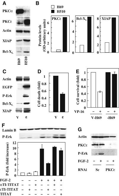
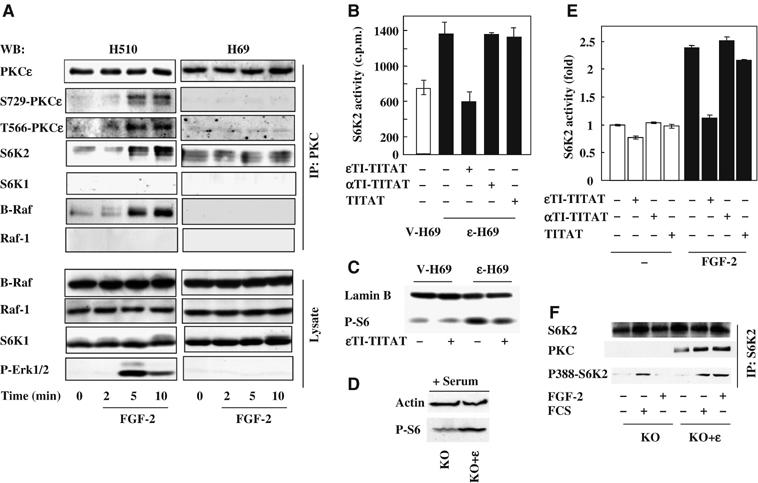
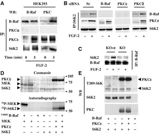
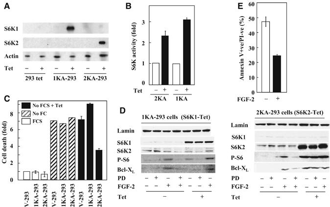
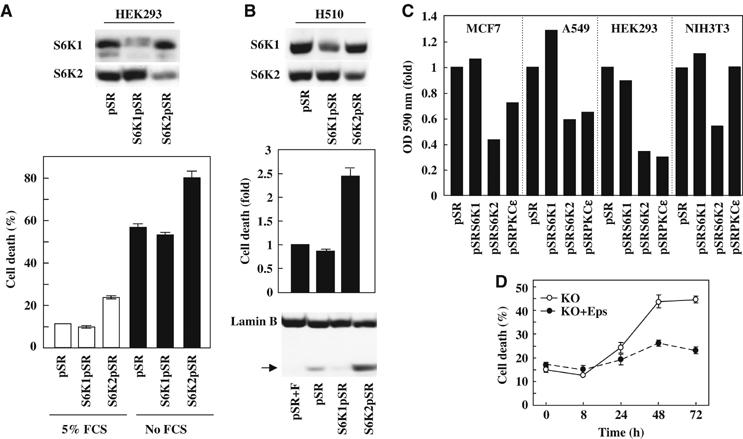
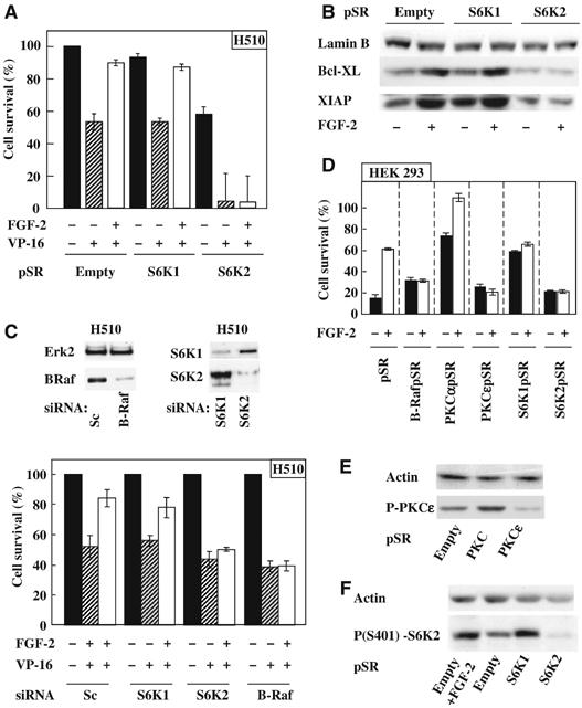
Patients with small cell lung cancer (SCLC) die because of chemoresistance. Fibroblast growth factor-2 (FGF-2) increases the expression of antiapoptotic proteins, XIAP and Bcl-X(L), and triggers chemoresistance in SCLC cells. Here we show that these effects are mediated through the formation of a specific multiprotein complex comprising B-Raf, PKCepsilon and S6K2. S6K1, Raf-1 and other PKC isoforms do not form similar complexes. RNAi-mediated downregulation of B-Raf, PKCepsilon or S6K2 abolishes FGF-2-mediated survival. In contrast, overexpression of PKCepsilon increases XIAP and Bcl-X(L) levels and chemoresistance in SCLC cells. In a tetracycline-inducible system, increased S6K2 kinase activity triggers upregulation of XIAP, Bcl-X(L) and prosurvival effects. However, increased S6K1 kinase activity has no such effect. Thus, S6K2 but not S6K1 mediates prosurvival/chemoresistance signalling.


Figures

Figure 1
Figure 1
PKCɛ levels correlate with XIAP and Bcl-XL expression and Erk phosphorylation in SCLC cells. (A) H69 and H510 cell lysates were Western blotted for the expression of PKCα, PKCɛ, XIAP, Bcl-XL and actin. (B) Representative blots from (A) were quantified by optical densitometry normalised for actin. (C) H69 cells transfected with empty (V) or a wt-PKCɛ-GFP-expressing vector (ɛ) were analysed for phospho-ERK, XIAP and Bcl-XL levels. (D) Baseline level cell death in V-H69 and ɛ-H69 cells growing in 10% FCS was determined by flow cytometry using Annexin V staining. (E) ɛ-H69 and V-H69 cells in SFM were treated with or without 0.1 μM etoposide (VP-16) and cell numbers determined 96 h later. Conditions were performed in quadruplicates and the average cell number±s.e.m. represented as fold over untreated. (F) H510 cells in SFM were treated with or without 40 μM ɛTI-TITAT, αTI-TITAT or TITAT for 4 h before stimulation for 5 min with or without FGF-2 (0.1 ng/ml). Cell lysates were Western blotted for biphospho-ERK. (F-lower panel) Results from three independent experiments were analysed by optical densitometry and represented as average±s.e.m. fold increase over control. (G) H510 cells transfected with PKCɛ or scrambled (sc) siRNA were stimulated with or without FGF-2. Lysates were analysed for PKCɛ levels and Erk phosphorylation. (A, C, F and G) Lamin B and actin immunodetection were used as loading controls.
Figure 2
Figure 2
PKCɛ forms a multiprotein complex with B-Raf and S6K2 in H510 cells following FGF-2 and regulates S6K2 activity. (A) H510 and H69 cells in SFM were treated with FGF-2 for the times indicated. Cell lysates were subjected to immunoprecipitation with a PKCɛ antibody before Western blotting (WB) for the molecules indicated. (A, lower panel) Total cell lysate was Western blotted as indicated. (B, E) S6K2 was immunoprecipitated from V-H69 and ɛ-H69 (B) or H510 cells (E) following 4 h treatment with or without ɛTI-TITAT, αTI-TITAT and TITAT. Immunoprecipitates were subjected to in vitro kinase assays with S6-peptide as a substrate and the results shown are average c.p.m.±s.e.m. from triplicates of a representative experiment. (C) The phosphorylation of the endogenous S6 protein from V-H69 and ɛ-H69 cells in SFM treated with or without ɛTI-TITAT was determined using a phospho-S6 antibody. (D, F) PKCɛ KO MEFs re-expressing (KO+ɛ) or not (KO) PKCɛ were (D) grown in 10% FCS and analysed for phospho-S6 levels or (F) stimulated with or without FGF-2 and FCS before S6K2 immunoprecipitation and Western blotted as indicated. (C and D) Lamin B and actin immunodetection were used as a loading control. (A–F) Results shown are representative of at least three independent experiments.
Figure 3
Figure 3
PKCɛ is required for B-Raf association with S6K2. (A) HEK293 cells were stimulated with FGF-2 and immunoprecipitates (IP) for the molecules indicated analysed by Western blotting (WB) for either B-Raf or PKCɛ. (B) HEK293 cells transfected with siRNAi for B-Raf, PKCα, PKCɛ, PKCδ or scramble control (sc) were stimulated with FGF-2 and S6K2 immunoprecipitates analysed by WB for B-Raf and PKCɛ. (C) MEFs from PKCɛ KO mice, re-expressing (KO+ɛ) or not (KO) PKCɛ were stimulated with or without FGF-2. B-Raf immunoprecipitates were analysed by WB for S6K2. (D, E) Recombinant PKCɛ, V600EB-Raf and S6K2 proteins were combined as indicated and subjected to in vitro kinase assay with 32P-γATP (D) or cold ATP (E). Recombinant GST-MEK was used as a positive control for V600EB-Raf activity. Samples were run on SDS–PAGE, Coomassie-stained (D, upper panel) or transferred to nitrocellulose (E), then exposed to an X-Ray film (D, lower panel) or subjected to WB for the molecules indicated (E). Results shown are representative of a minimum of three independent experiments.
Figure 4
Figure 4
Specific induction of S6K2 kinase activity in HEK293 cells increase cell viability and upregulates Bcl-XL. HEK293-Tet clones transfected with an inducible vector for kinase active S6K1 (1KA), S6K2 (2KA) or empty vector were treated with tetracycline for 6 h before (A) Western blotting (WB) as indicated or (B) S6K1 or 2 kinase assay using an S6 peptide as substrate. (C) V-, 1KA- and 2KA-293 cells were incubated in the absence of tetracycline with (white bars) or without (hatched bars) serum for 18 h or with tetracycline in the absence of serum (black bars) and the proportion of apoptotic cells determined using annexin V staining. (D) Cell lysates from 2KA and 1KA-293 cells treated with or without tetracycline, FGF-2 and PD098059 were analysed for Bcl-XL expression and S6 phosphorylation. (E) HEK293 cells incubated with or without FGF-2 (4 h) before serum deprivation (18 h) were analysed by annexin V staining. (A, D) Actin and lamin B immunodetections were used as a loading control. (B, C, E) Results represent the average of triplicates±s.e.m. (A–E) Results are representative of at least three independent experiments.
Figure 5
Figure 5
S6K2 and PKCɛ downregulation decreases cell viability and clonogenic cell growth in mammalian cells. (A) HEK293 cells were transfected with empty-vector (pSR), or pSR encoding for S6K1 (S6K1pSR) or S6K2 (S6K2pSR) RNAi sequences. Cells were grown in 5% FCS or serum-free medium for 18 h and cell viability was determined by trypan blue exclusion. (B) Lysates from H510 cells expresssing pSR, S6K1pSR or S6K2pSR were Western blotted as indicated (upper panel). The baseline cell death in 10% FCS was determined by Annexin V staining (middle panel). Lamin B cleavage was used as readout for caspase 3 activity (lower panel). pSR cells treated with FGF-2 (pSR+F) were used as negative control. (C) MCF-7, A549, HEK293 and NIH3T3 cells were transfected with the indicated pSR shRNAi constructs and grown in 5% FCS for 10 days. The OD of crystal violet-stained colonies was determined at 590 nm. For each cell line, results were normalised for absorbance found in pSR empty-vector cells. (D) KO or KO+ɛ MEFs were plated in the absence of FCS for the times indicated and the proportion of Trypan blue-positive cells determined. For (A, lower panel) and (B, middle panel) results represent the average of triplicates±s.e.m. For (A–C), the results shown are representative of at least three independent experiments.
Figure 6
Figure 6
S6K2, but not S6K1, downregulation prevents FGF-2-mediated survival of H510 and HEK293 cells. (A–C) H510 cells were subjected to downregulation of the indicated proteins either by pSR RNAi retroviral vectors (A, B) or oligonucleotide RNAi (C). (A) Cells were preincubated with or without FGF-2 before etoposide treatment (VP-16). (B) Lysate from H510 cells infected with the indicated vectors and treated as shown were Western blotted for XIAP and Bcl-XL. (C) H510 cells treated with oligonucleotide RNAis were (upper panel) lysed and Western blotted as indicated or (lower panel) treated as described in (A). (D–F) HEK293 cells were subjected to downregulation of the indicated proteins by transfection of RNAi-encoding pSR vectors. (D) Cells were preincubated with or without FGF-2 before serum depletion. (A and D) Survival was determined by trypan blue exclusion. (E) HEK293 cells transfected as indicated were Western blotted for phosphoS729-PKCɛ (P-PKCɛ). (F) Transfected HEK293 cells incubated with or without FGF-2 were Western blotted for phosphoS412-S6K2. (A, C, D) Results are averages±s.e.m. of quadruplicates and (A, C) normalised to pSR (A) or Sc (C). (B, E, F) LaminB or actin immunodetection were used as loading control. (A–F) Results are representative of at least three independent experiments.

Similar articles

Cited by

References

    1. Calipel A, Lefevre G, Pouponnot C, Mouriaux F, Eychene A, Mascarelli F (2003) Mutation of B-Raf in human choroidal melanoma cells mediates cell proliferation and transformation through the MEK/ERK pathway. J Biol Chem 278: 42409–42418 - PubMed
    1. Cenni V, Doppler H, Sonnenburg ED, Maraldi N, Newton AC, Toker A (2002) Regulation of novel protein kinase C epsilon by phosphorylation. Biochem J 363: 537–545 - PMC - PubMed
    1. Cheng JJ, Wung BS, Chao YJ, Wang DL (2001) Sequential activation of protein kinase C (PKC)-alpha and PKC-epsilon contributes to sustained Raf/ERK1/2 activation in endothelial cells under mechanical strain. J Biol Chem 276: 31368–31375 - PubMed
    1. Davies SP, Reddy H, Caivano M, Cohen P (2000) Specificity and mechanism of action of some commonly used protein kinase inhibitors. Biochem J 351: 95–105 - PMC - PubMed
    1. Dillon TJ, Karpitski V, Wetzel SA, Parker DC, Shaw AS, Stork PJ (2003) Ectopic B-Raf expression enhances extracellular signal-regulated kinase (ERK) signaling in T cells and prevents antigen-presenting cell-induced anergy. J Biol Chem 278: 35940–35949 - PubMed

Publication types

MeSH terms

Substances