

Simulations of congenital septal defect closure and reactivity testing in patient-specific models of the pediatric pulmonary vasculature: A 3D numerical study with fluid-structure interaction
- PMID: 16813447
- PMCID: PMC4050970
- DOI: 10.1115/1.2206202
Simulations of congenital septal defect closure and reactivity testing in patient-specific models of the pediatric pulmonary vasculature: A 3D numerical study with fluid-structure interaction
Abstract
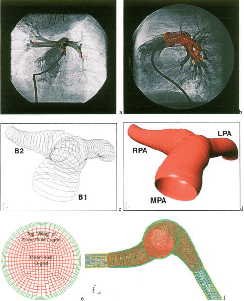
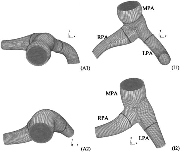
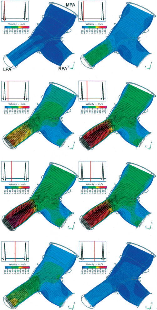
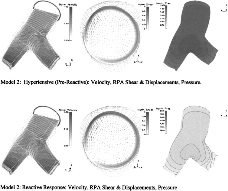
Clinical imaging methods are highly effective in the diagnosis of vascular pathologies, but they do not currently provide enough detail to shed light on the cause or progression of such diseases, and would be hard pressed to foresee the outcome of surgical interventions. Greater detail of and prediction capabilities for vascular hemodynamics and arterial mechanics are obtained here through the coupling of clinical imaging methods with computational techniques. Three-dimensional, patient-specific geometric reconstructions of the pediatric proximal pulmonary vasculature were obtained from x-ray angiogram images and meshed for use with commercial computational software. Two such models from hypertensive patients, one with multiple septal defects, the other who underwent vascular reactivity testing, were each completed with two sets of suitable fluid and structural initial and boundary conditions and used to obtain detailed transient simulations of artery wall motion and hemodynamics in both clinically measured and predicted configurations. The simulation of septal defect closure, in which input flow and proximal vascular stiffness were decreased, exhibited substantial decreases in proximal velocity, wall shear stress (WSS), and pressure in the post-op state. The simulation of vascular reactivity, in which distal vascular resistance and proximal vascular stiffness were decreased, displayed negligible changes in velocity and WSS but a significant drop in proximal pressure in the reactive state. This new patient-specific technique provides much greater detail regarding the function of the pulmonary circuit than can be obtained with current medical imaging methods alone, and holds promise for enabling surgical planning.
Figures

References
-
- Malek AM, Alper SL, Izumo S. Hemodynamic Shear Stress and Its Role in Artherosclerosis. JAMA, J Am Med Assoc. 1999;282:2035–2042. - PubMed
-
- Topper JN, Cai J, Falb D, Gimbrone MA. Identification of Vascular Endothelial Genes Differentially Responsive to Fluid Mechanical Stimuli: Cycoloxgenase-2, Manganese Superoxide Dismutase, and Endothelial Cell Nitric Oxide Synthase are Selectively Up-Regulated by Steady Laminar Shear Stress. Proc Natl Acad Sci USA. 1996;93:10417–10422. - PMC - PubMed
-
- Ziegler T, Bouzourène K, Harrison VJ, Brunner HR, Hayoz D. Influence of Oscillatory and Unidirectional Flow Environments on the Expression of Endothelin and Nitric Oxide Synthase in Cultured Endothelial Cells. Arterioscler, Thromb, Vasc Biol. 1998;18:686–692. - PubMed
-
- Weinberg CE, Hertzberg JR, Ivy DD, Kirby KS, Chan KC, Valdes-Cruz L, Shandas R. Extraction of Pulmonary Vascular Compliance, Pulmonary Vascular Resistance, and Right Ventricular Work From Single-Pressure and Doppler Flow Measurements in Children With Pulmonary Hypertension: A New Method for Evaluating Reactivity—In Vitro and Clinical Studies. Circulation. 2005;110(17):2609–2617. - PubMed
Publication types
MeSH terms
Grants and funding
LinkOut - more resources
Full Text Sources
Medical
Research Materials
Miscellaneous
