Antimicrobial barrier of an in vitro oral epithelial model
- PMID: 16815238
- PMCID: PMC2376809
- DOI: 10.1016/j.archoralbio.2006.05.007
Antimicrobial barrier of an in vitro oral epithelial model
Abstract
Objective: Oral epithelia function as a microbial barrier and are actively involved in recognizing and responding to bacteria. Our goal was to examine a tissue engineered model of buccal epithelium for its response to oral bacteria and proinflammatory cytokines and compare the tissue responses with those of a submerged monolayer cell culture.
Design: The tissue model was characterized for keratin and beta-defensin expression. Altered expression of beta-defensins was evaluated by RT-PCR after exposure of the apical surface to oral bacteria and after exposure to TNF-alpha in the medium. These were compared to the response in traditional submerged oral epithelial cell culture.
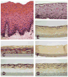
Results: The buccal model showed expression of differentiation specific keratin 13, hBD1 and hBD3 in the upper half of the tissue; hBD2 was not detected. hBD1 mRNA was constitutively expressed, while hBD2 mRNA increased 2-fold after exposure of the apical surface to three oral bacteria tested and hBD3 mRNA increased in response to the non-pathogenic bacteria tested. In contrast, hBD2 mRNA increased 3-600-fold in response to bacteria in submerged cell culture. HBD2 mRNA increased over 100-fold in response to TNF-alpha in the tissue model and 50-fold in submerged cell culture. Thus, the tissue model is capable of upregulating hBD2, however, the minimal response to bacteria suggests that the tissue has an effective antimicrobial barrier due to its morphology, differentiation, and defensin expression.
Conclusions: The oral mucosal model is differentiated, expresses hBD1 and hBD3, and has an intact surface with a functional antimicrobial barrier.
Figures


Similar articles
-
Differential and coordinated expression of defensins and cytokines by gingival epithelial cells and dendritic cells in response to oral bacteria.BMC Immunol. 2010 Jul 9;11:37. doi: 10.1186/1471-2172-11-37. BMC Immunol. 2010. PMID: 20618959 Free PMC article.
-
Modulation of host antimicrobial peptide (beta-defensins 1 and 2) expression during gastritis.Gut. 2002 Sep;51(3):356-61. doi: 10.1136/gut.51.3.356. Gut. 2002. PMID: 12171956 Free PMC article.
-
Inducible expression of human beta-defensin 2 by Fusobacterium nucleatum in oral epithelial cells: multiple signaling pathways and role of commensal bacteria in innate immunity and the epithelial barrier.Infect Immun. 2000 May;68(5):2907-15. doi: 10.1128/IAI.68.5.2907-2915.2000. Infect Immun. 2000. PMID: 10768988 Free PMC article.
-
Role of Toll-like receptor 2 in mediating the production of cytokines and human beta-defensins in oral mucosal epithelial cell response to Leptospiral infection.Asian Pac J Allergy Immunol. 2019 Dec;37(4):198-204. doi: 10.12932/AP-100518-0308. Asian Pac J Allergy Immunol. 2019. PMID: 30118246
-
Differential expression of human beta defensin 2 and 3 in gastric mucosa of Helicobacter pylori-infected individuals.Helicobacter. 2013 Feb;18(1):6-12. doi: 10.1111/hel.12000. Epub 2012 Aug 28. Helicobacter. 2013. PMID: 23067102 Review.
Cited by
-
A 3D Model of Human Buccal Mucosa for Compatibility Testing of Mouth Rinsing Solutions.Pharmaceutics. 2023 Feb 21;15(3):721. doi: 10.3390/pharmaceutics15030721. Pharmaceutics. 2023. PMID: 36986582 Free PMC article.
-
Streptococcus mutans strains recovered from caries-active or caries-free individuals differ in sensitivity to host antimicrobial peptides.Mol Oral Microbiol. 2011 Jun;26(3):187-99. doi: 10.1111/j.2041-1014.2011.00607.x. Epub 2011 Jan 31. Mol Oral Microbiol. 2011. PMID: 21545696 Free PMC article.
-
Three-Dimensional In Vitro Oral Mucosa Models of Fungal and Bacterial Infections.Tissue Eng Part B Rev. 2020 Oct;26(5):443-460. doi: 10.1089/ten.TEB.2020.0016. Epub 2020 Apr 7. Tissue Eng Part B Rev. 2020. PMID: 32131719 Free PMC article. Review.
-
Caspase-12 silencing attenuates inhibitory effects of cigarette smoke extract on NOD1 signaling and hBDs expression in human oral mucosal epithelial cells.PLoS One. 2014 Dec 11;9(12):e115053. doi: 10.1371/journal.pone.0115053. eCollection 2014. PLoS One. 2014. PMID: 25503380 Free PMC article.
-
Engineered Tissue Models to Decode Host-Microbiota Interactions.Adv Sci (Weinh). 2025 Jun;12(23):e2417687. doi: 10.1002/advs.202417687. Epub 2025 May 14. Adv Sci (Weinh). 2025. PMID: 40364768 Free PMC article. Review.
References
-
- Ali RS, Falconer A, Ikram M, Bissett CE, Cerio R, Quinn AG. Expression of the peptide antibiotics human beta defensin-1 and human beta defensin-2 in normal human skin. J Invest Dermatol. 2001;117(1):106–111. - PubMed
-
- Chadebech P, Goidin D, Jacquet C, Viac J, Schmitt D, Staquet MJ. Use of human reconstructed epidermis to analyze the regulation of beta-defensin hBD-1, hBD-2, and hBD-3 expression in response to LPS. Cell Biol Toxicol. 2003;19(5):313–324. - PubMed
-
- Chronnell CM, Ghali LR, Ali RS, Quinn AG, Holland DB, Bull JJ, Cunliffe WJ, McKay IA, Philpott MP, Muller-Rover S. Human beta defensin-1 and -2 expression in human pilosebaceous units: upregulation in acne vulgaris lesions. J Invest Dermatol. 2001;117(5):1120–1125. - PubMed
-
- Chung WO, Hansen SR, Rao D, Dale BA. Protease -activated signaling of hBD-2 expression in oral epithelial cells. J Immunol. 2004;173:5165–5170. - PubMed
Publication types
MeSH terms
Substances
Grants and funding
LinkOut - more resources
Full Text Sources
Molecular Biology Databases
Research Materials

